ELECTROCARDIOGRAPHY
- An electrocardiogram (ECG) is a simple, non-invasive test that records the electrical activity of the heart.
- The electrical potential generated by the cardiac muscle can be detected on the surface of the body and a record of such potential is called electrocardiogram (ECG or EKG). The machine used for recording this potential is called an electrocardiograph.
- The purposes of ECG test:
- An ECG can help diagnose certain heart conditions, including abnormal heart rhythms and coronary heart disease (heart attack and angina).
- A doctor may recommend an ECG if you are experiencing symptoms like chest pain, breathlessness, dizziness, fainting or a feeling of your heart racing, fluttering, thumping or pounding in your chest (palpitations).
- An ECG can also help monitor how treatments for a heart condition, like medicines or implantable cardiac devices, are working.
1. Instruments
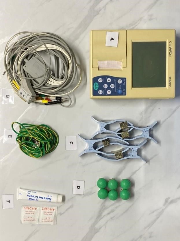
A: A standard 12-lead electrocardiograph and ECG paper
B: Patient’s cable
C: Limb electrodes
D: Chest electrodes
E: Potential equalization cable
F: Electrode gel and alcohol swab
2. How to perform electrocardiography
- Apply electrode gel on the electrodes before placement.
- Limb leads - place the electrodes on the right forearm, left forearm, right leg and left leg.
- Chest leads - place the electrodes in the appropriate places for V1 to V6 and connect them to the six wires.
- When all the electrodes are properly positioned and connected to the electrocardiograph, recording can be started. Machines will record all 12 leads automatically when the start button is pressed.
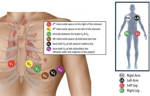
The chest leads are:-
1. V1 (+ ve electrode on 4th. RICS, sternal margin)
2. V2 (+ ve electrode on 4th. LICS, sternal margin)
3. V3 (+ ve electrode midway between V2 and V4)
4. V4 (+ ve electrode on 5th. LICS - mid clavicular line)
5. V5 (+ ve electrode on 5th. LICS, anterior axillary line)
6. V6 (+ ve electrode on 5th. LICS - mid axillary line)
(R = right, L = left and ICS = Intercostal space).
You can also watch the video from this link:
https://www.youtube.com/watch?v=1k4B_fIX_t0
DISCLAIMER: This video is for educational purpose only.
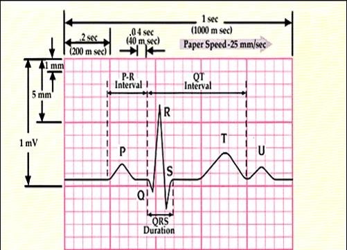
3. Identify the various waves and intervals.
4. Calculating the heart rate
Paper speed = 25mm/sec
1. If the QRS complexes are at regular interval;
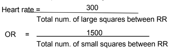
2. If the QRS complexes are at irregular interval;
Calculate how many QRS complexes in 6 seconds (30 big boxes) = A
Heart rate = A x 10