Eye: M766/90
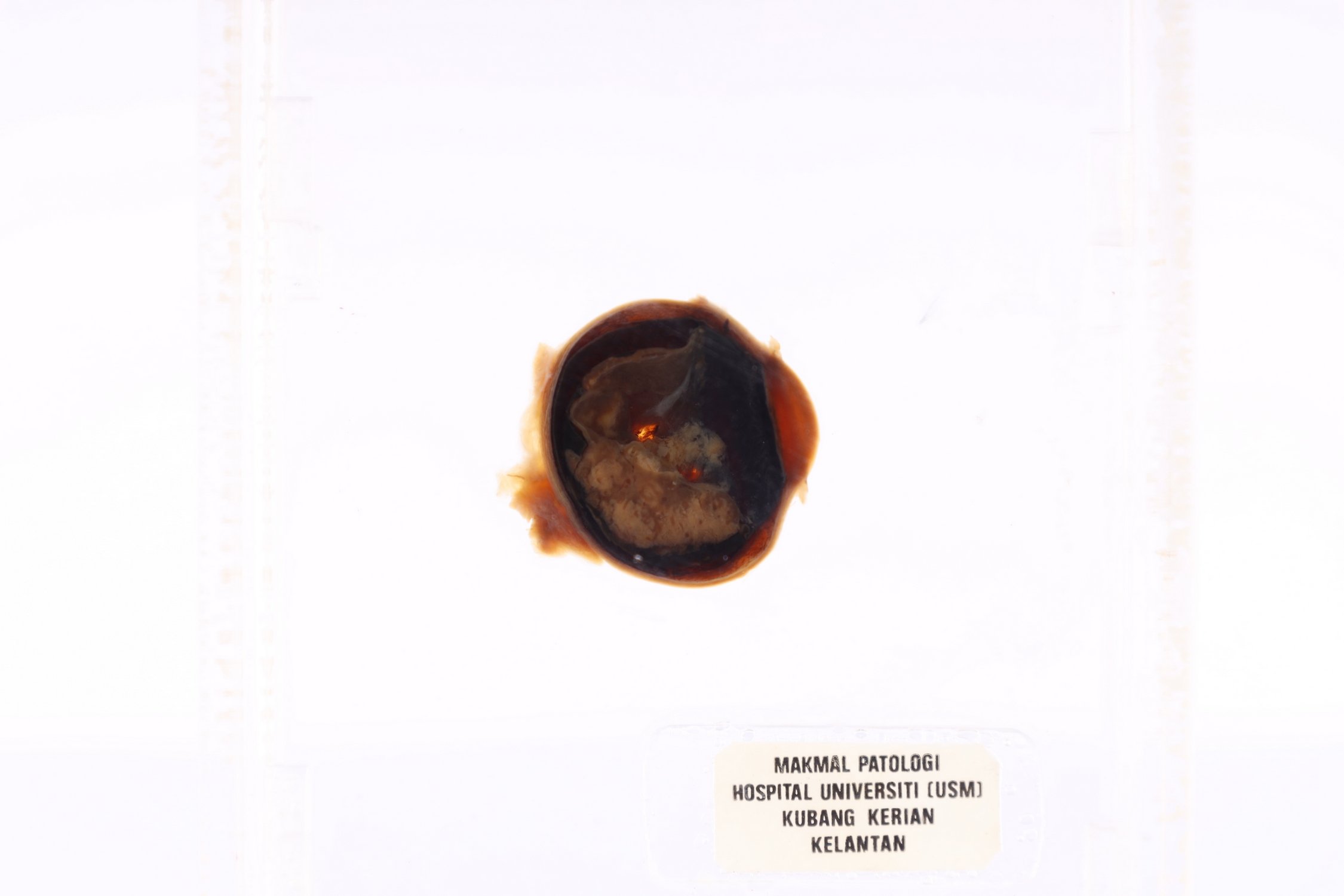
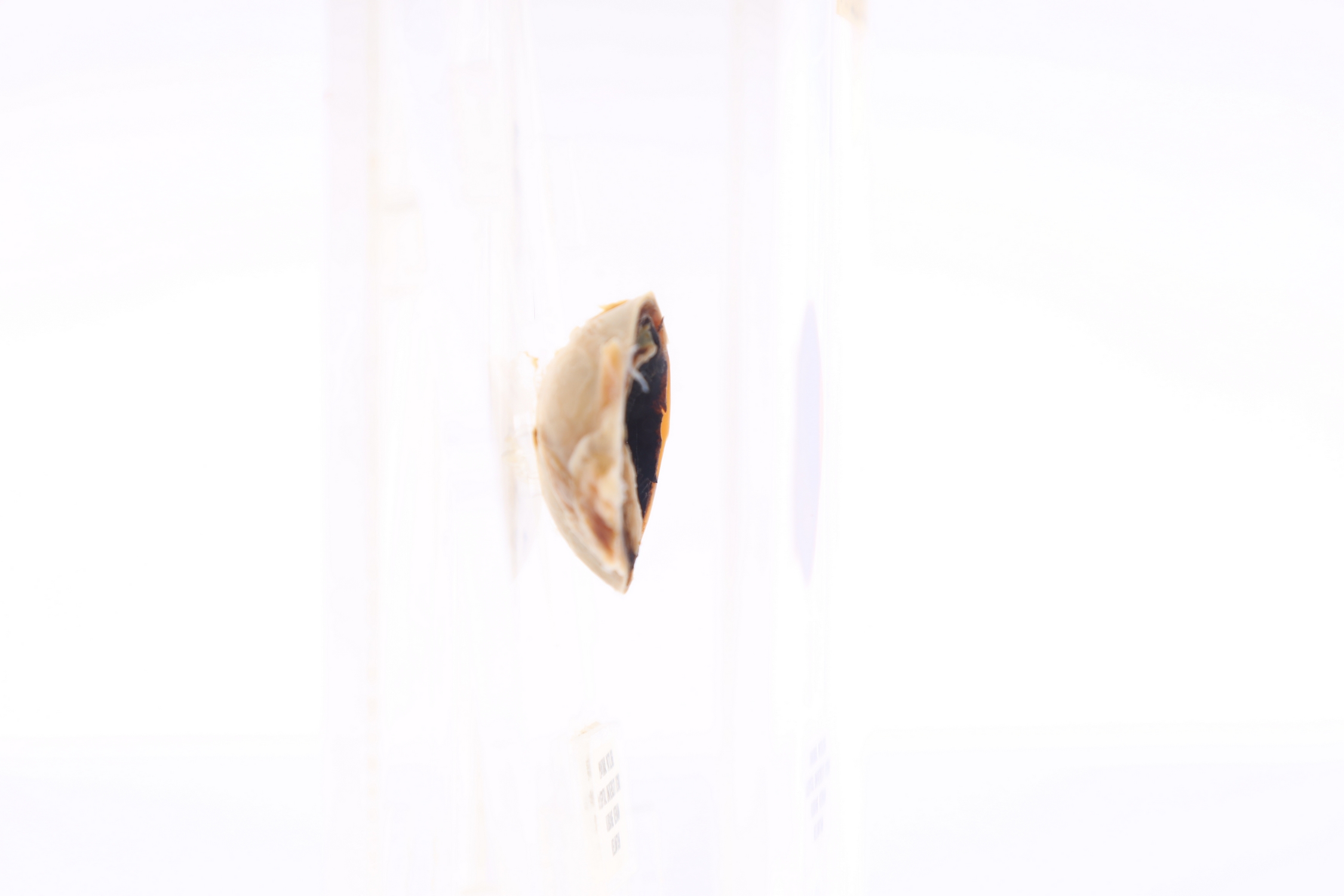
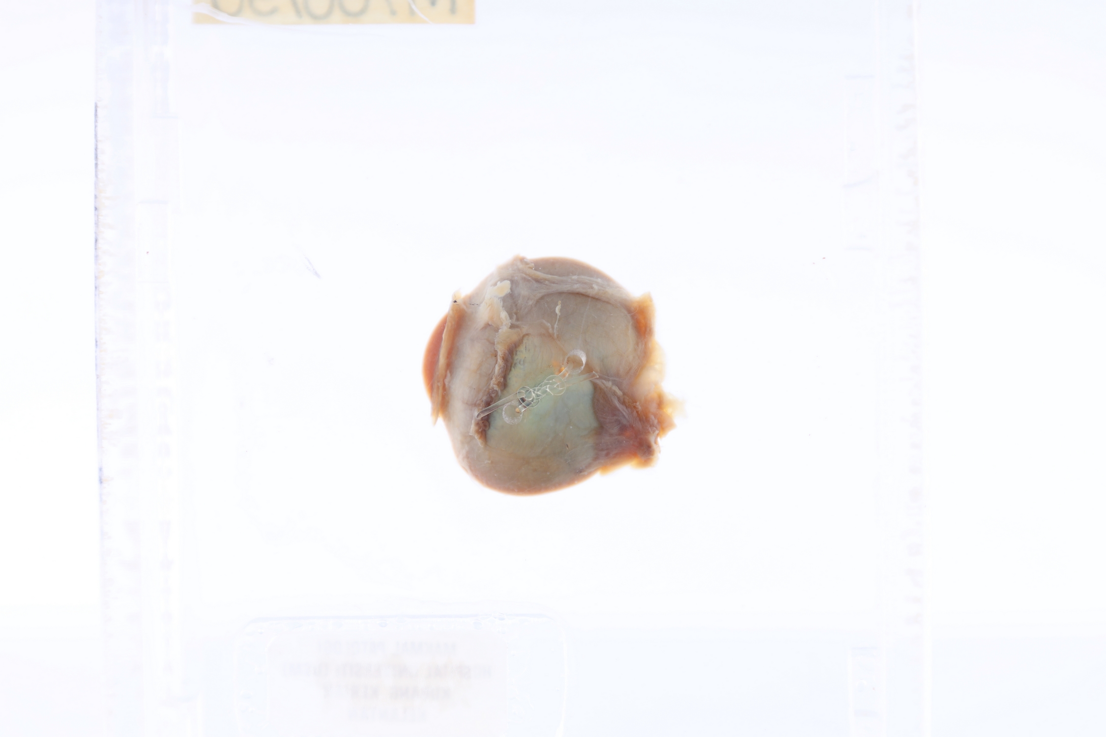
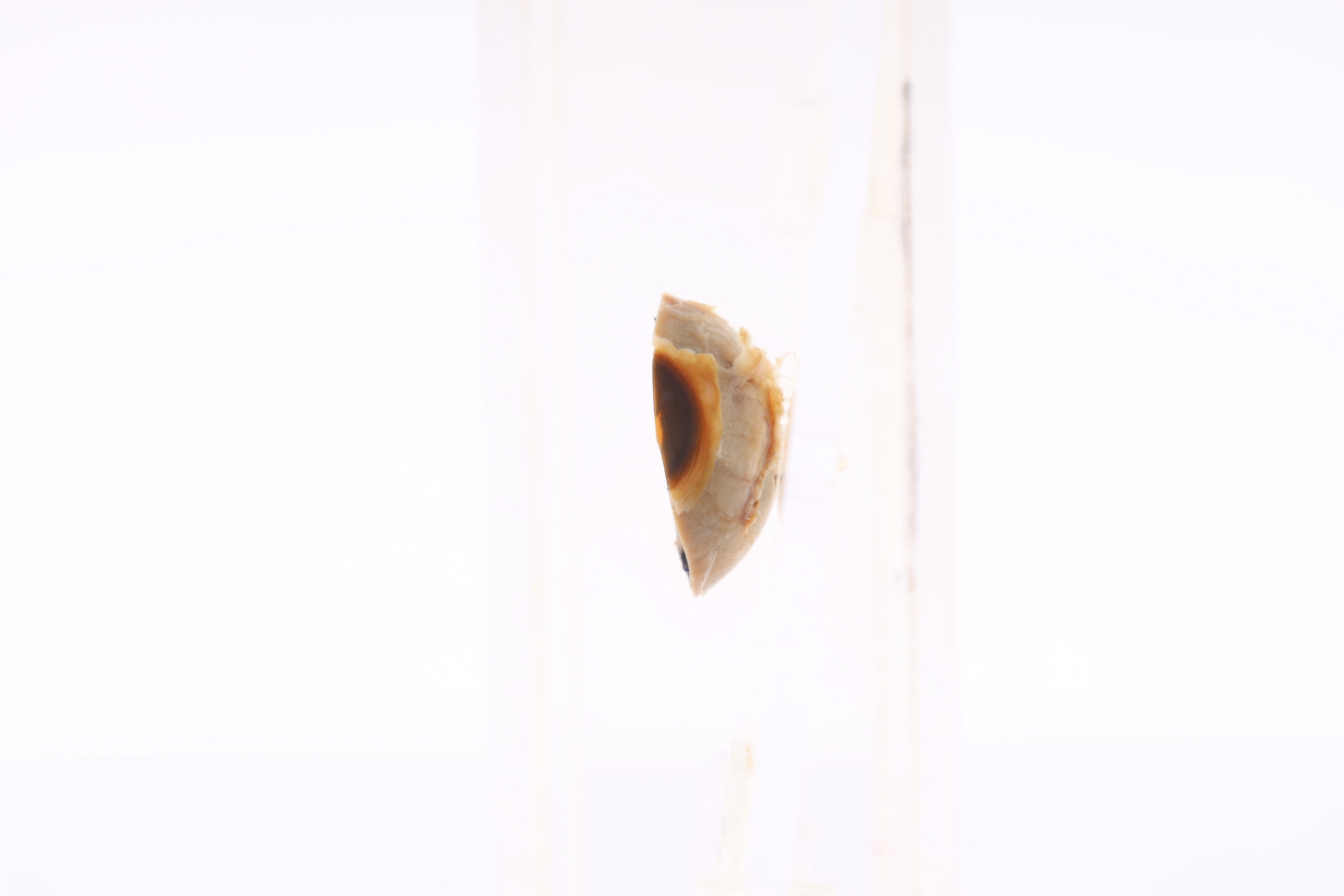
EYE
SPICES No. M766/90
-
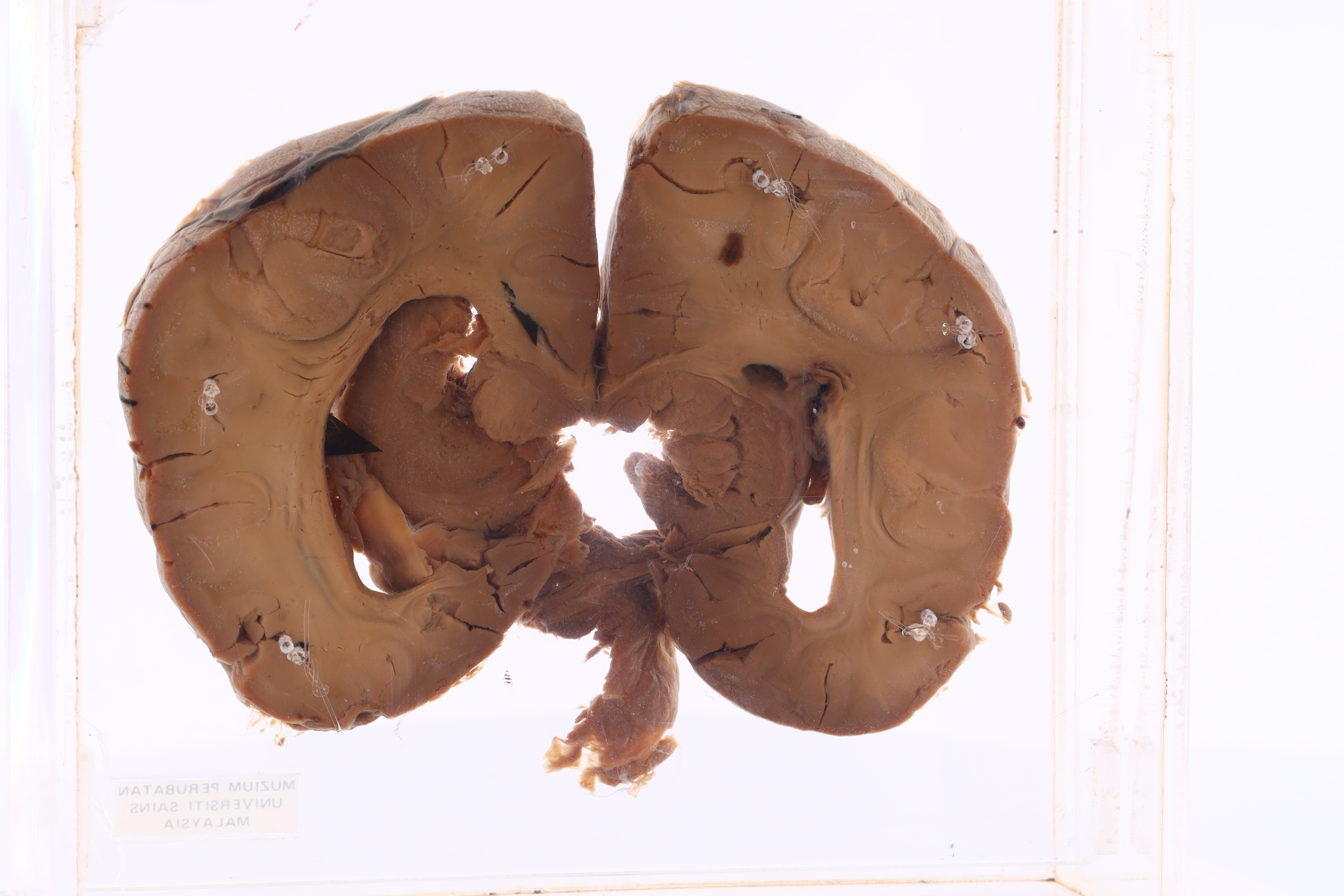
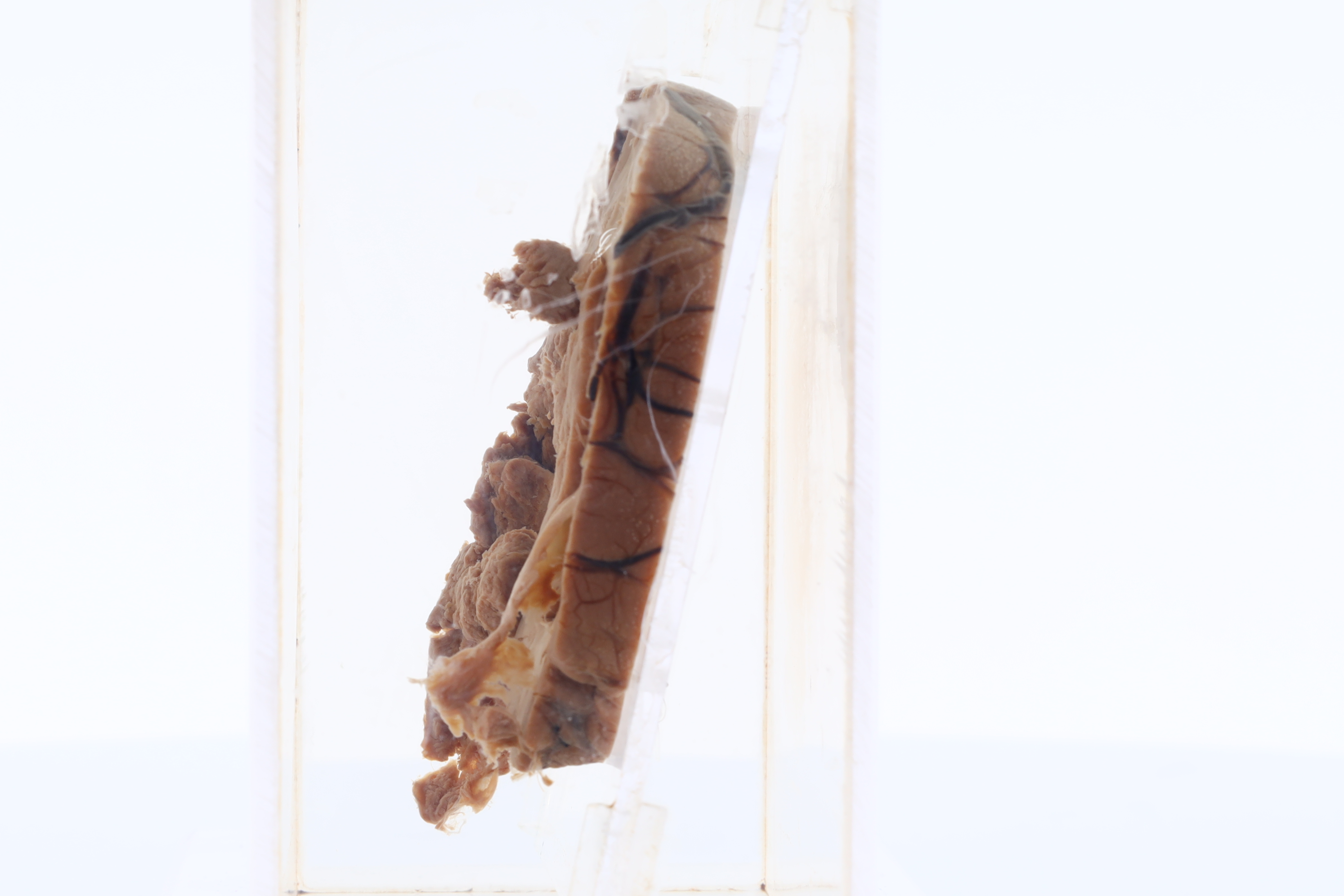
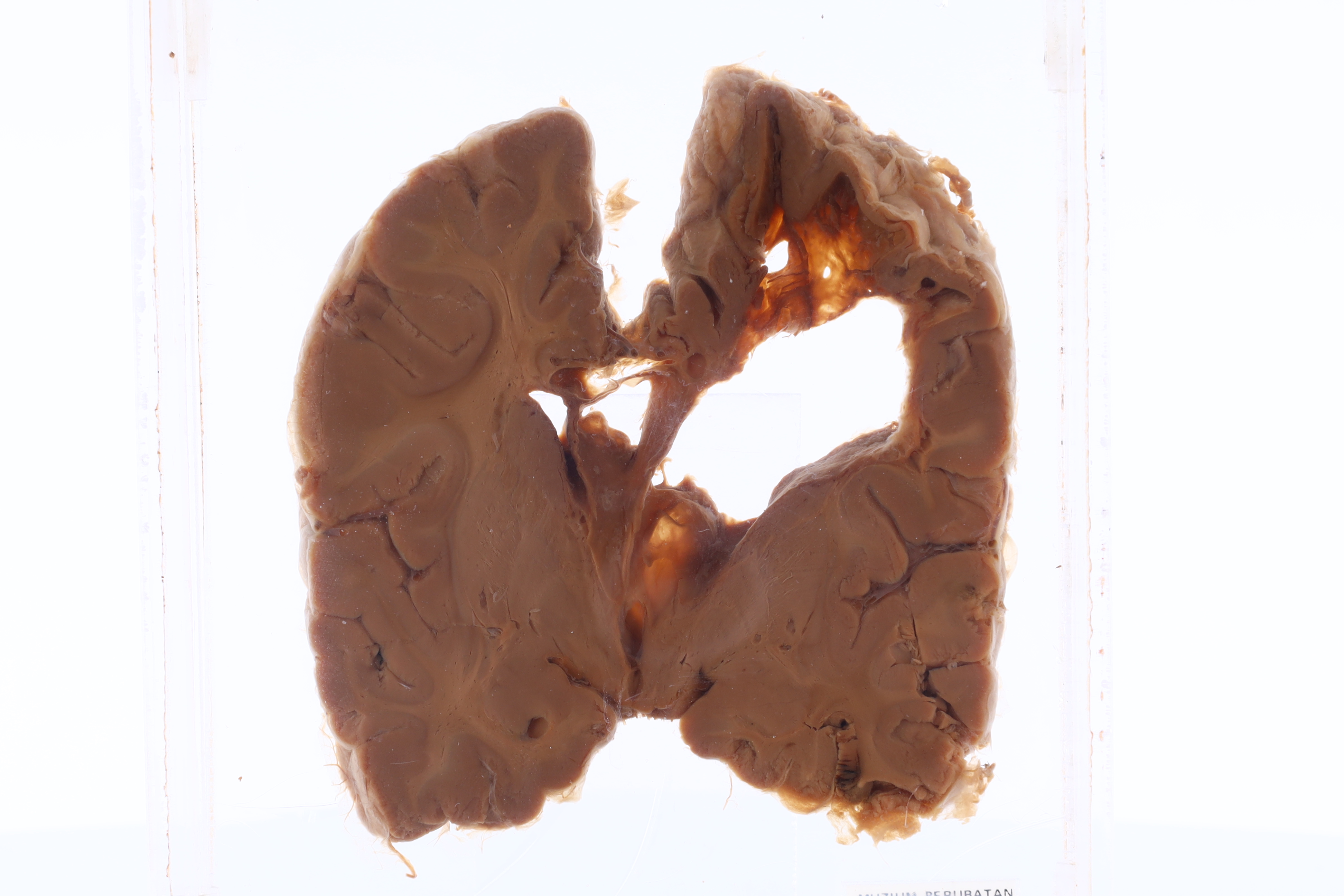
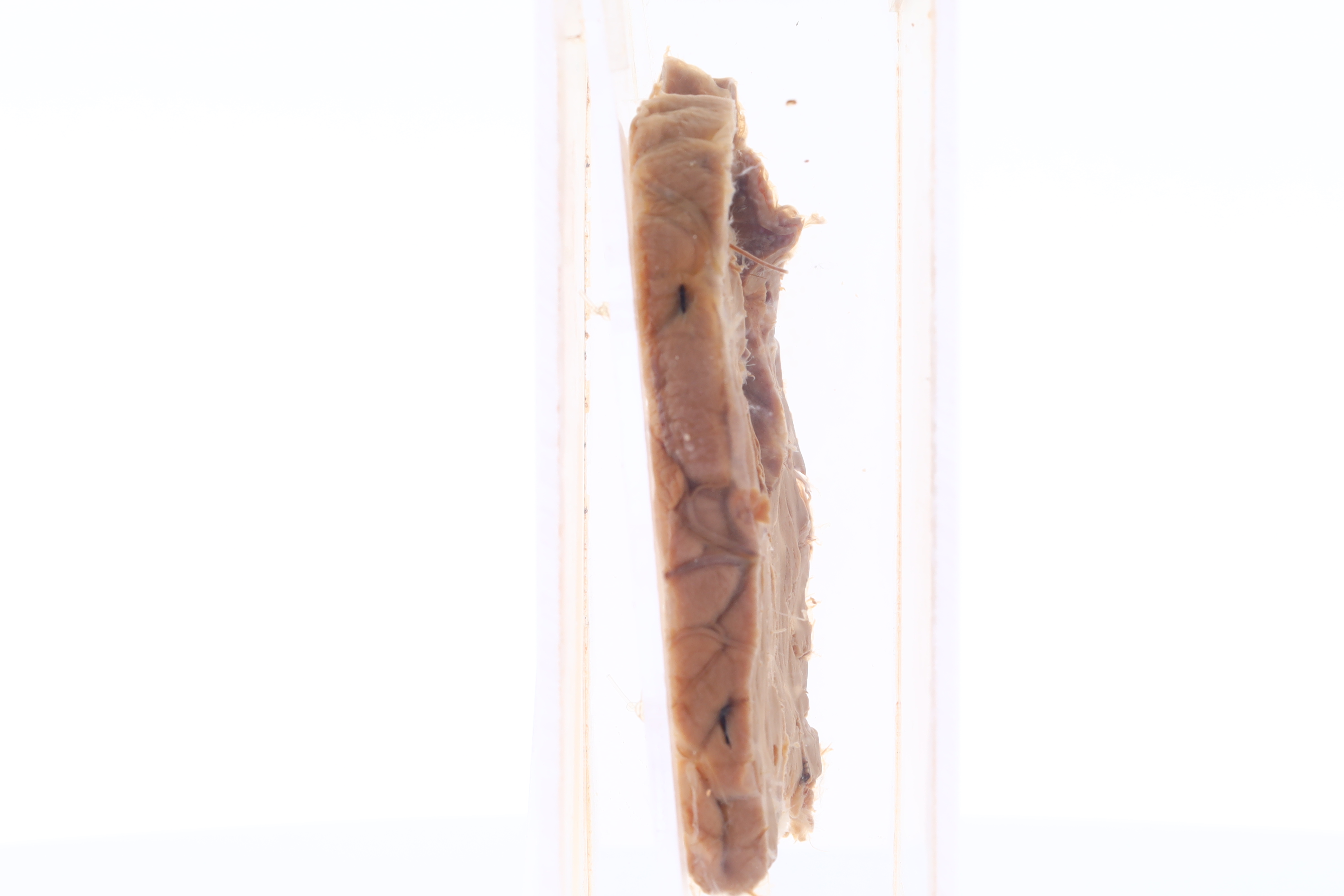
GROSS DESCRIPTION
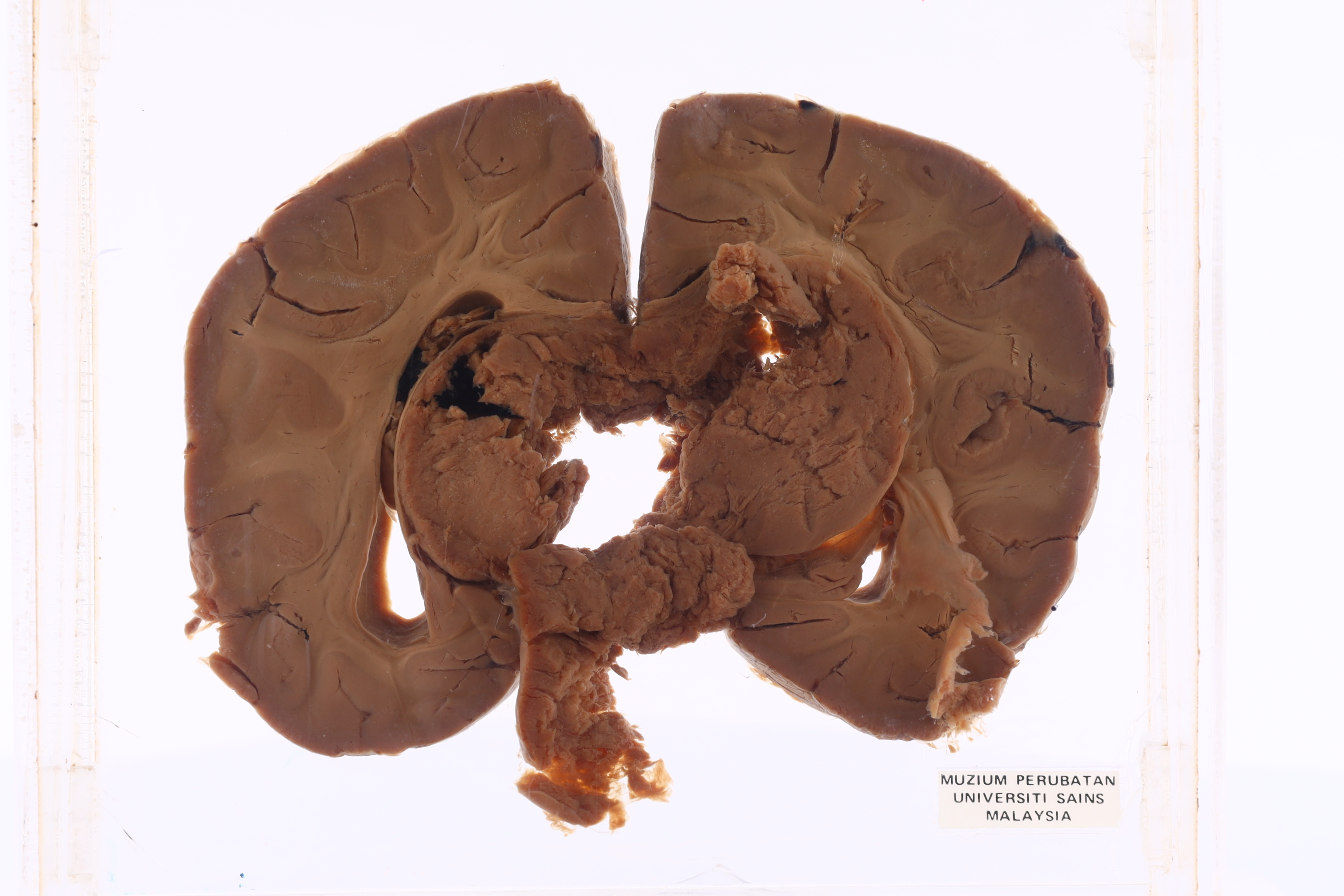
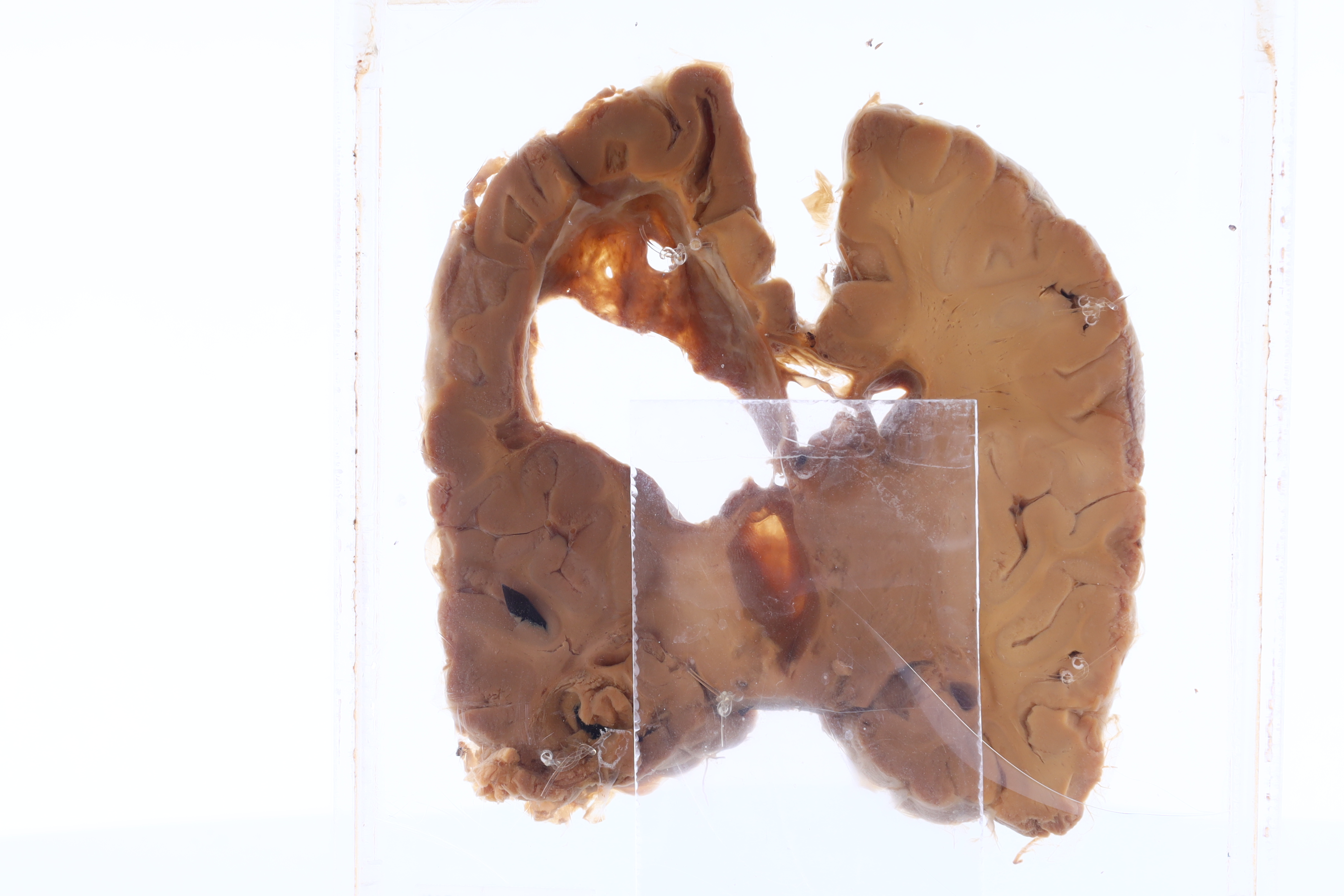
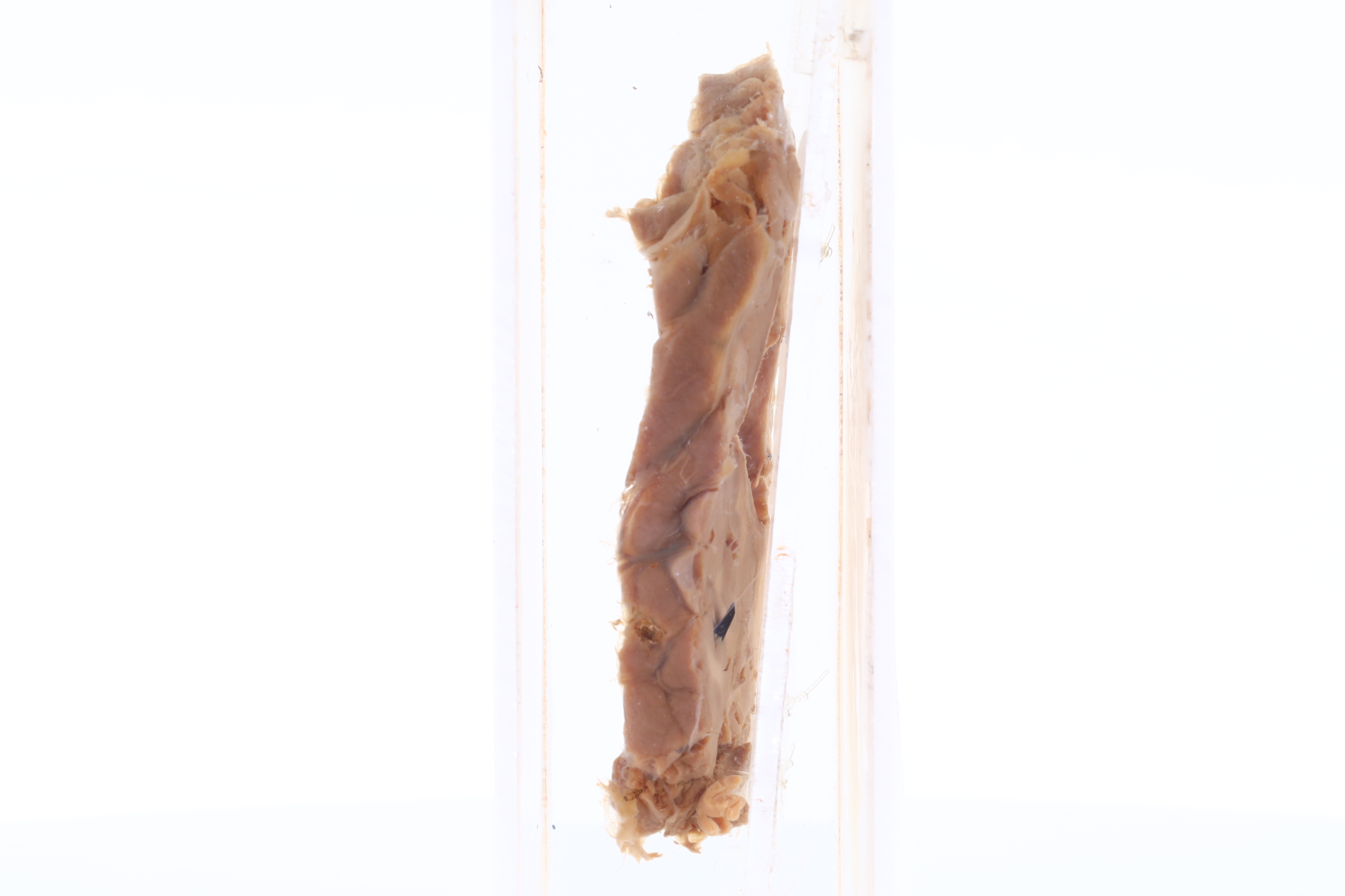
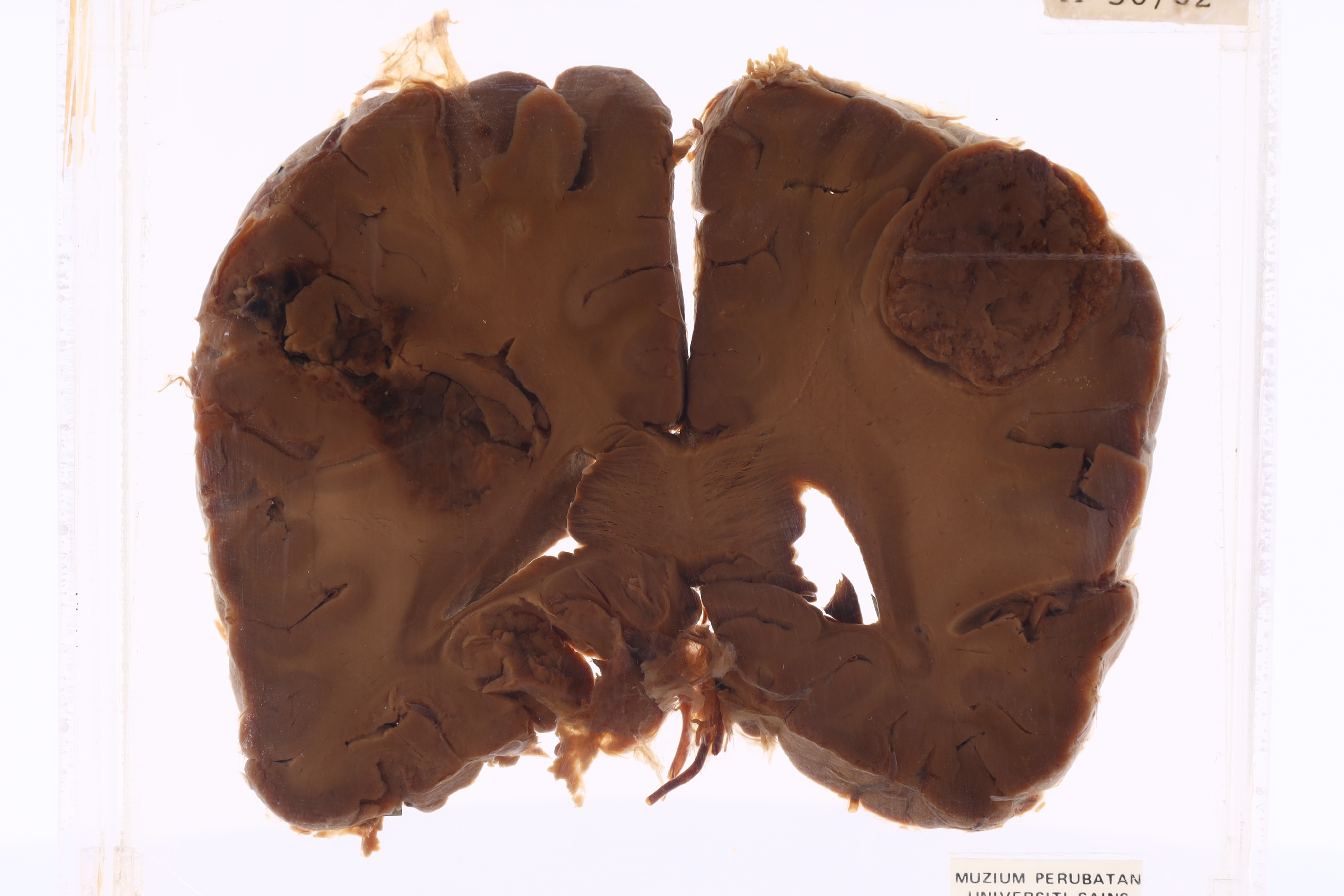
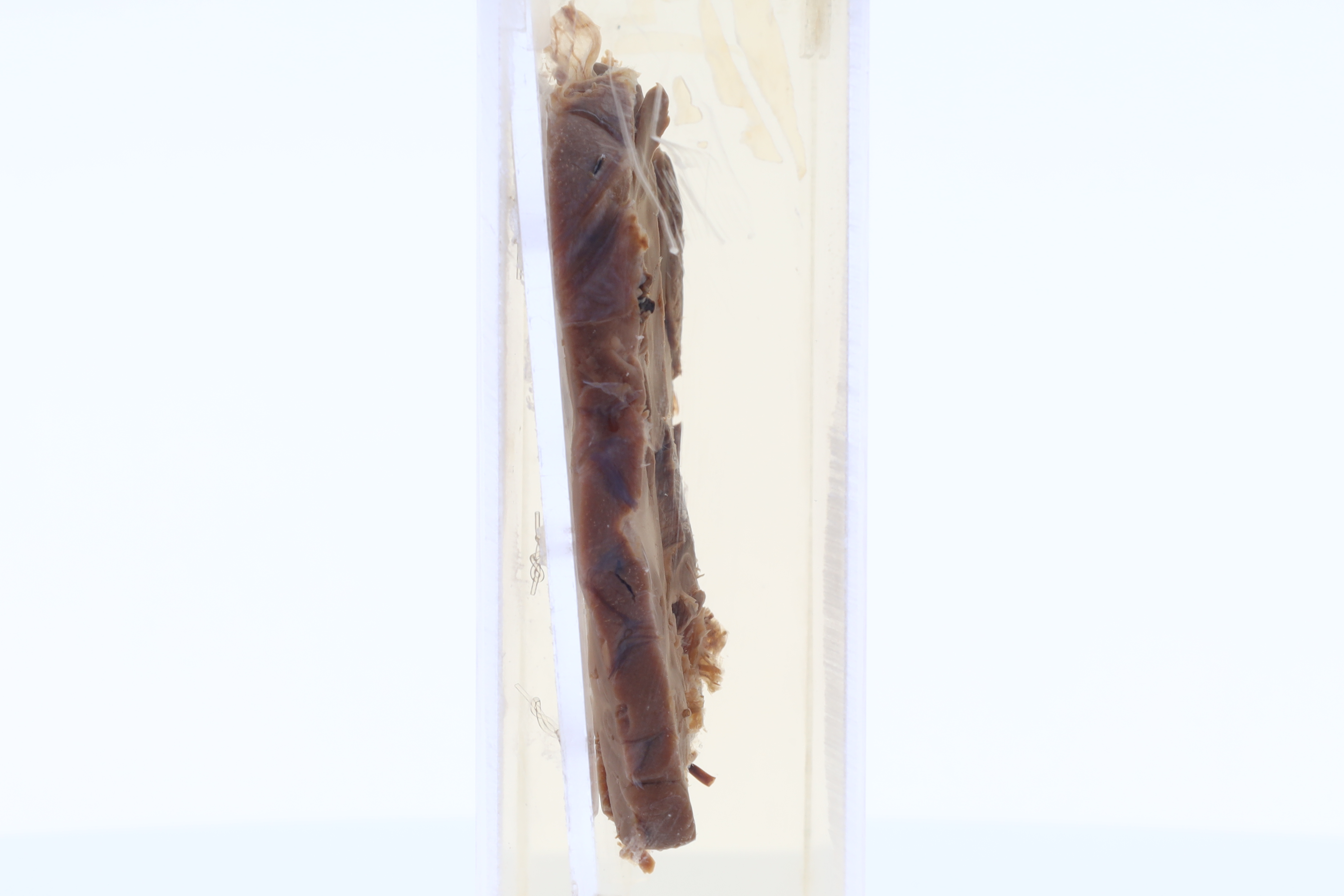
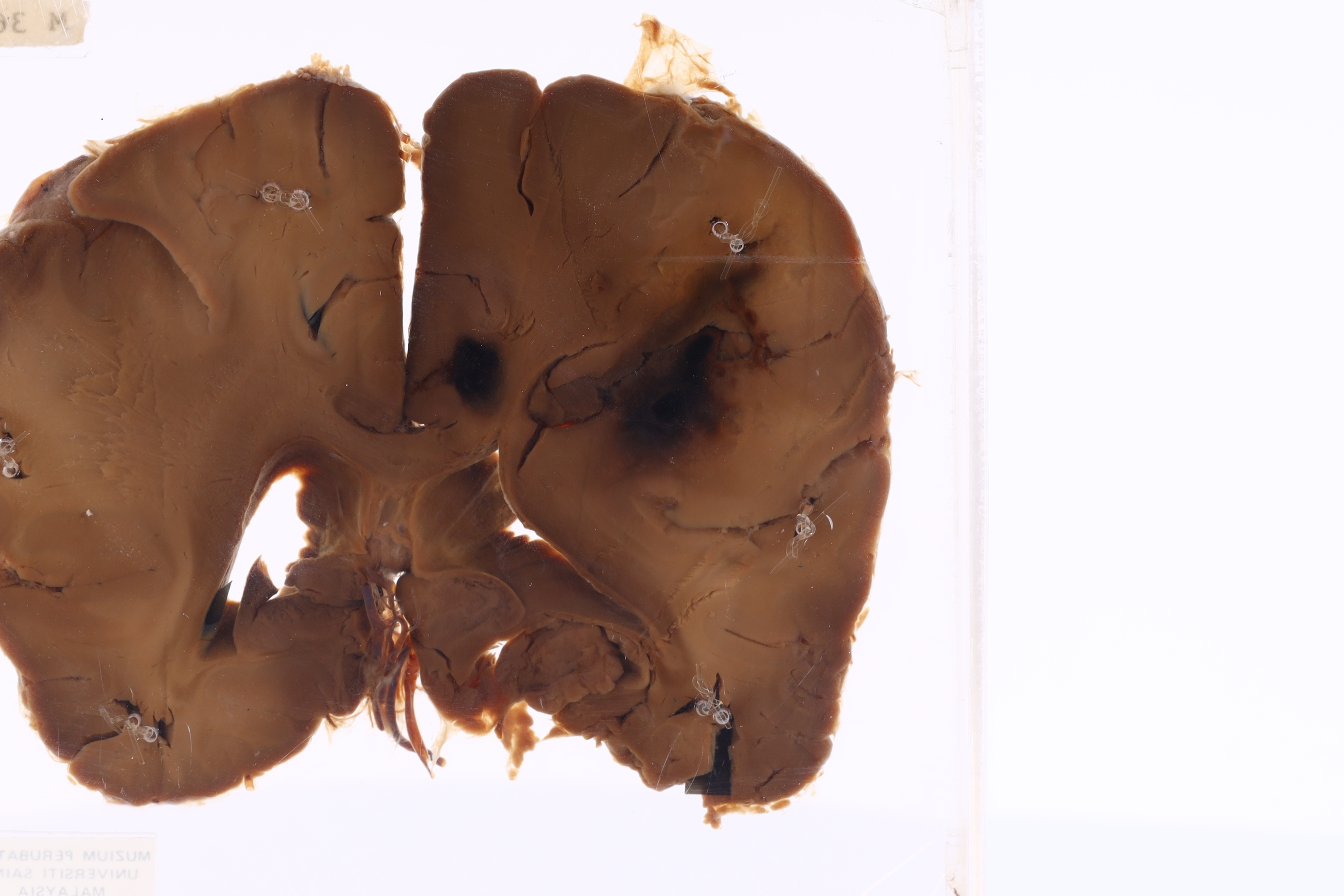
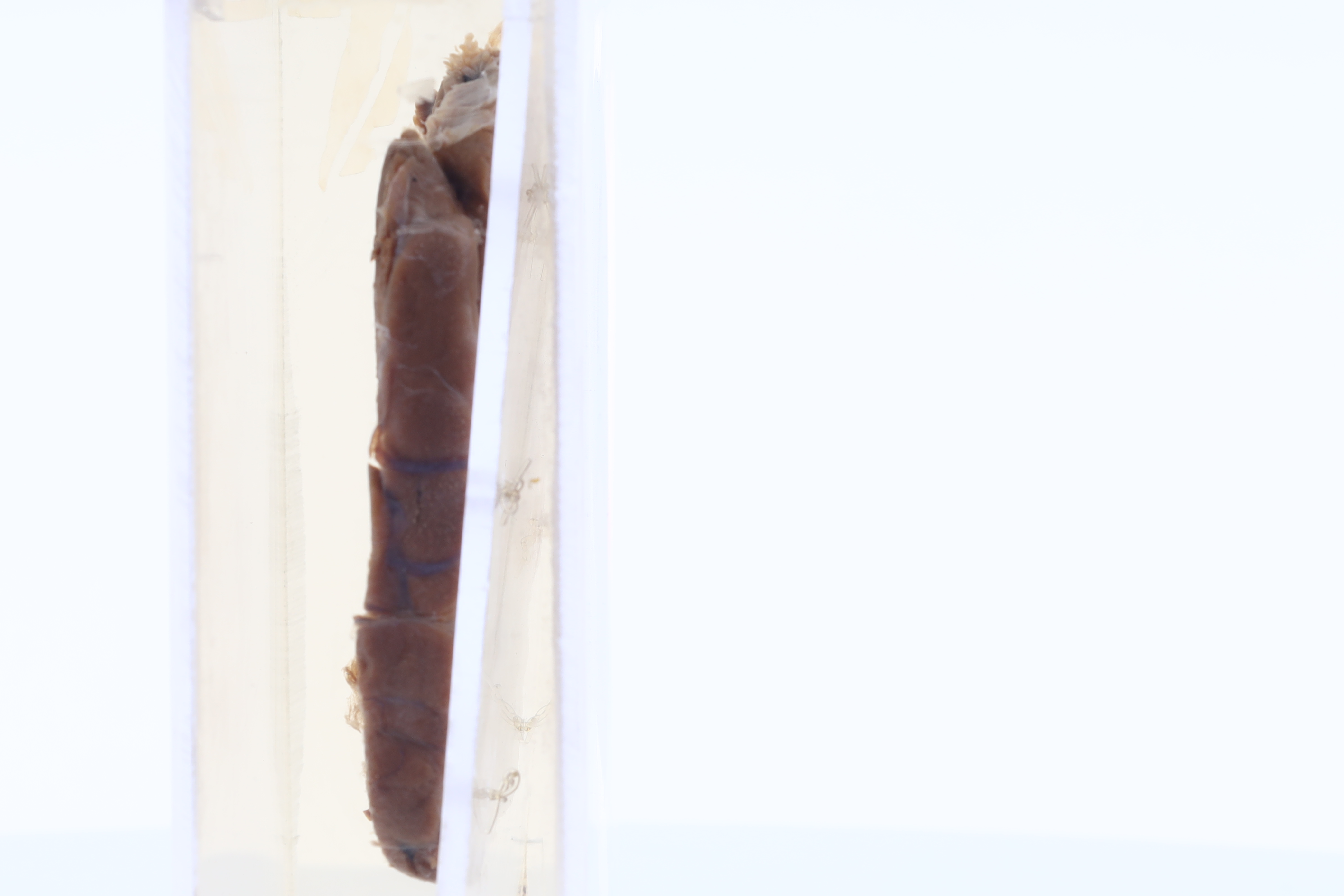
This is exhibit displays eye with retinoblastoma. There is malignant tumor arising from the retina growing into subretinal spaces and vitreous.
-
HISTOPATHOLOGY DESCRIPTION
Retinoblastoma is show highly cellular tumours arranged in sheets and some in trabeculae pattern. The cells are small, round and blue with scanty cytoplasm. Flexner-Wintersteiner rosettes is seen. Calcification and perivascular necrosis also appreciated.
-
DIAGNOSIS
Retinoblastoma

