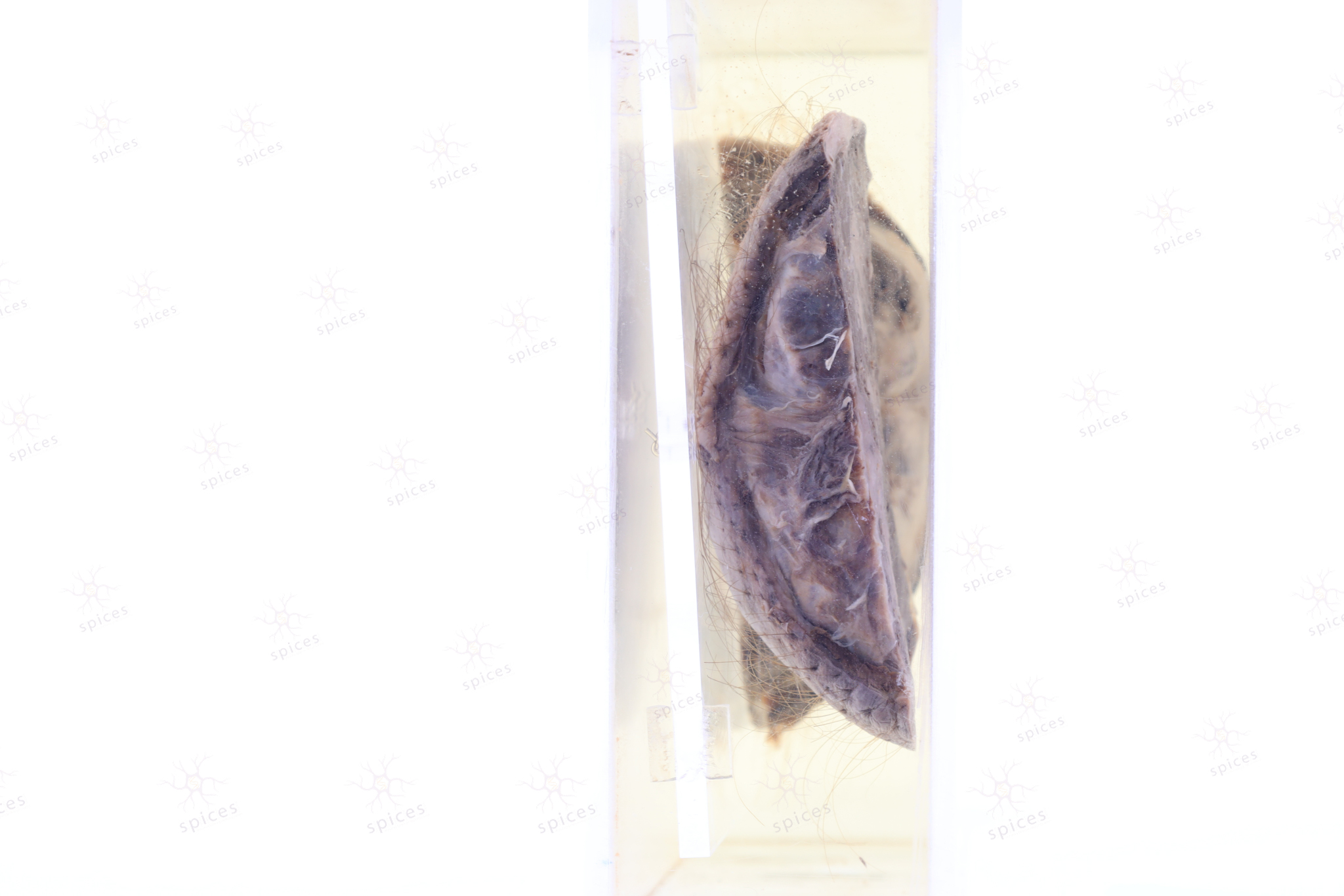
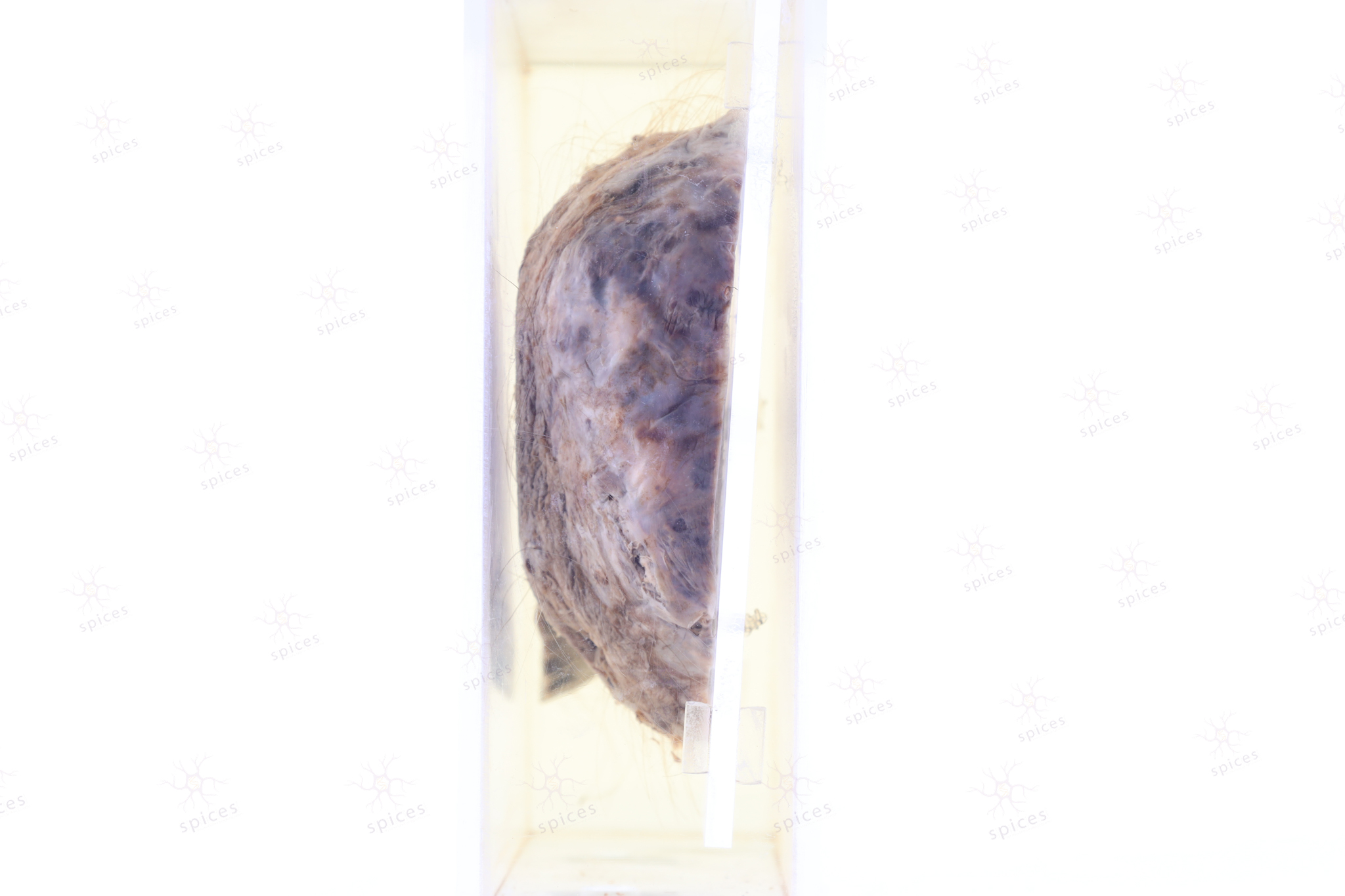
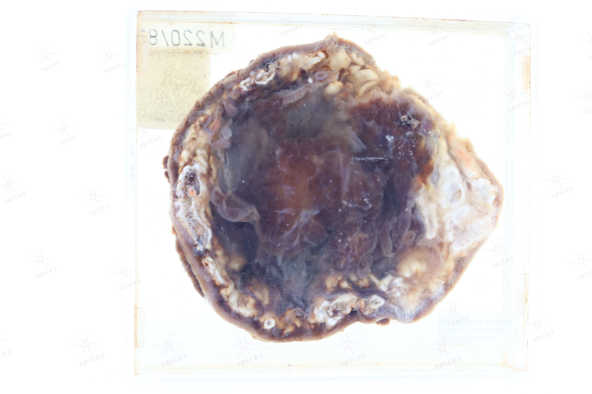
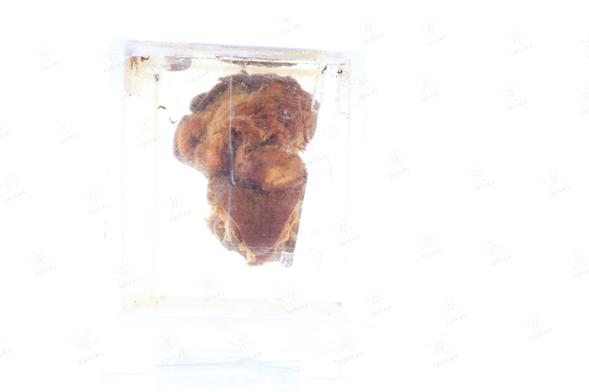
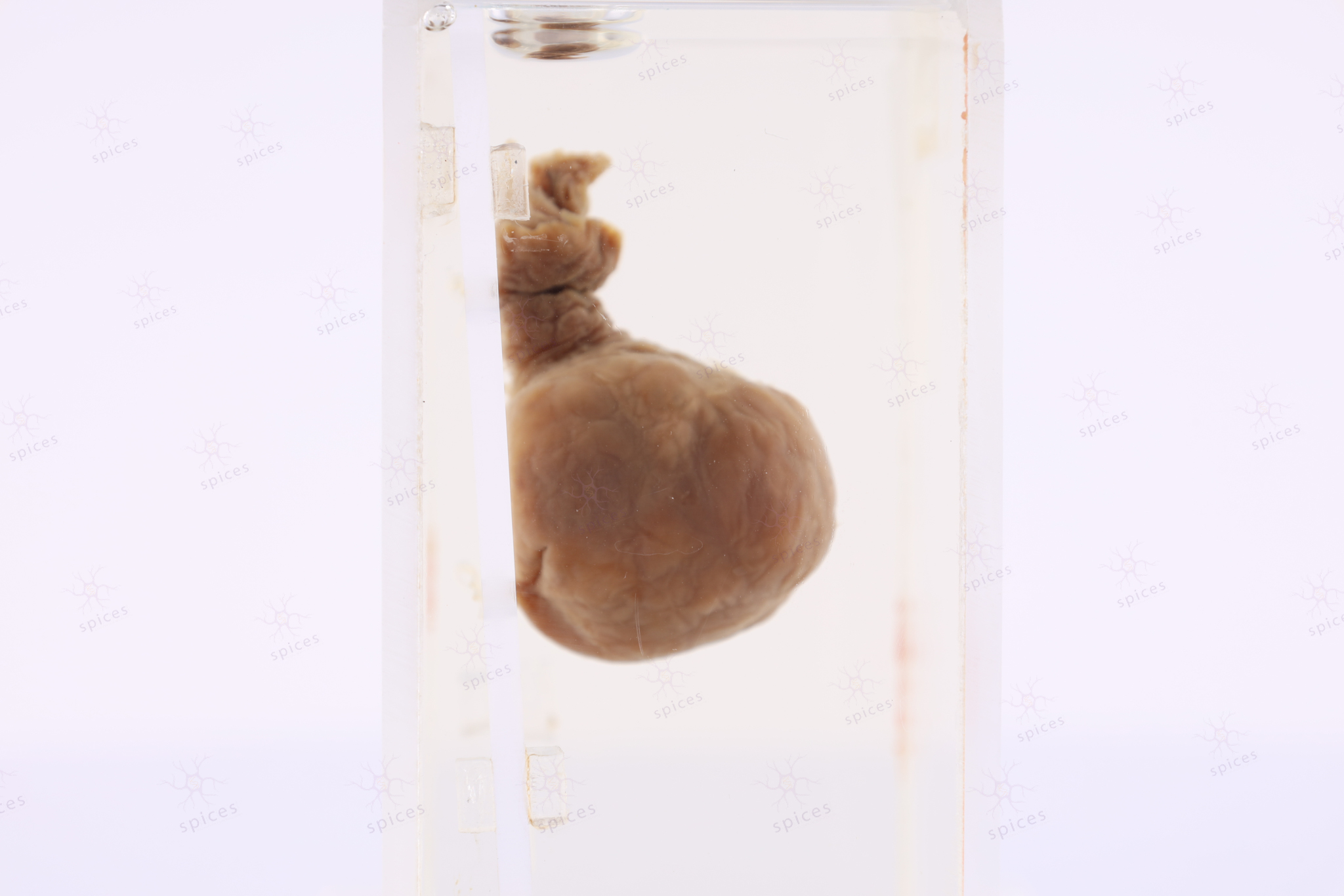
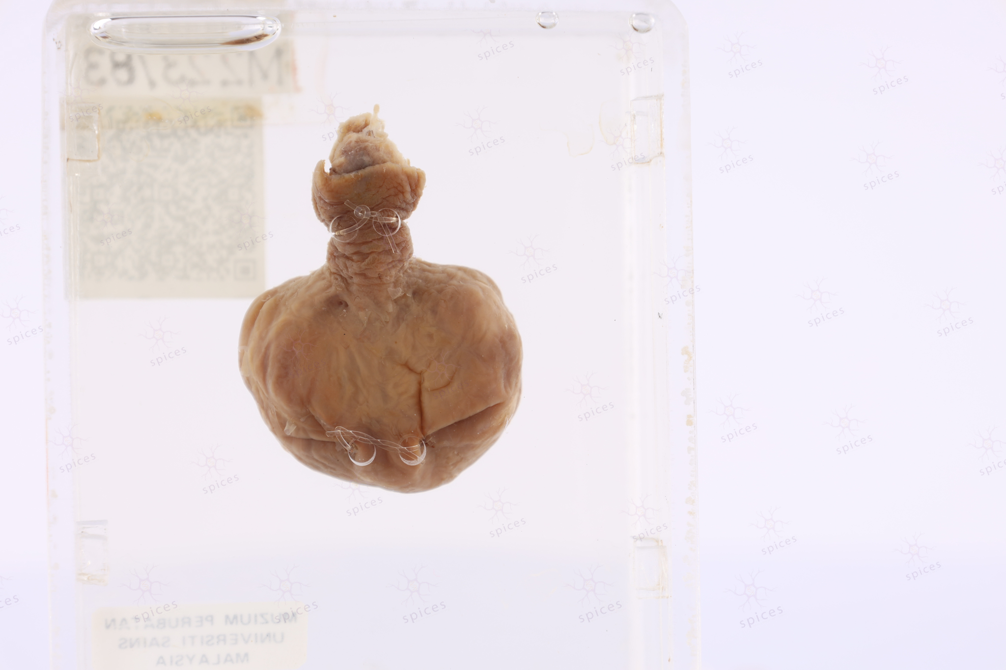
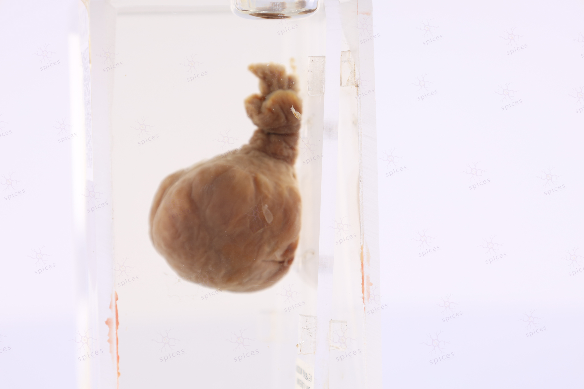
Swelling anterior : M129/82
Swelling anterior
Spices No: M129/82
-
GROSS DESCRIPTION
-
HISTOPATHOLOGY DESCRIPTION
-
DIAGNOSIS
Epidermoid cyst
-
QUIZ
-
BAHASA MELAYU
Epidermoid cyst
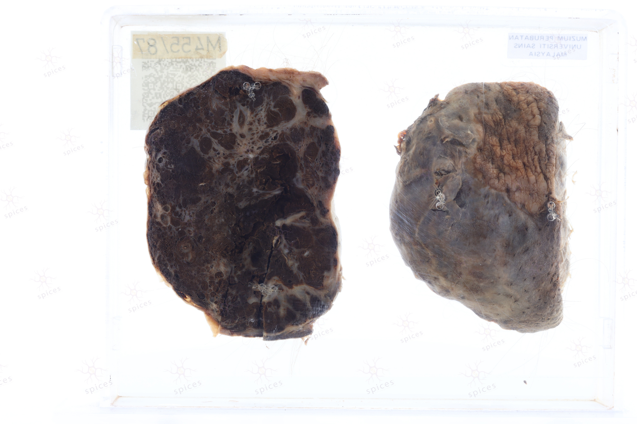
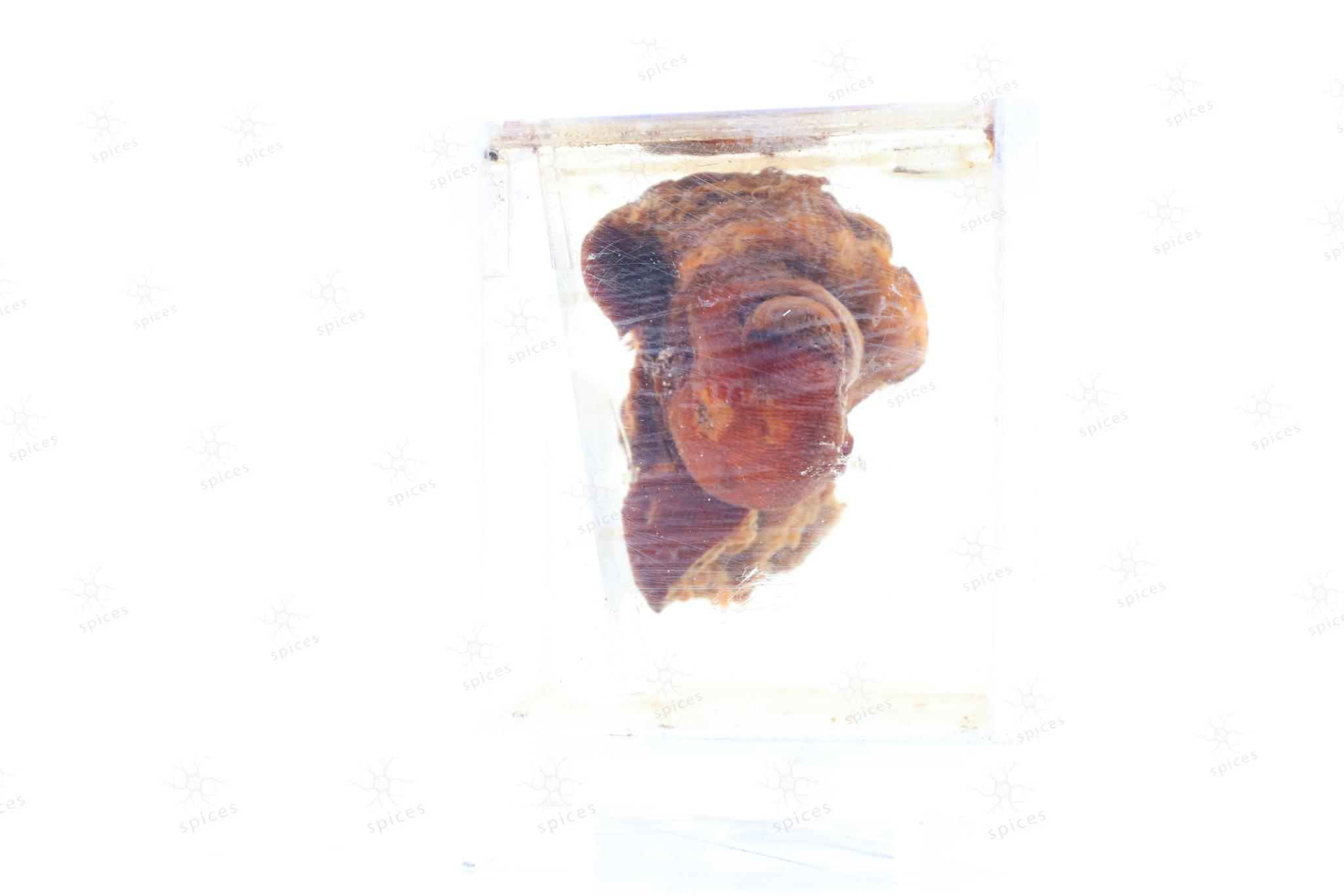
The exhibit displays hemangioma with a classical cut surface showing red brown colour. The vascular spaces filled by red blood cell can be appreciated.
Capillary hemangioma composed of proliferation of small sized of blood vessels arranged in lobules separated by fibrous septae.
Capillary hemangioma
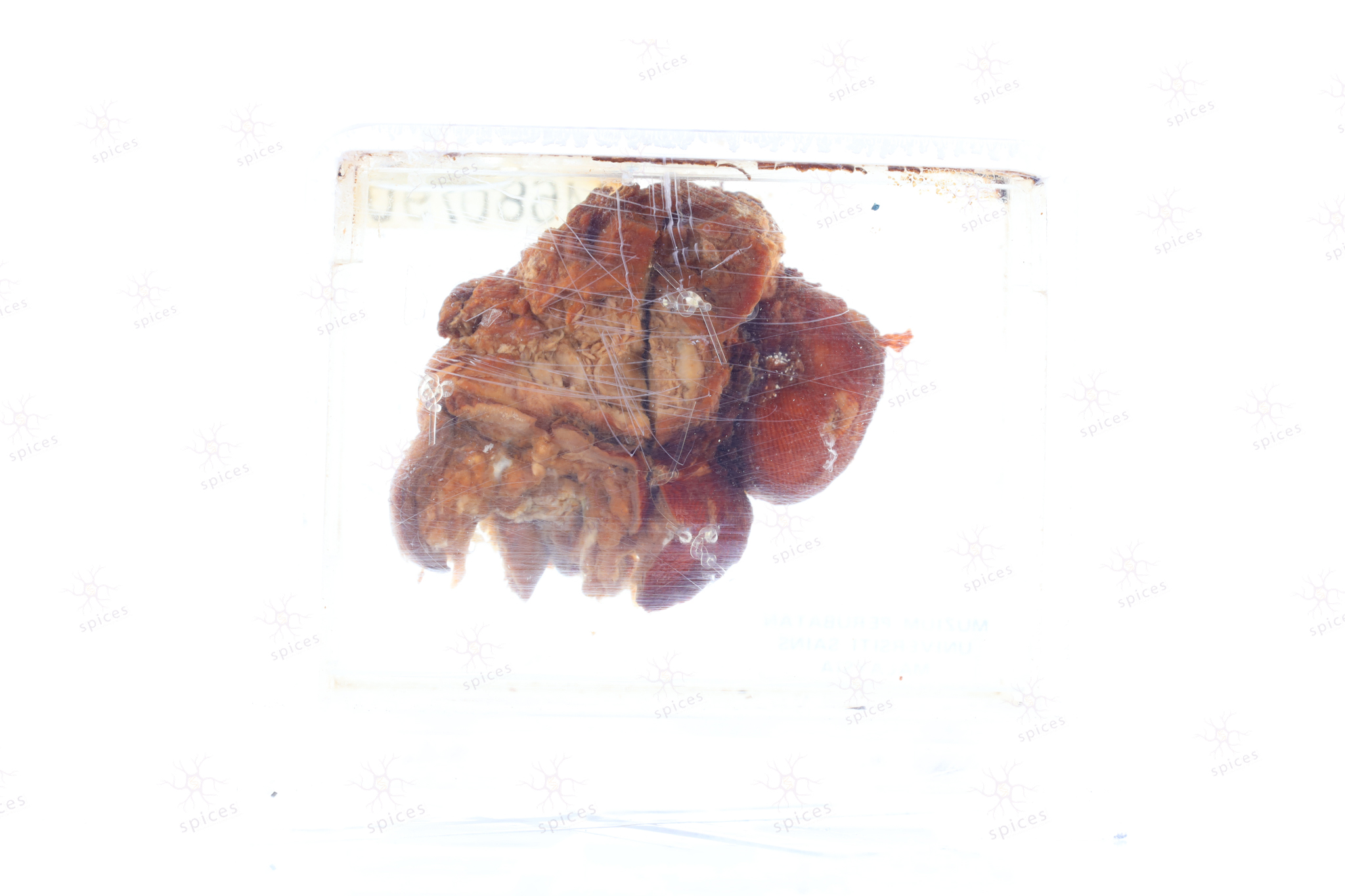
The exhibit displays exophytic mass arising from the skin. The mass shows irregular hyperkeratotic scaly plaque surface.
Squamous cell carcinoma show malignant epithelium with evidence of squamous differentiation. These include the presence of individual keratinization, intercellular bridges, and keratin pearl.
Squamous cell carcinoma
The exhibit displays a toe with presence of exophytic fungating malignant lesion arising from the skin. The mass shows irregular hyperkeratotic scaly plaque surface.
Squamous cell carcinoma show malignant epithelium with evidence of squamous differentiation. These include the presence of individual keratinization, intercellular bridges, and keratin pearl.
Squamous cell carcinoma
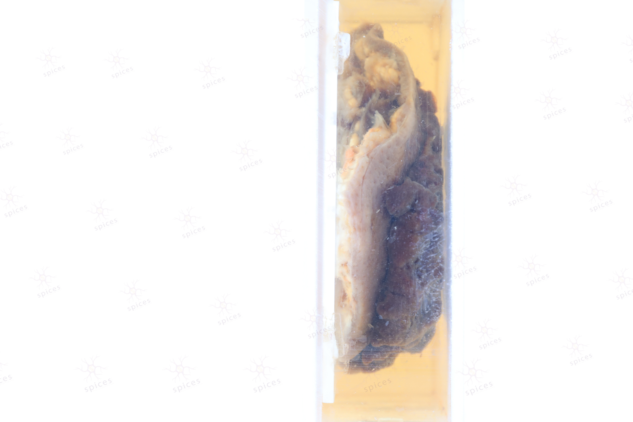
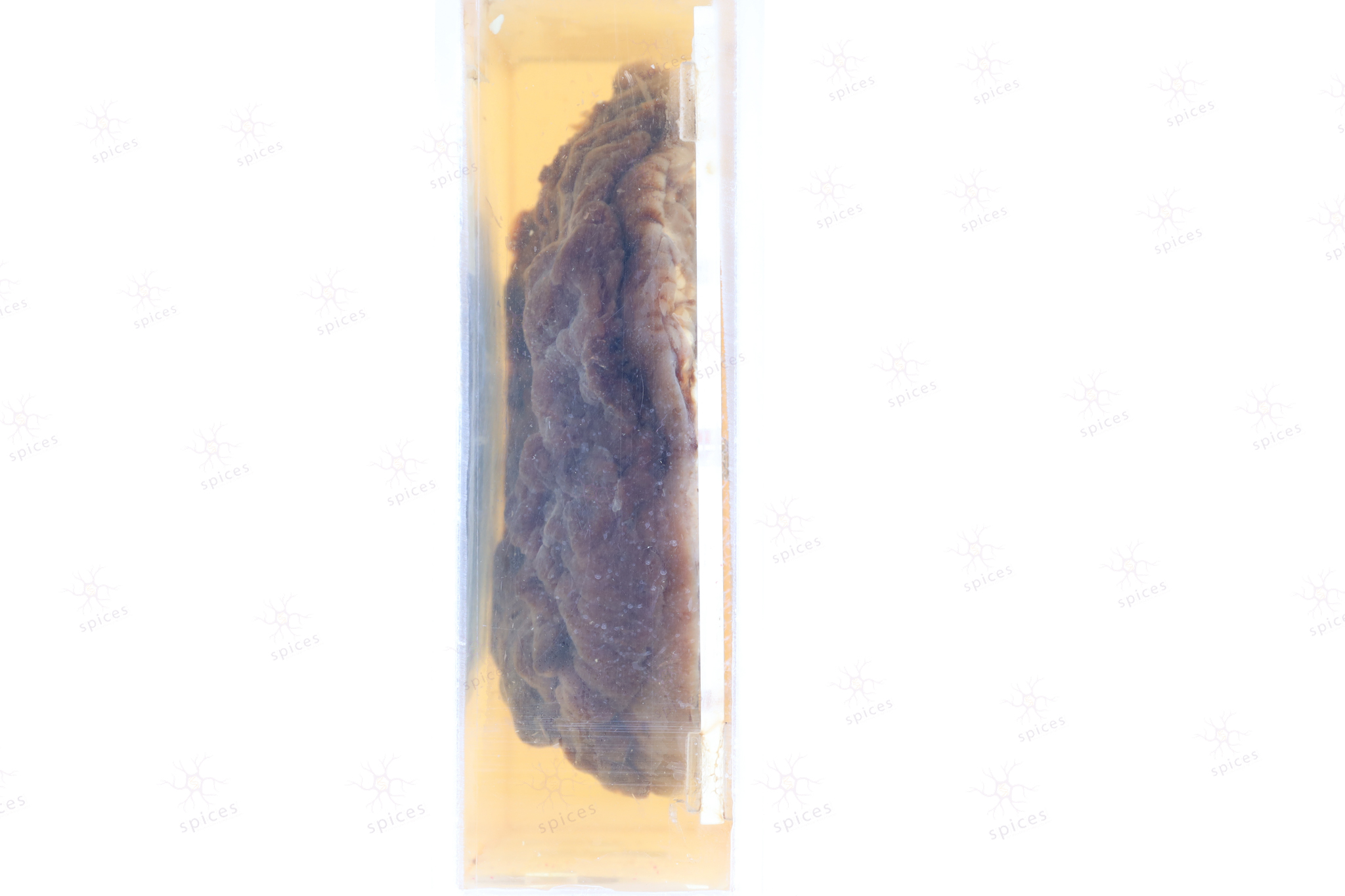
The exhibit displays cystic structurecovered by unremarkable overlying skin
Sebaceous adenoma shows well circumscribed, nodular growth of lobules consist of admixture of basaloid cells and mature sebocytes. Some lobules may communicate directly with the surface epithelium. Basaloid cells are usually located at the periphery of lobules and sebaceous cells with intracytoplasmic lipid vacuoles, which are usually located at the center of lobules. They are composed of expanded germinative layer, with more than the normal 2 cell layers seen in mature sebaceous glands or sebaceous hyperplasia but still less than 50% of the tumor volume.
Sebaceous adenoma of montgomery gland of breast