Descending Colon : S018/2024
Descending colon
SPICES No: S018/2024

-
Clinical information
A 31-year-old lady presented with abdominal pain and altered bowel habit for a few months associated with symptomatic anemia, requiring a blood transfusion. The biopsy confirmed adenocarcinoma. Left hemicolectomy with en-block resection of the anterior abdominal wall was done.
-
Gross description
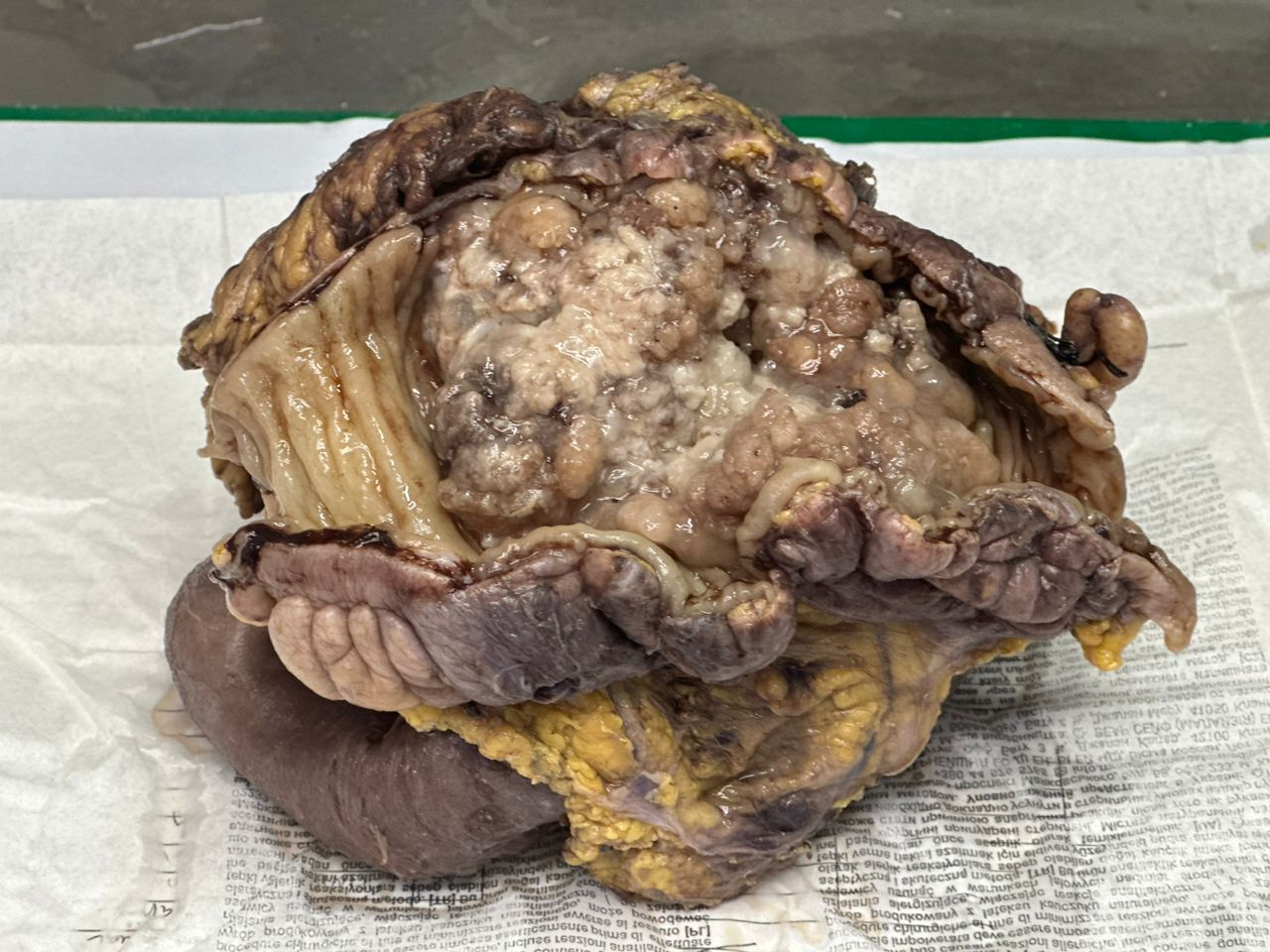
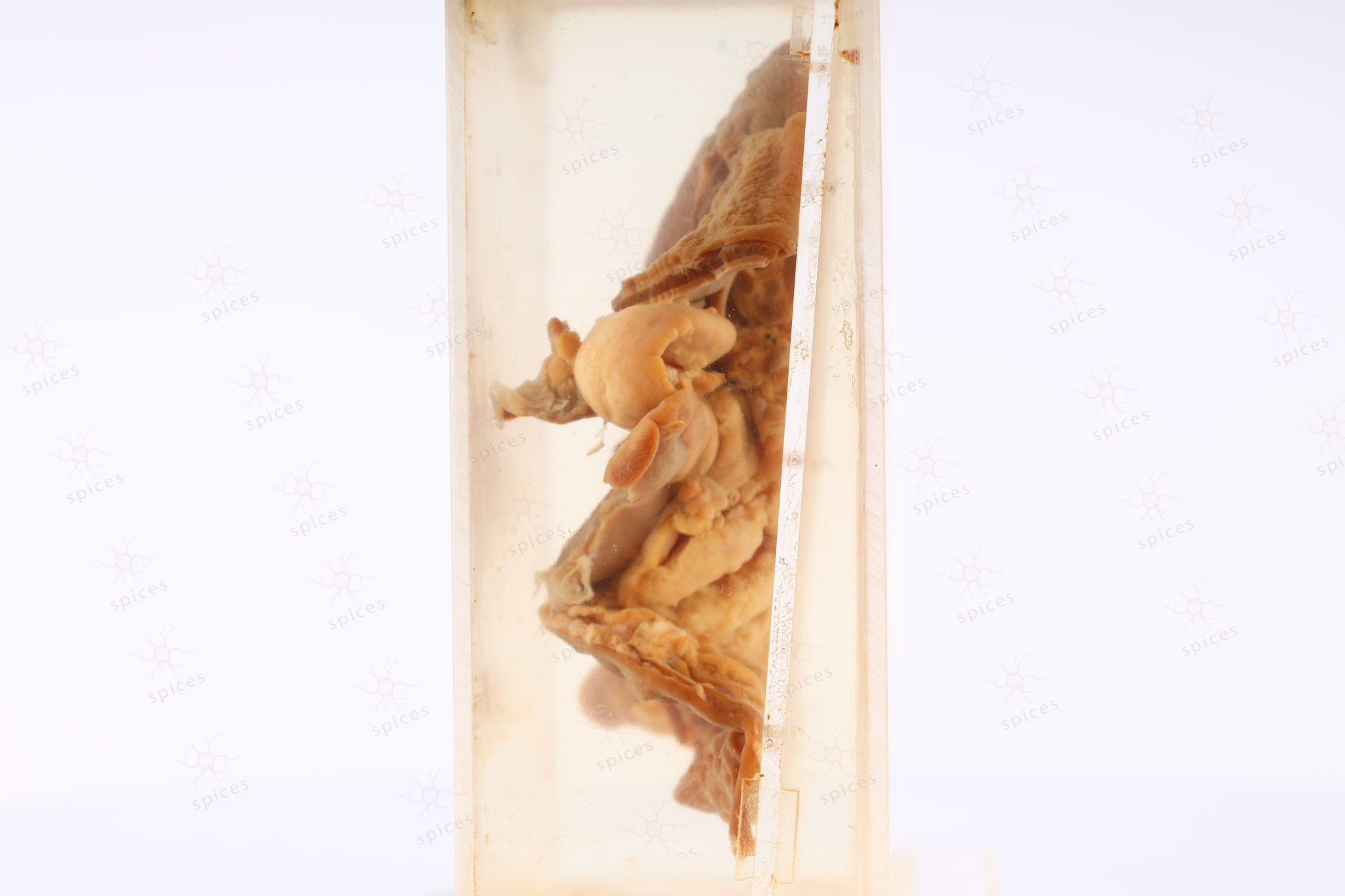
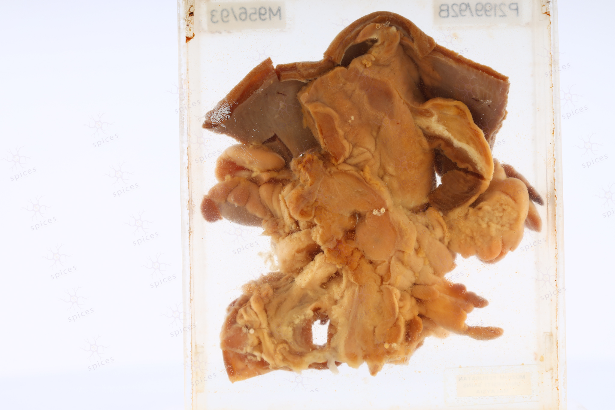
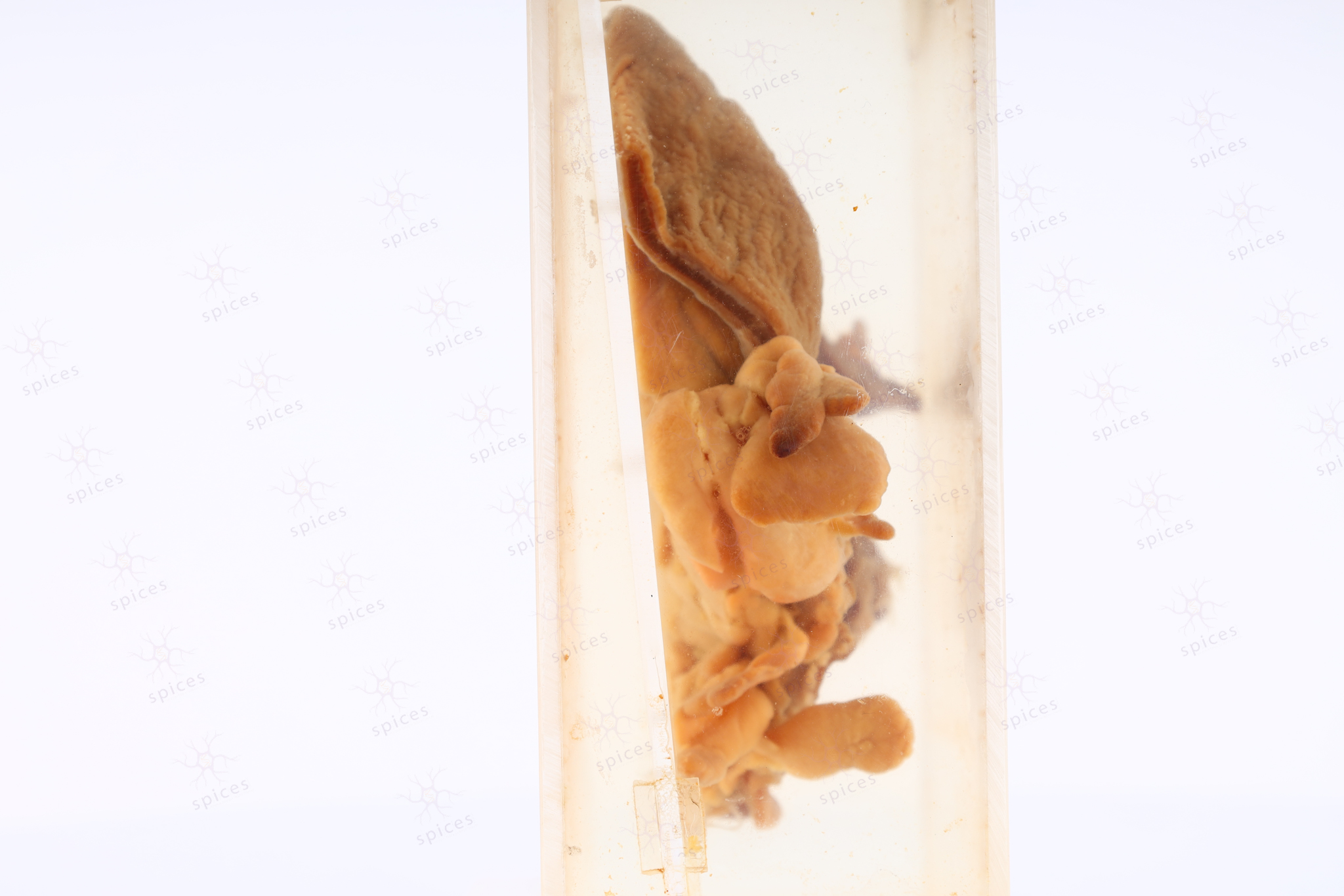
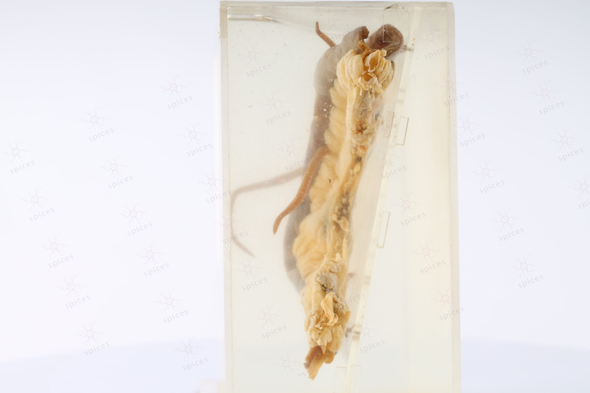
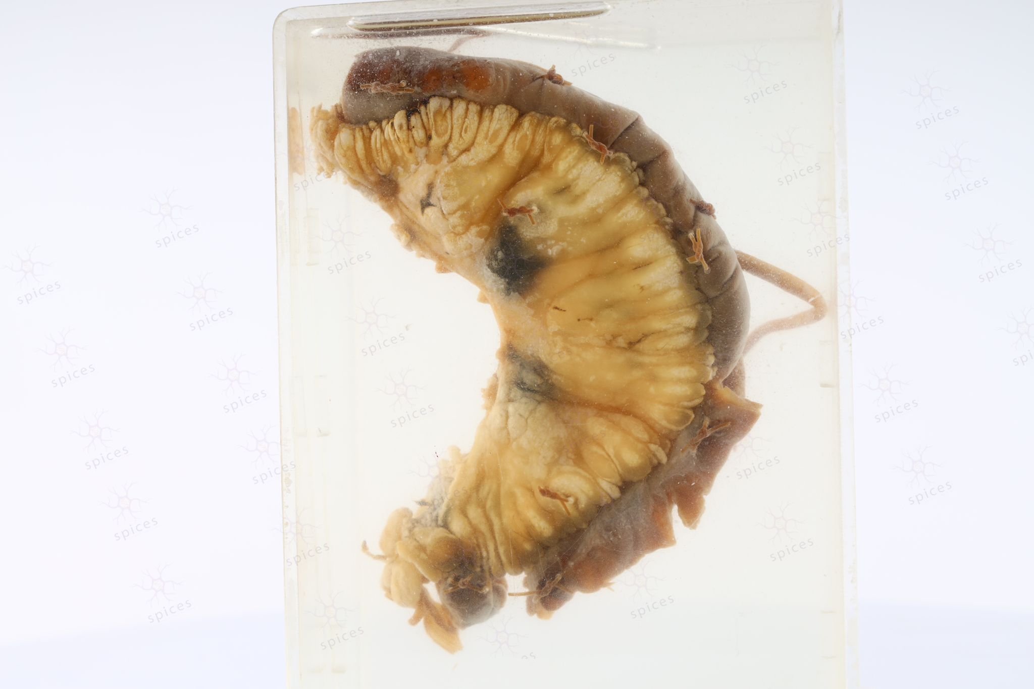
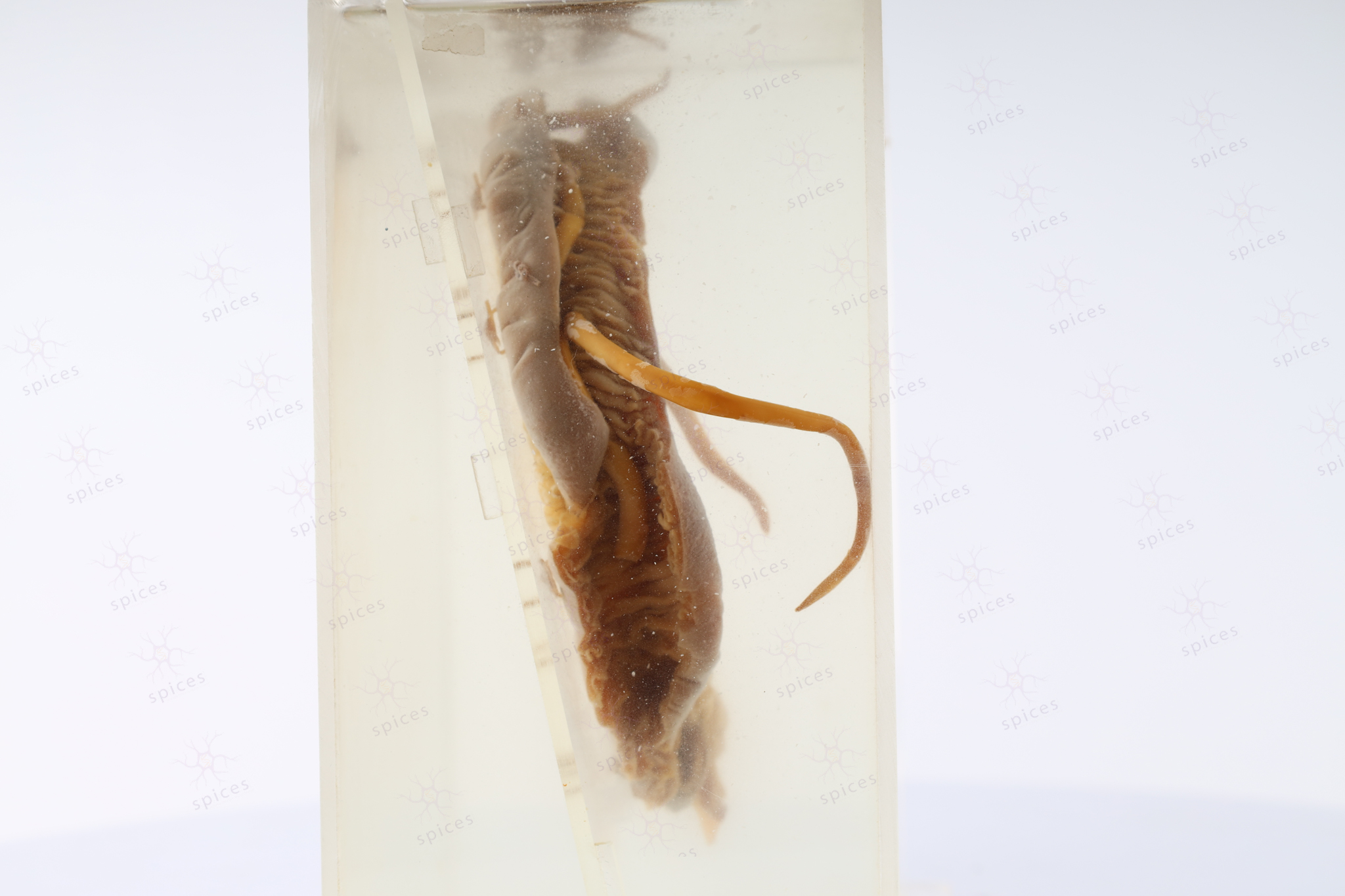
The picture shows en-block specimen before grossing.
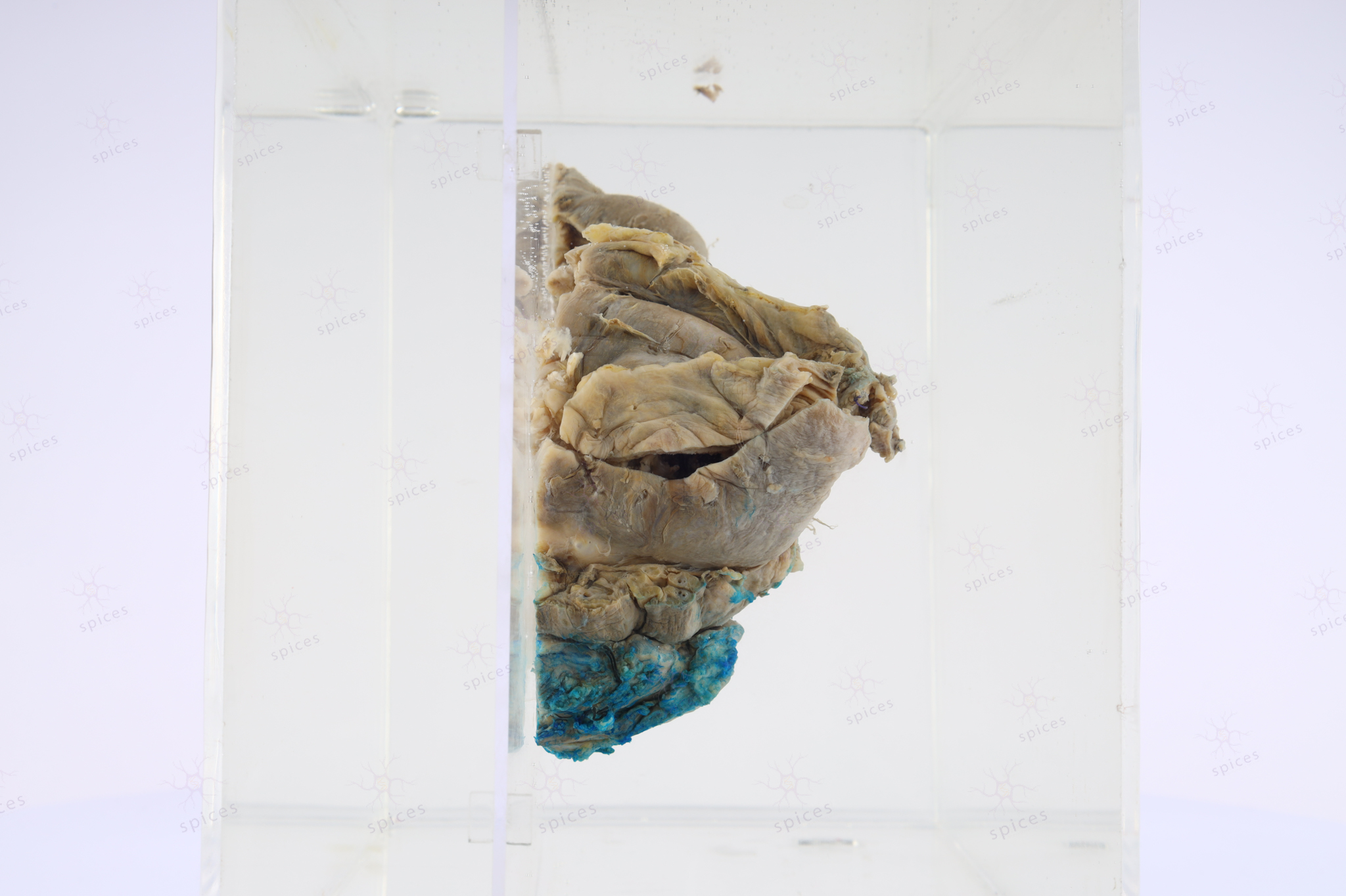
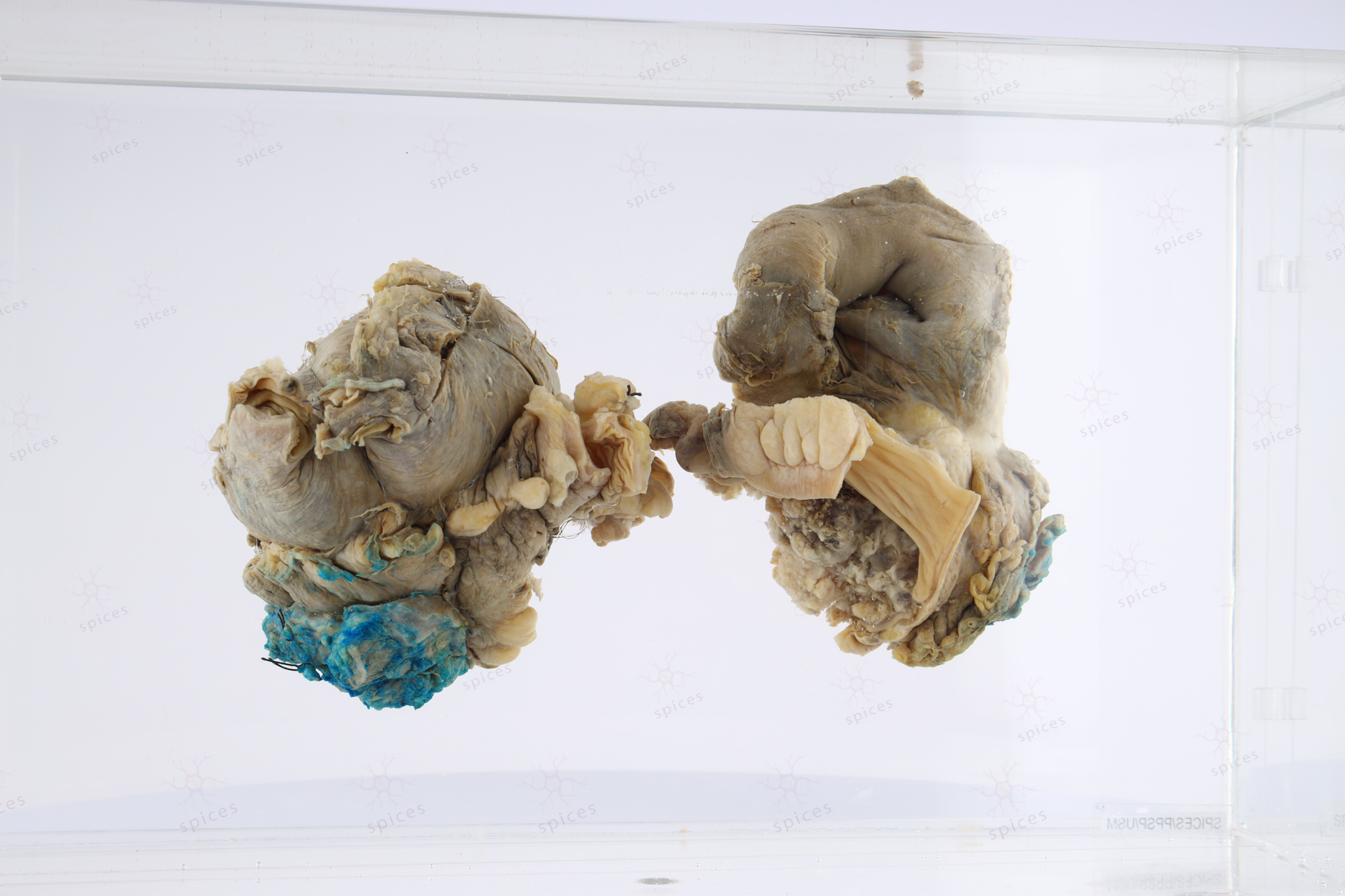
The specimen consists of a surgically opened segment of the colon with an adherent small bowel. There is a fungating and ulcerating mass measuring 105x90x65mm. The floor shows the presence of a slough. The mass is tan, firm to friable with mucoid material noted. There are focal areas of hemorrhage and necrosis. The tumour extends into the muscularis propria, and serosa and directly invades into the small bowel wall. The tumour completely occludes the colonic lumen. The small bowel shows 90% luminal occlusion by the tumour mass. There is no polyp, ulcer, or diverticulum seen in the adjacent mucosa of both the colon and the small bowel.
-
HISPATHOLOGY DESCRIPTION
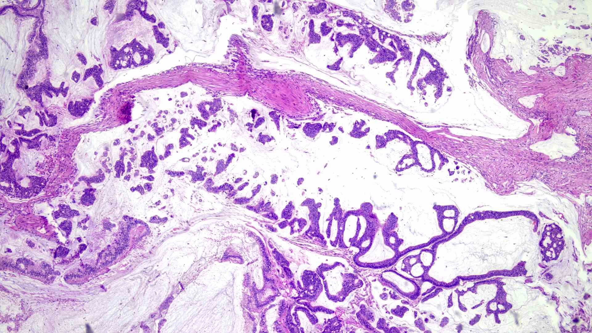
H&Ex4: Malignant glands are floating within the pools of extracellular mucin.
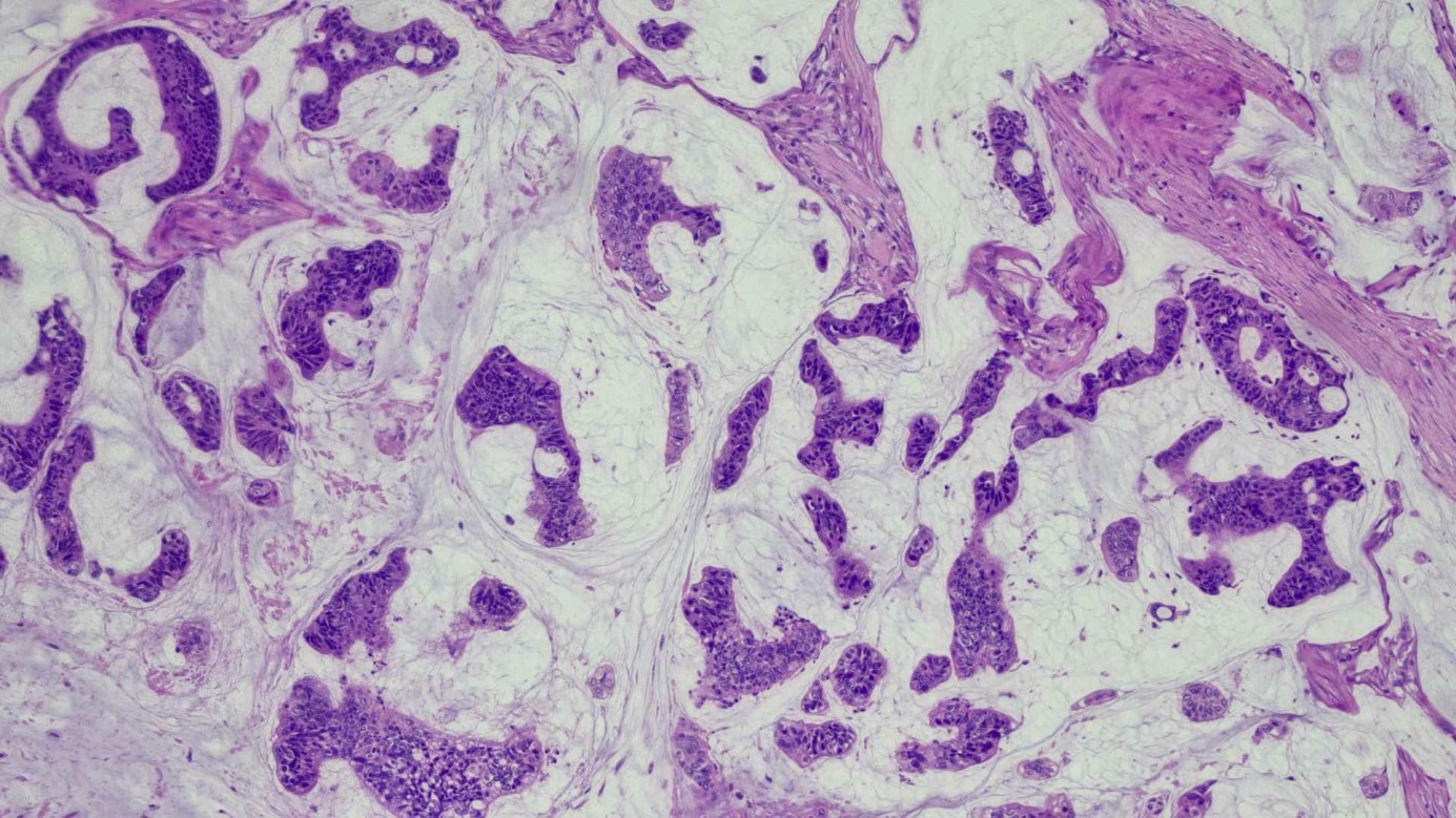
H&E x10:Malignant glands and tumour clusters are seen within the pools of extracellular mucin.
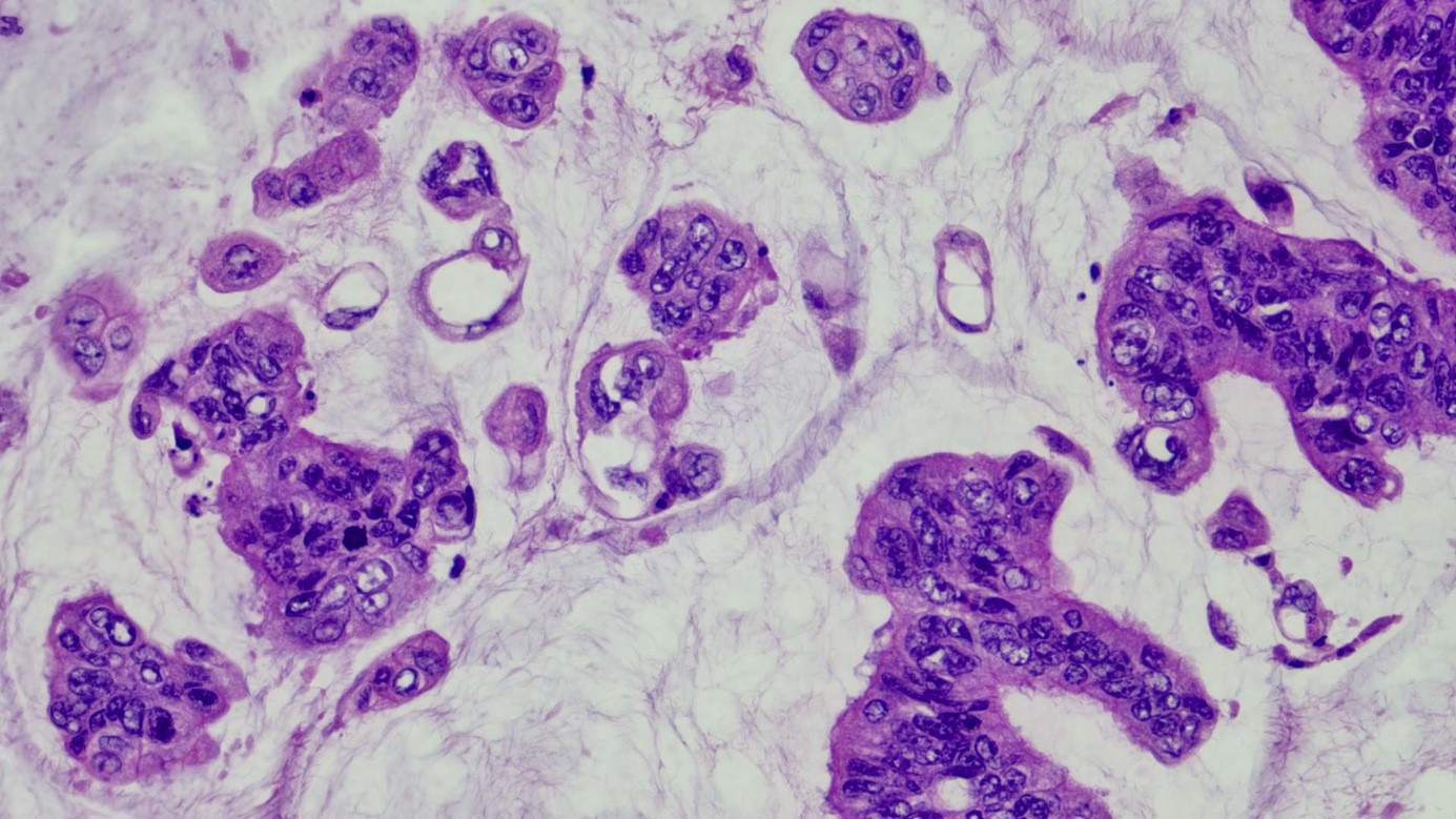
H&E x 40: Signet ring cells are seen.
Sections from the colon show infiltrative malignant glands arising from the colonic mucosa extending beyond the muscularis propria, breaching the serosal layer and directly invading the small intestine. These malignant cells are arranged in nests, singly, in irregular clusters and glandular patterns, and are seen floating in large extracellular mucin pools (>50%). Occasional signet ring cells are seen. The malignant cells exhibit moderate nuclear pleomorphism, having hyperchromatic to vesicular nuclei and prominent nucleoli. Mitotic figures are seen including aberrant forms. The surrounding stroma is fibrotic and shows a desmoplastic reaction. The stroma is moderately to markedly infiltrated by mixed inflammatory cells.
-
DIAGNOSIS
Moderately differentiated Mucinous Adenocarcinoma with direct invasion to the small bowel.
-
Bahasa Melayu
Pesakit merupakan seorang wanita berusia 31 tahun mengalami sakit perut dan mengalami perubahan tabiat buang air besar selama beberapa bulan dan anemia simptomatik, yang memerlukan pemindahan darah. Biopsi mengesahkan kanser kolon. Pembedahan telah dilakukan.
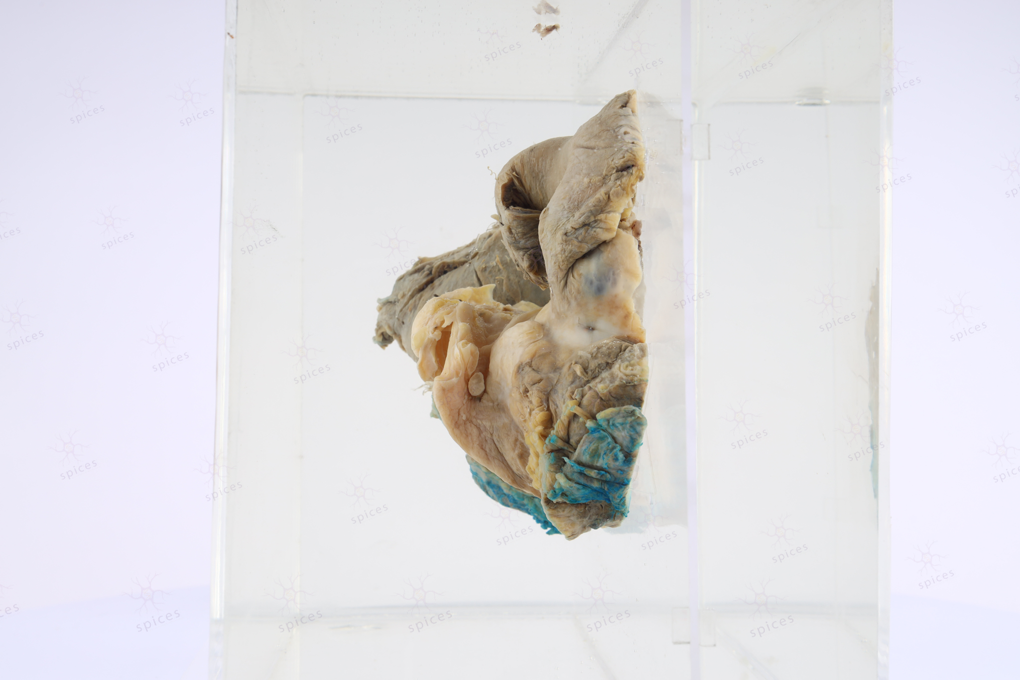
Kanser kolon didapati telah merebak ke usus kecil. Spesimen di dalam jar ini menunjukkan kanser colon yang memenuhi kolon dan telah merebak ke usus kecil.