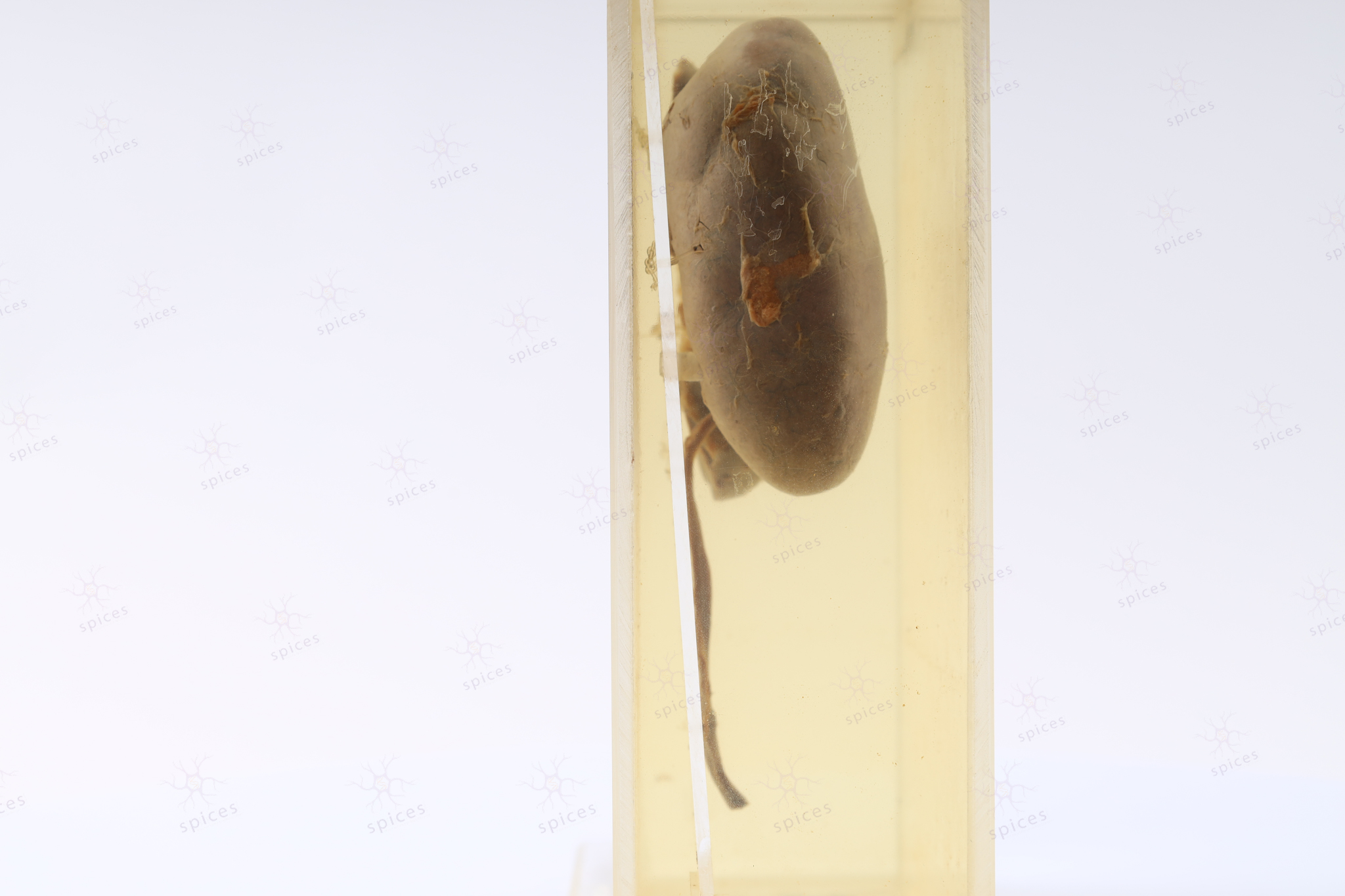
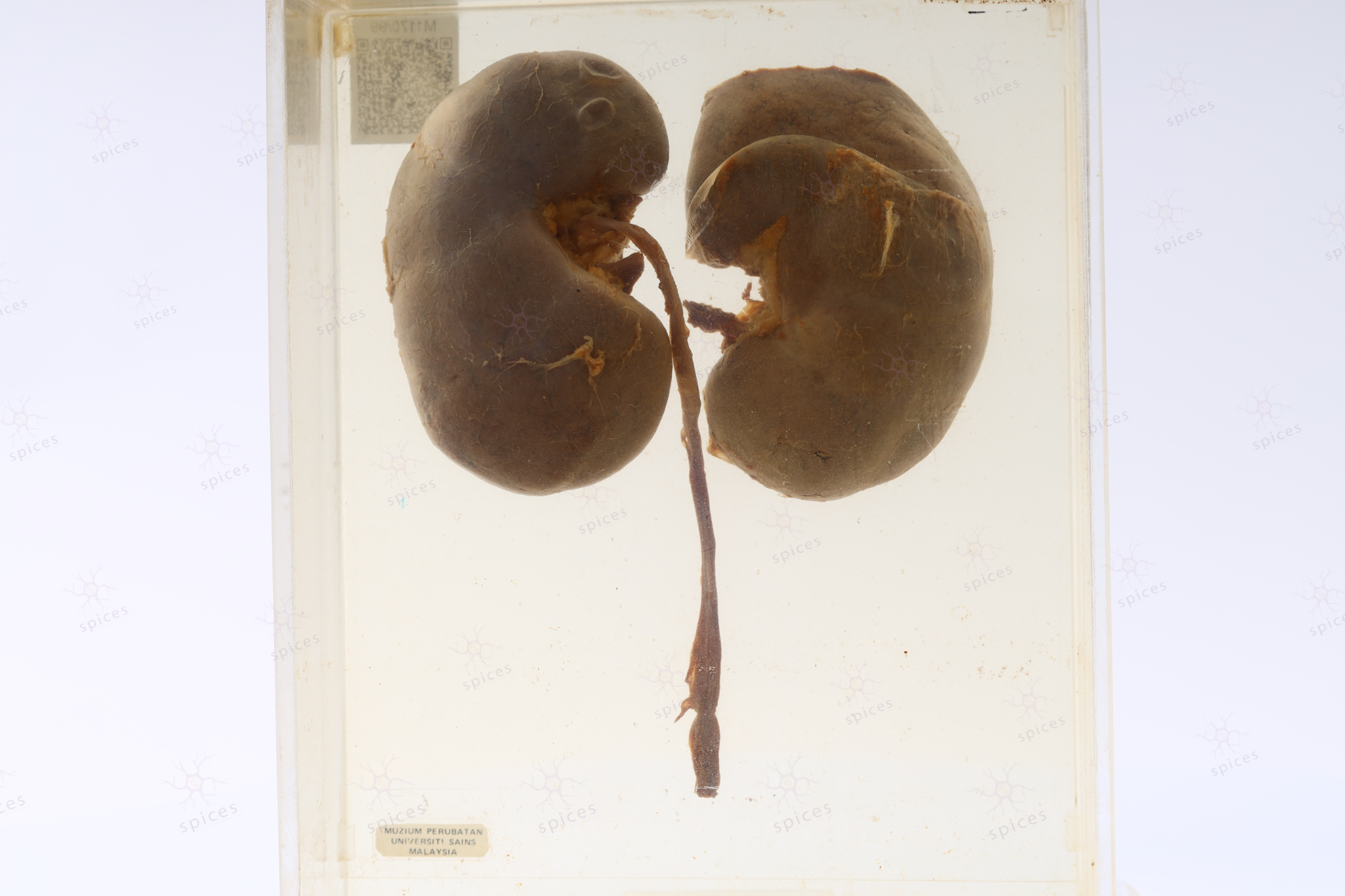
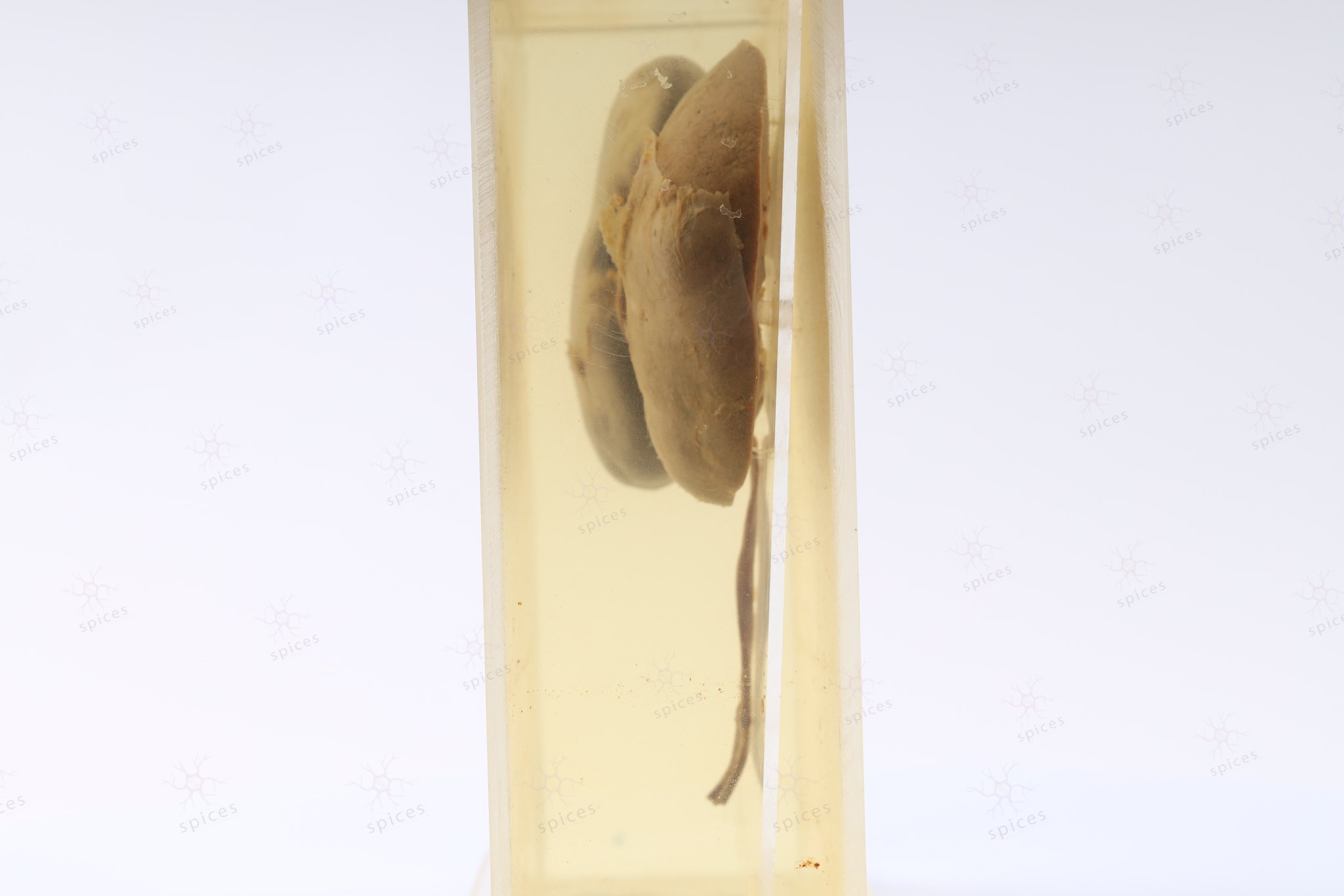
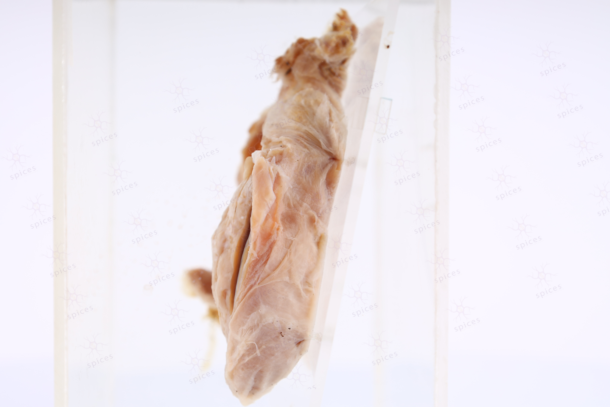
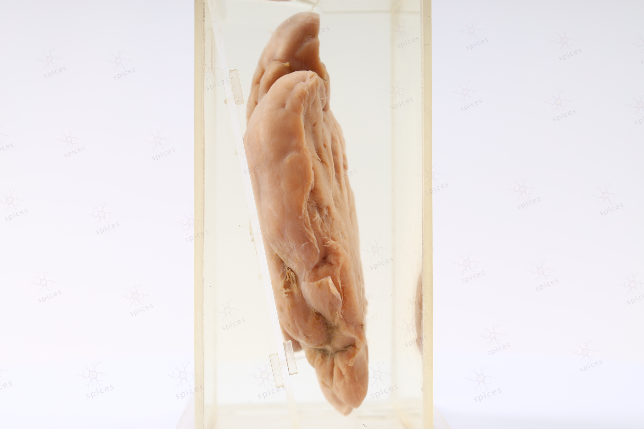
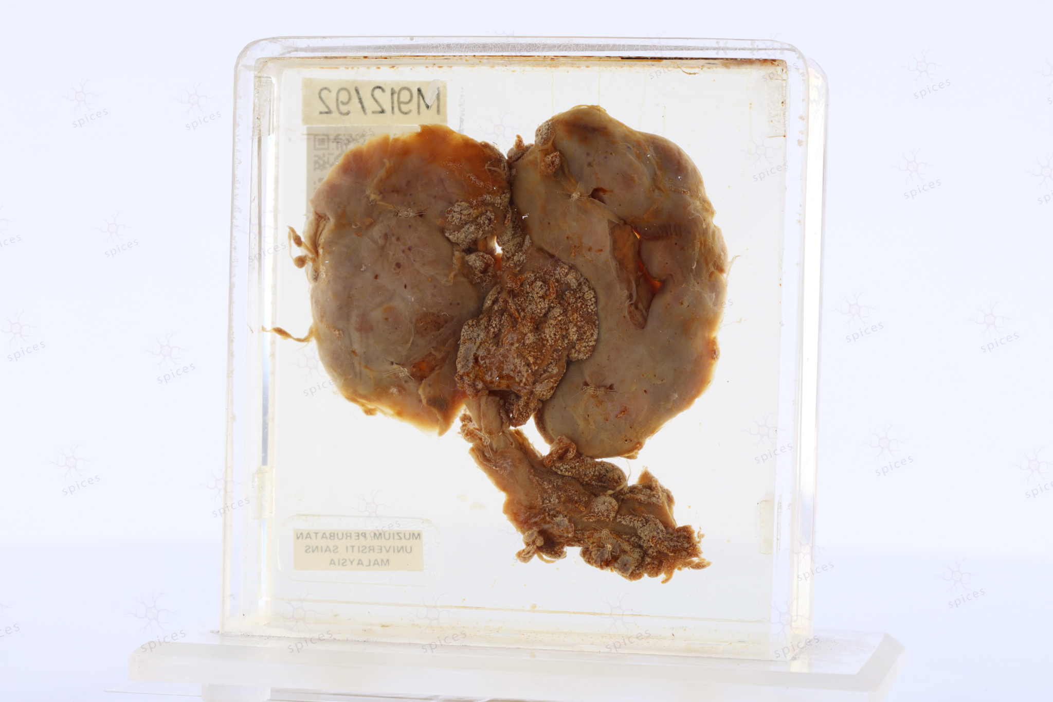
SPICES, School of Medical Sciences  |  Health Campus, USM

Kidney : M912/92

KIDNEY

Spices No: M912/92

  • Front

  • Left

  • BACK

  • RIGHT

  • GROSS DESCRIPTION

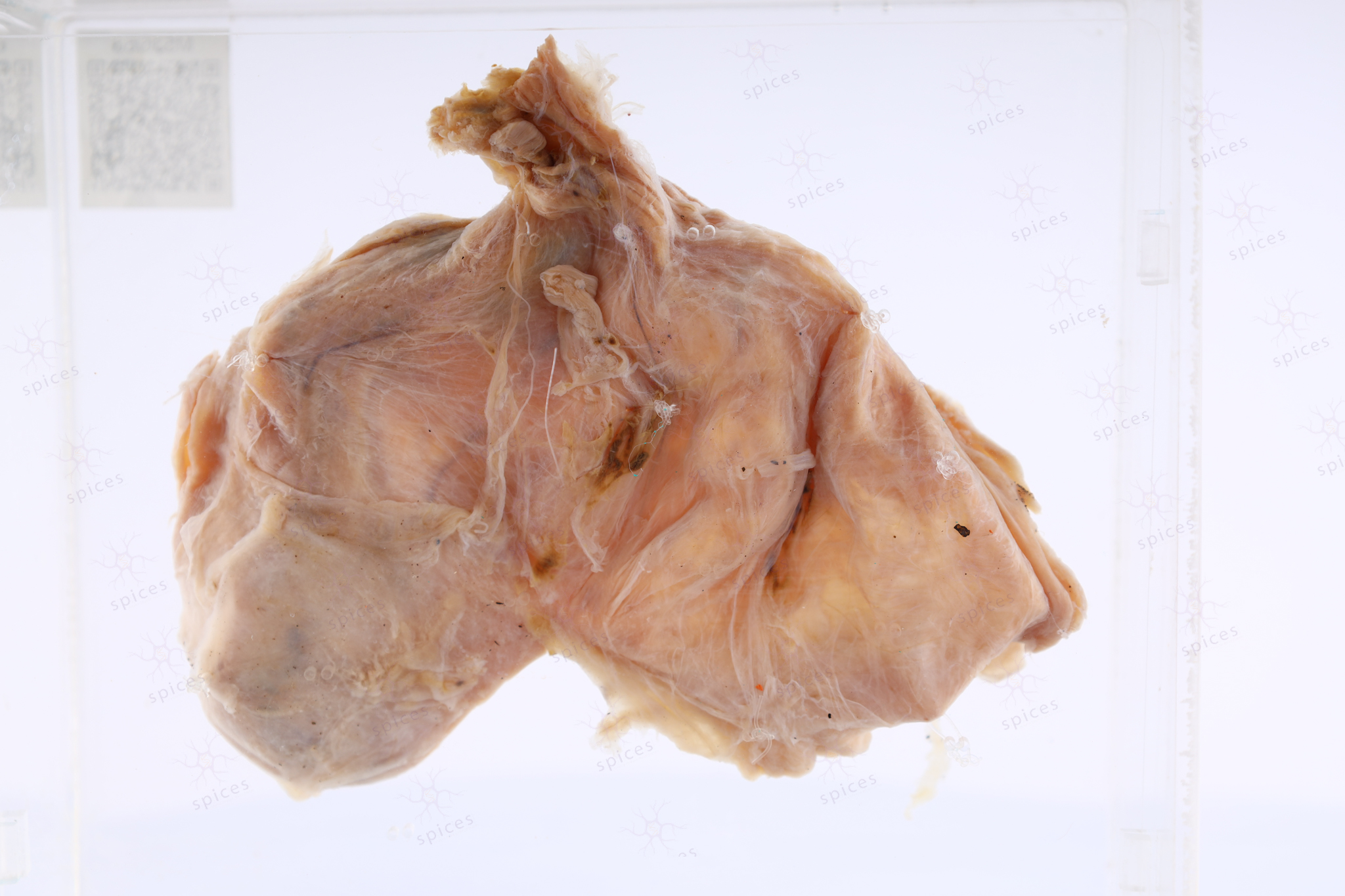
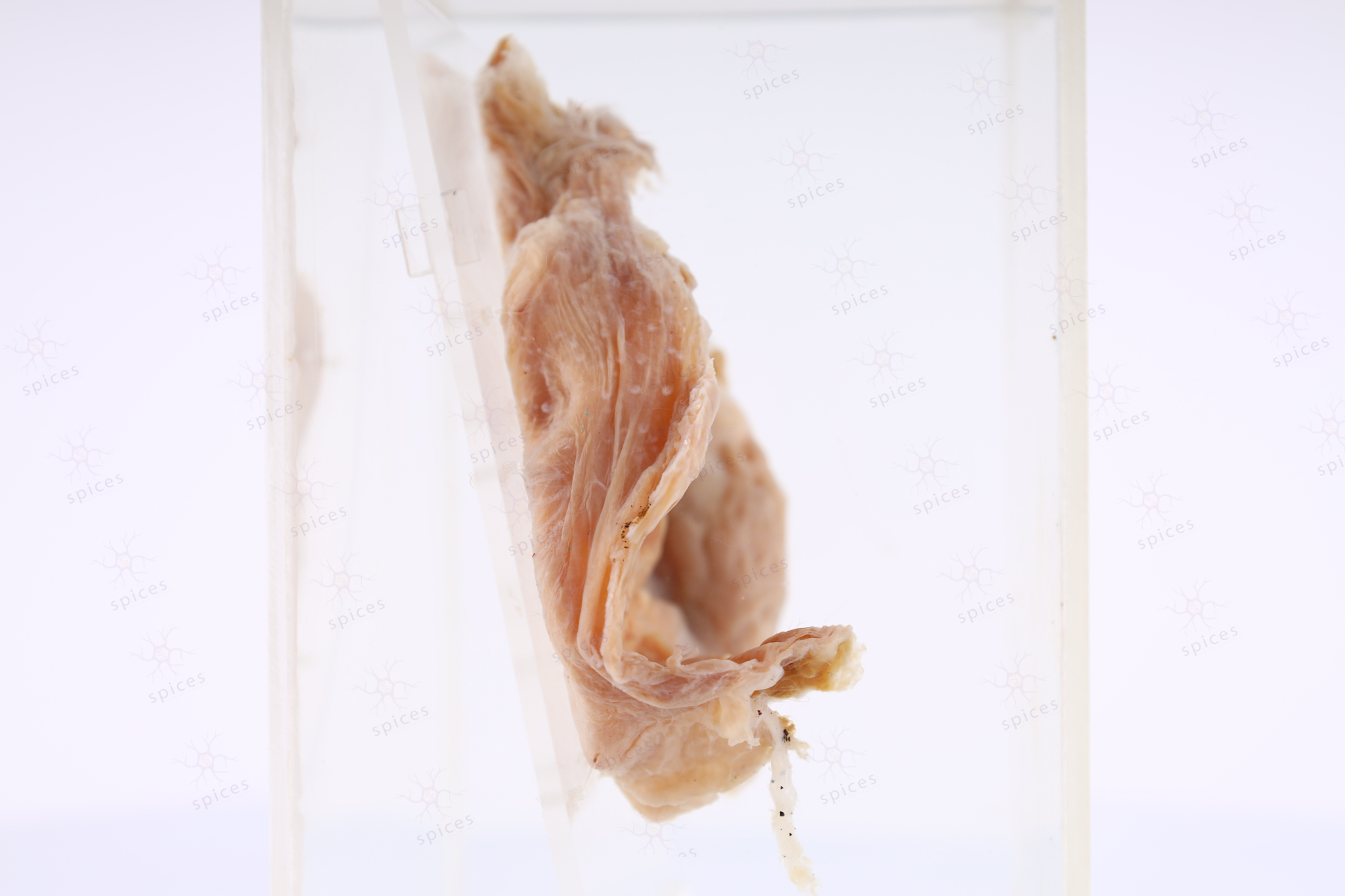
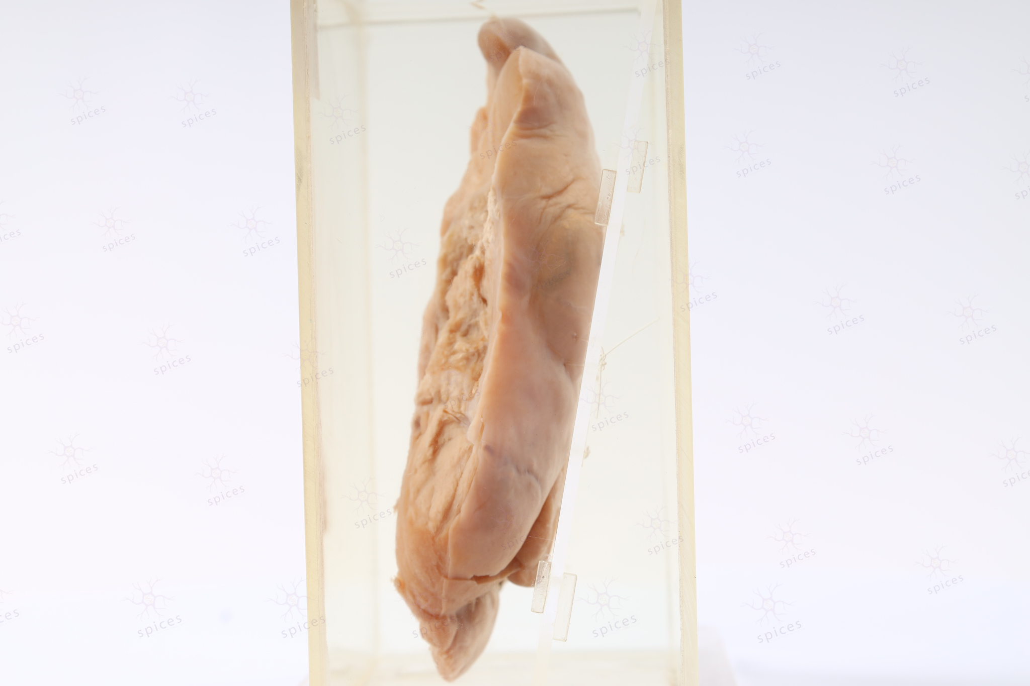
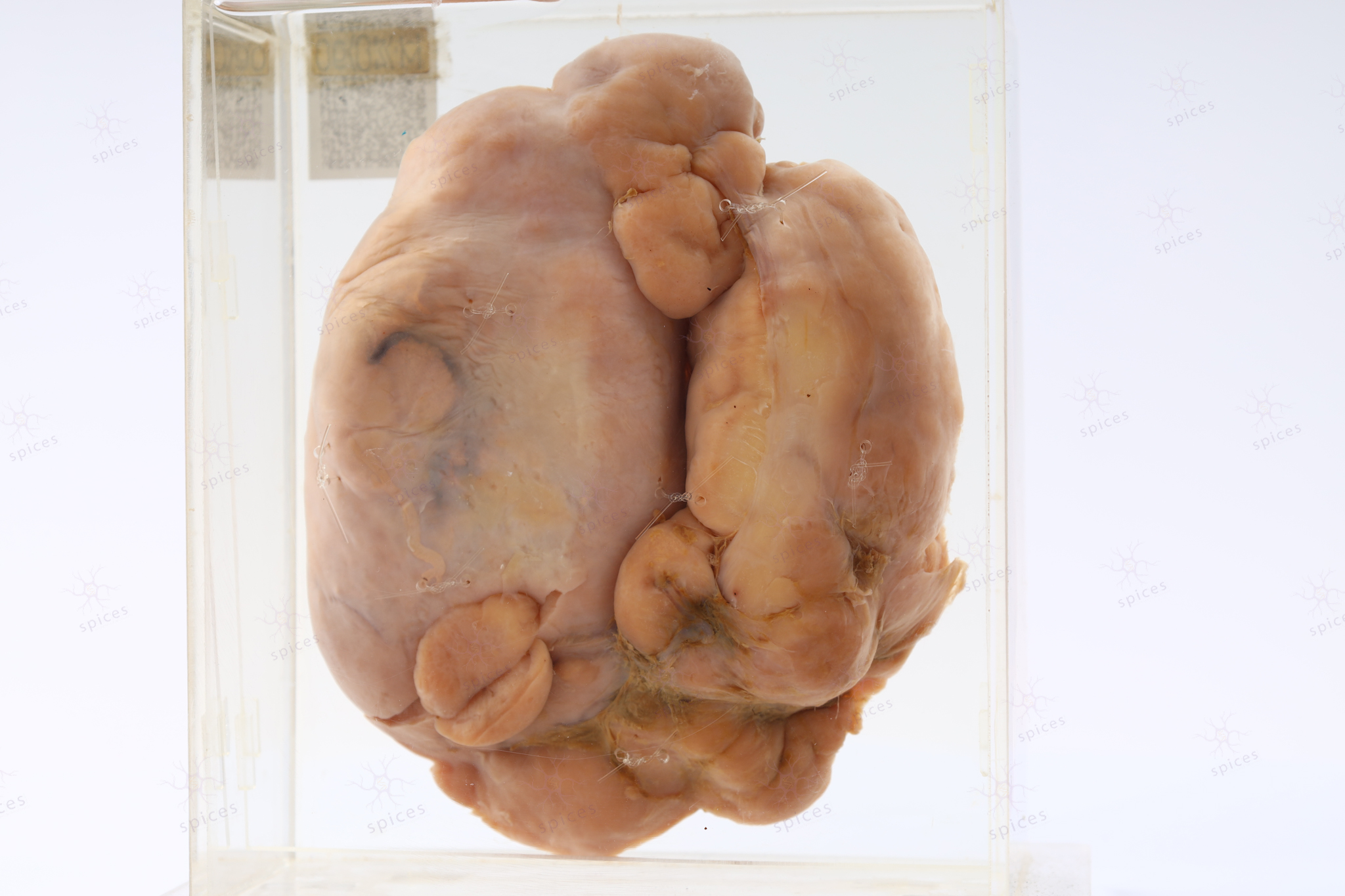
    The exhibit displays kidney with presence of multiple cystic spaces.

  • HISTOPATHOLOGY DESCRIPTION

    Renal parenchyma in chronic pyelonephritis shows tubular atrophy and interstitial and periglomerular fibrosis. Lymphoplasmacytic infiltration can be observed.

  • DIAGNOSIS

    Chronic pyelonephritis

  • BAHASA MELAYU

    Penerangan kasar: Spesimen menunjukkan ginjal dengan ruang sistik yang banyak.

Kidney : M683/90

KIDNEY

Spices No: M683/90

  • GROSS DESCRIPTION

    The exhibit displays an irregular mass ocupying renal parenchymal composed of varigated surface of tan brownish in calour associated with necrosis and haemorraghe

  • HISTOPATHOLOGY DESCRIPTION

    Renal cell carcinoma shows solid growth with nests of the tumour dells composed of polygonal shape cells with sharply defined cytoplasmic border. The nuclei are irregular, wrinkled some showing binucleation. Perinuclear halos are prominent. The cytoplasm is moderate in amount with predominant granular eosinophilic cytoplasm. Mitosis is seen.

  • DIAGNOSIS

    Renal cell carcinoma - chromophobe

  • BAHASA MELAYU

Kidney : M1170/99

KIDNEY

Spices No: M1170/99

  • Front

  • Left

  • BACK

  • RIGHT

  • GROSS DESCRIPTION

    The exhibit displays a normal kidney. The kidney is a bean-shaped organ with the concave surface directed medially. The parenchyma composed of lobes made up of a medullary pyramid and cortex.

  • HISTOPATHOLOGY DESCRIPTION

    Normal kidney shows unremarkable renal pelvycalyceal structure.

  • DIAGNOSIS

    Normal kidney

  • BAHASA MELAYU

    Penerangan kasar: Spesimen memaparkan sebuah ginjal yang normal. Ginjal adalah organ berbentuk kacang dengan permukaan cekung yang mengarah ke tengah. Parenkim ginjal terdiri daripada lobus yang terdiri daripada piramid medulari dan korteks.

    Penerangan histopatologi: Ginjal normal menunjukkan struktur pelvi-kaliseal yang tidak menonjol.

Testis : M536/88

TESTIS

Spices No: M536/88

  • Front

  • Left

  • BACK

  • RIGHT

  • GROSS DESCRIPTION

    The exhibit displays testis with presence of thin-walled cystic space around. This cystic space is due to accumulation of fluid between visceral and parietal layers of tunica vaginalis.

  • HISTOPATHOLOGY DESCRIPTION

    Hydrocele is characterized by loose connective tissue lined by a single layer of cuboidal or flattened mesothelial cells

  • DIAGNOSIS

    Hydrocele

  • BAHASA MELAYU

    Penerangan kasar: Spesimen menunjukkan testis dengan ruang sistik (cystic) berdinding nipis di sekelilingnya. Ruang sistik ini disebabkan oleh penumpukan cecair di antara lapisan viseral dan lapisan parietal tunika vaginalis.

    Penerangan histopatologi: Ciri-ciri hydrocele menunjukkan jaringan tisuu penghubung yang longgar dan dilapisi oleh lapisan tunggal sel mesothelial kuboidal atau pipih.

Testis : M740/90

TESTIS

Spices No: M740/90

  • Front

  • Left

  • BACK

  • RIGHT

  • GROSS DESCRIPTION

    The exhibit displays a solid tumour occupying the whole testis with. It show homogenous tan-yellowish glistening surface. Nephroblastoma are usually solitary, rounded, multinodular masses and sharply demarcated from the adjacent renal parenchym by a peritumoural fibrus pseudocapsule.

  • HISTOPATHOLOGY DESCRIPTION

    Seminoma shows tumour cells with characteristically pale to clear cytoplasm and polygonal nuclei. They are often arranged in sheet and lobules with lymphocytes infiltrates.

  • DIAGNOSIS

    Seminoma

  • BAHASA MELAYU

    Penerangan kasar: Spesimen menunjukkan ketumbuhan pejal yang mengisi seluruh testis. Ia menunjukkan permukaan homogen yang berkilat berwarna coklat kekuningan. Nefroblastoma (barah ginjal) biasanya padat, berbentuk bulat dan bersisik bersebelahan dengan parenkim ginjal yang berdekatan oleh pseudokapsul serat peritumoral.