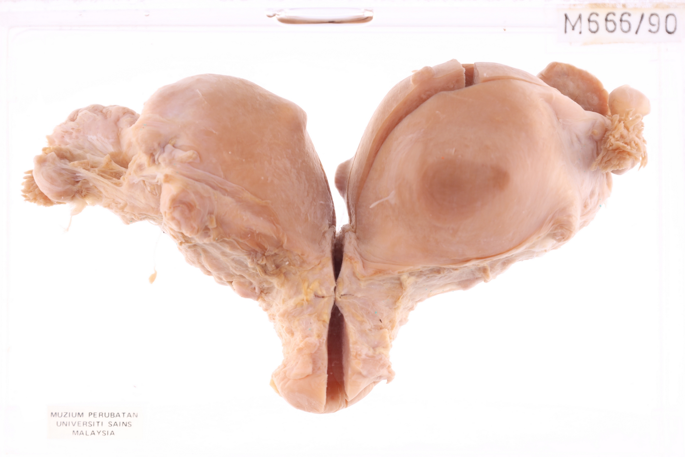
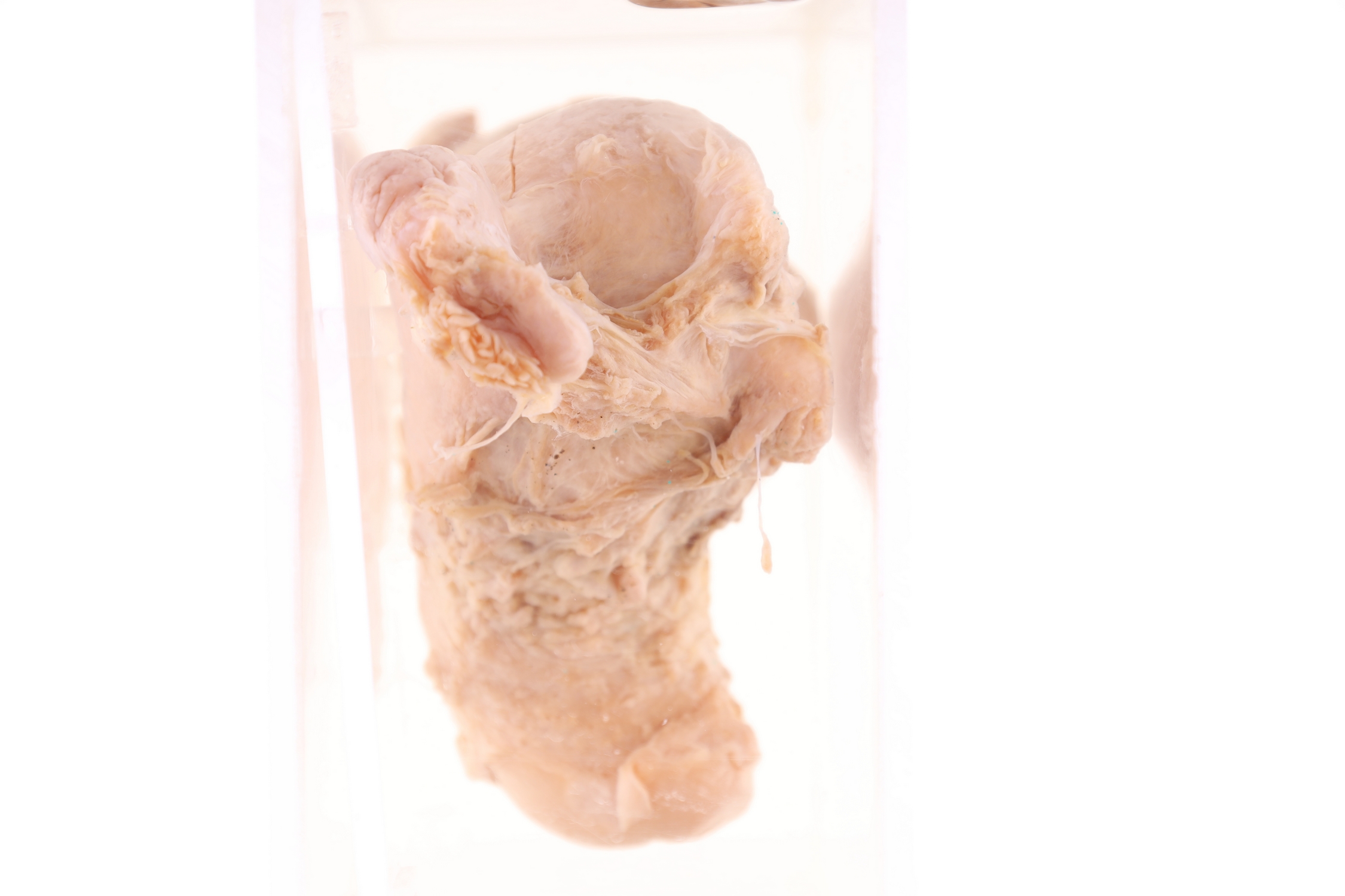
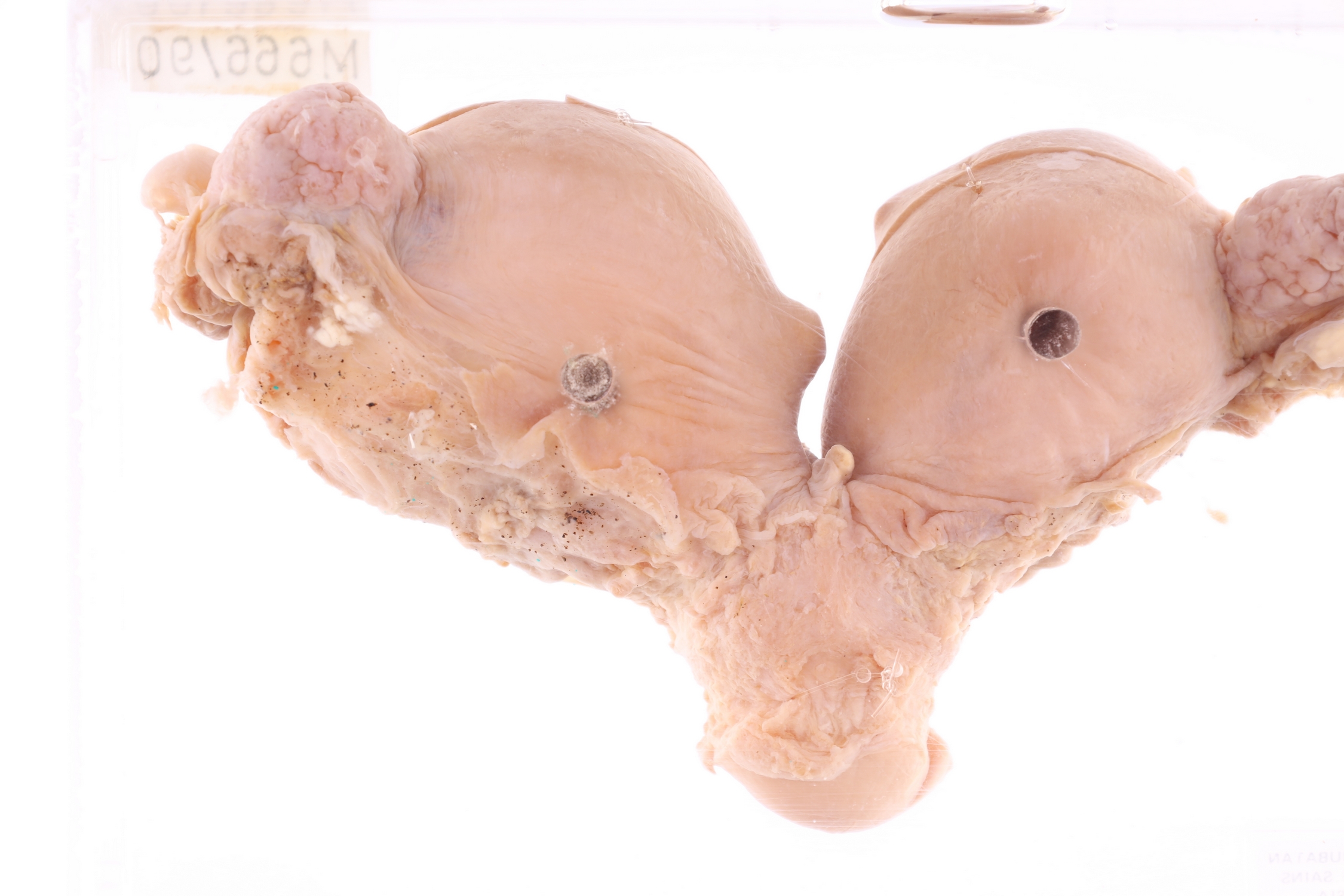
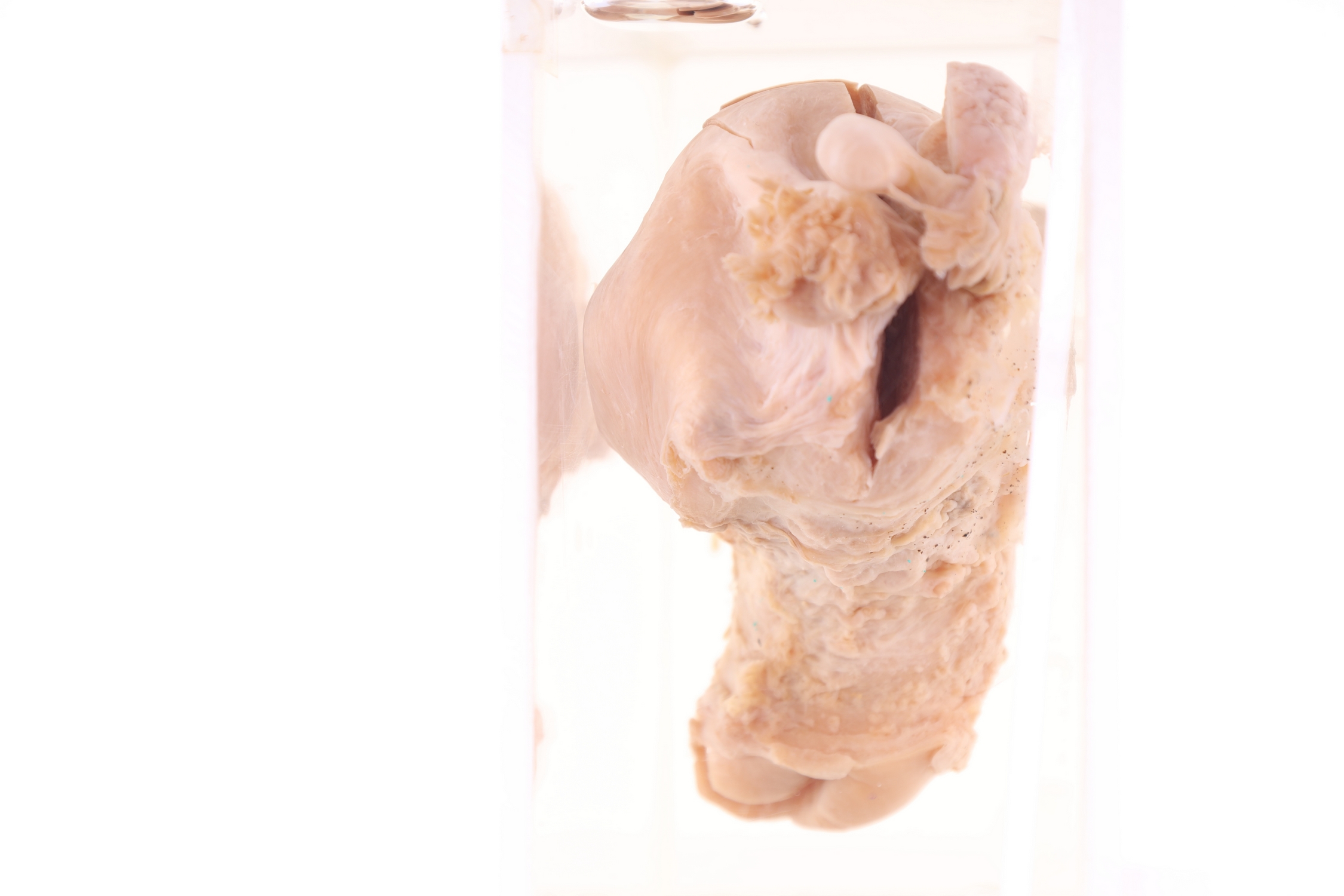
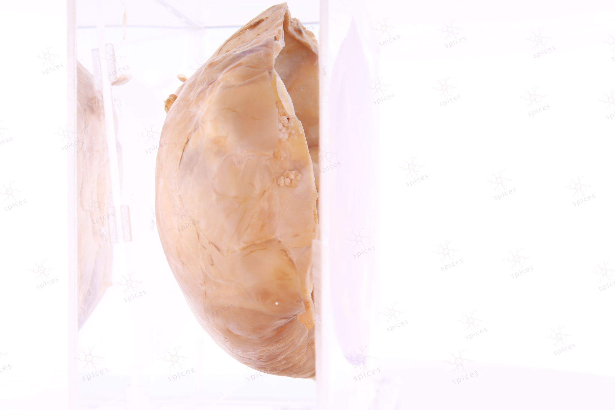
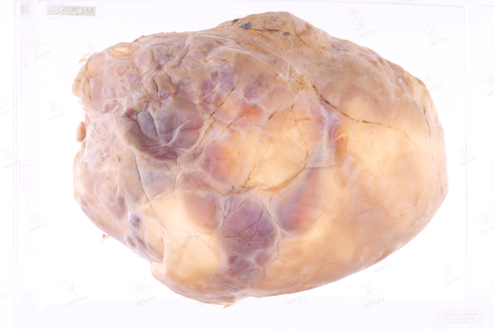
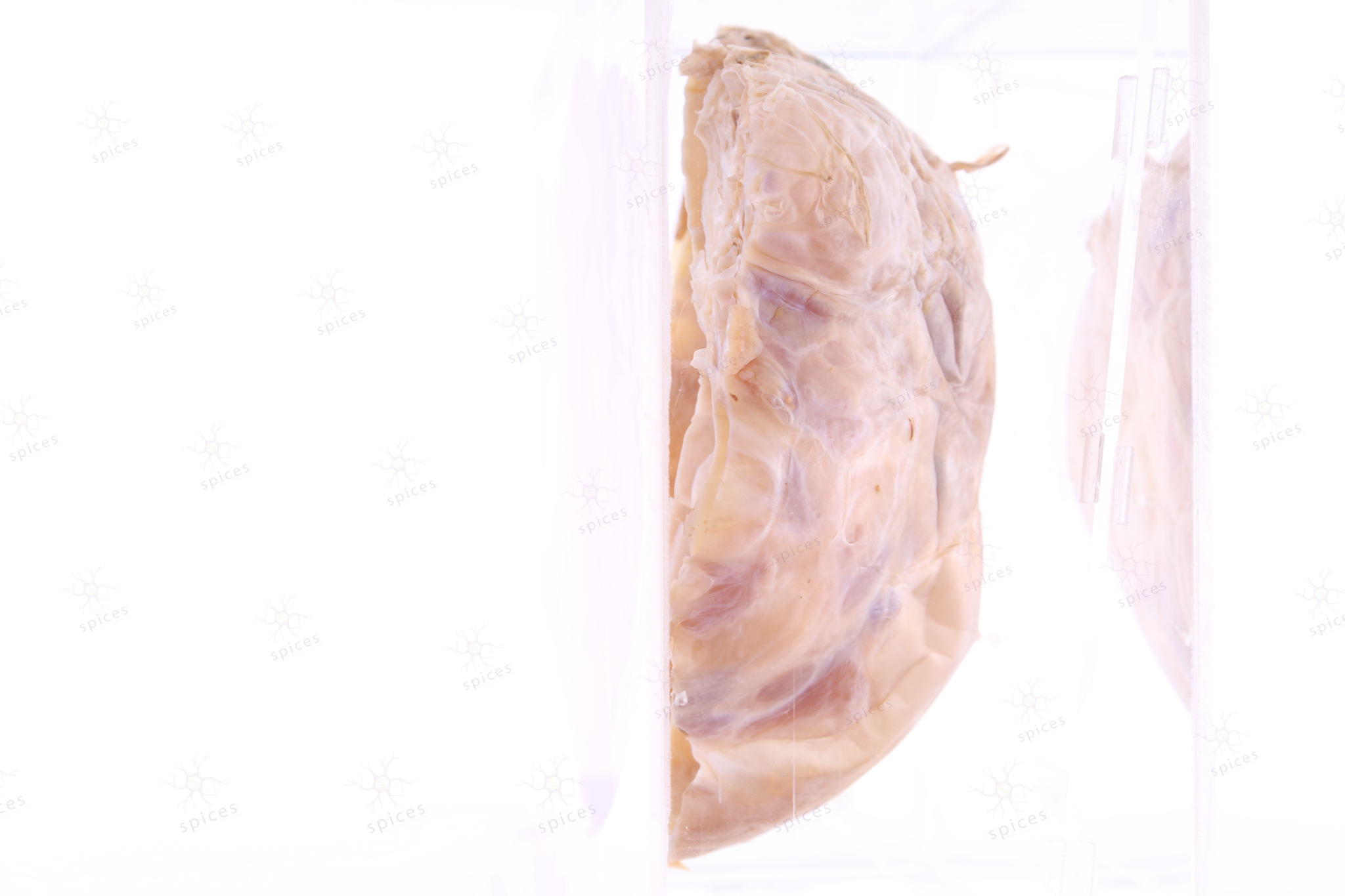
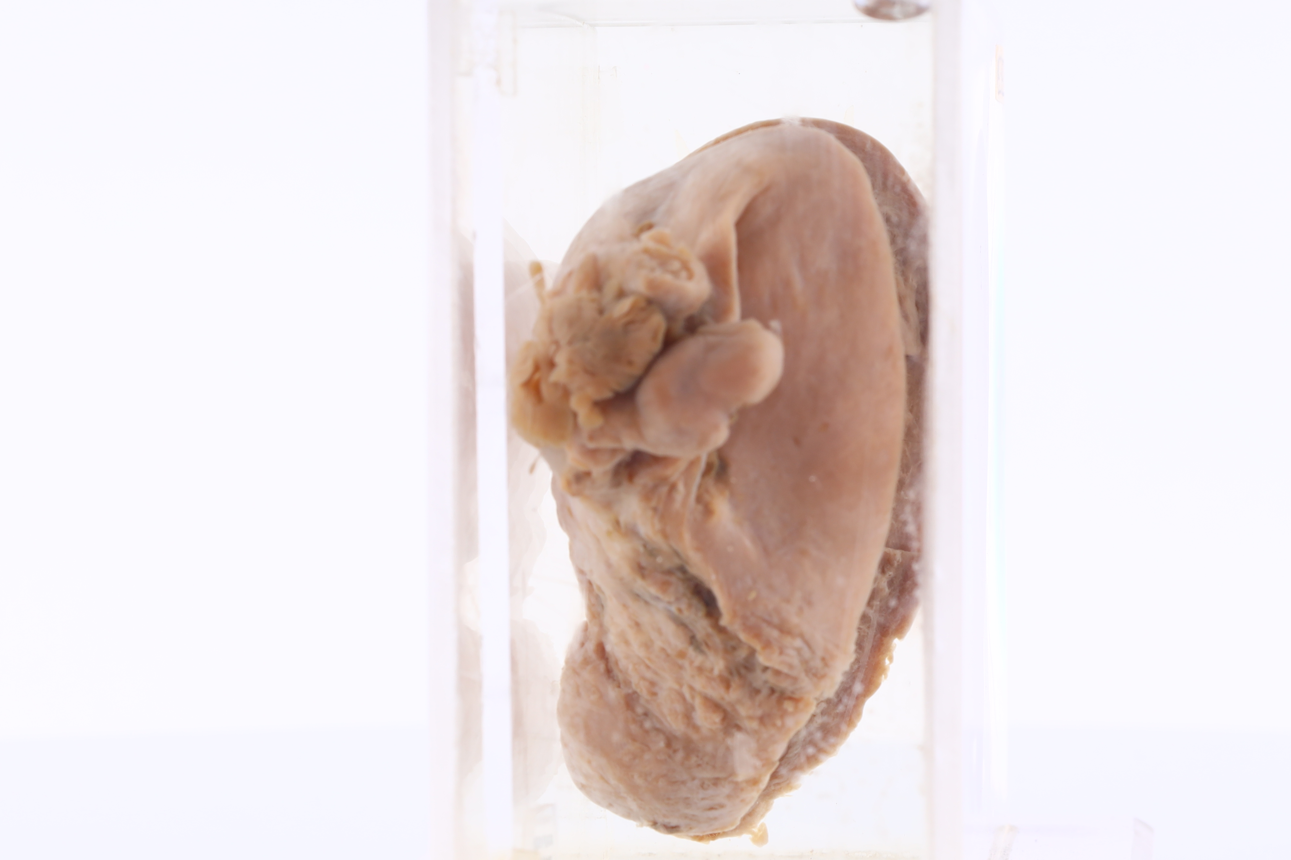
Ovary : M425/86B
Ovary
Spices No : M425/86B

-
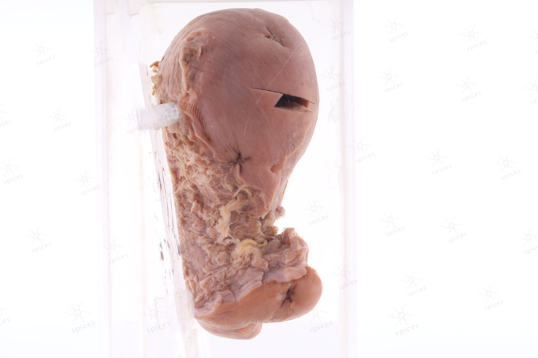
GROSS DESCRIPTION
The exhibit displays a cystic mass with multiloculation and septation. The wall is thin, however there is thick areas with foci of simple papillary structure project into the lumen. Cystadenoma exhibit smooth outer and inner surface.
-
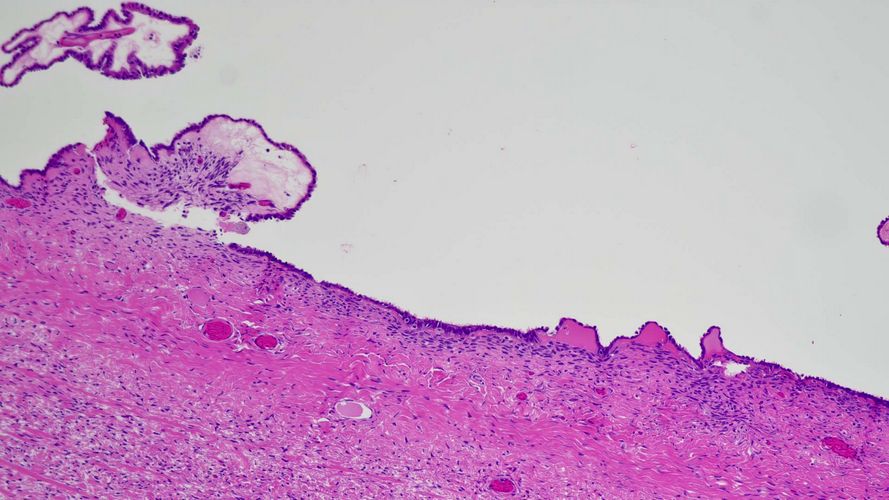
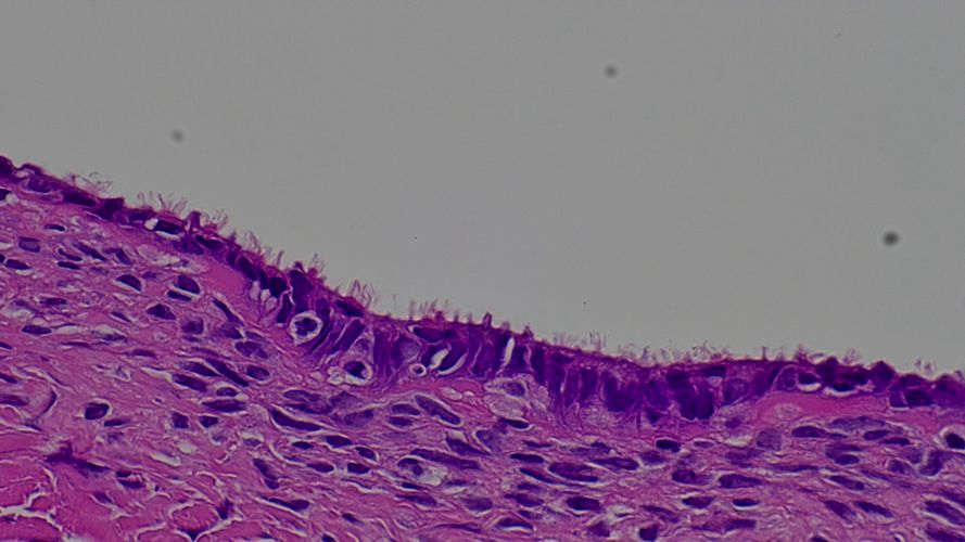
HISTOPATHOLOGY DESCRIPTION
Serous cystadenoma show fibrocollagenous wall lined by single layer of bland tubal epithelium. Epithelial proliferation should be less than 10%.
-
DIAGNOSIS
Serous cystadenoma
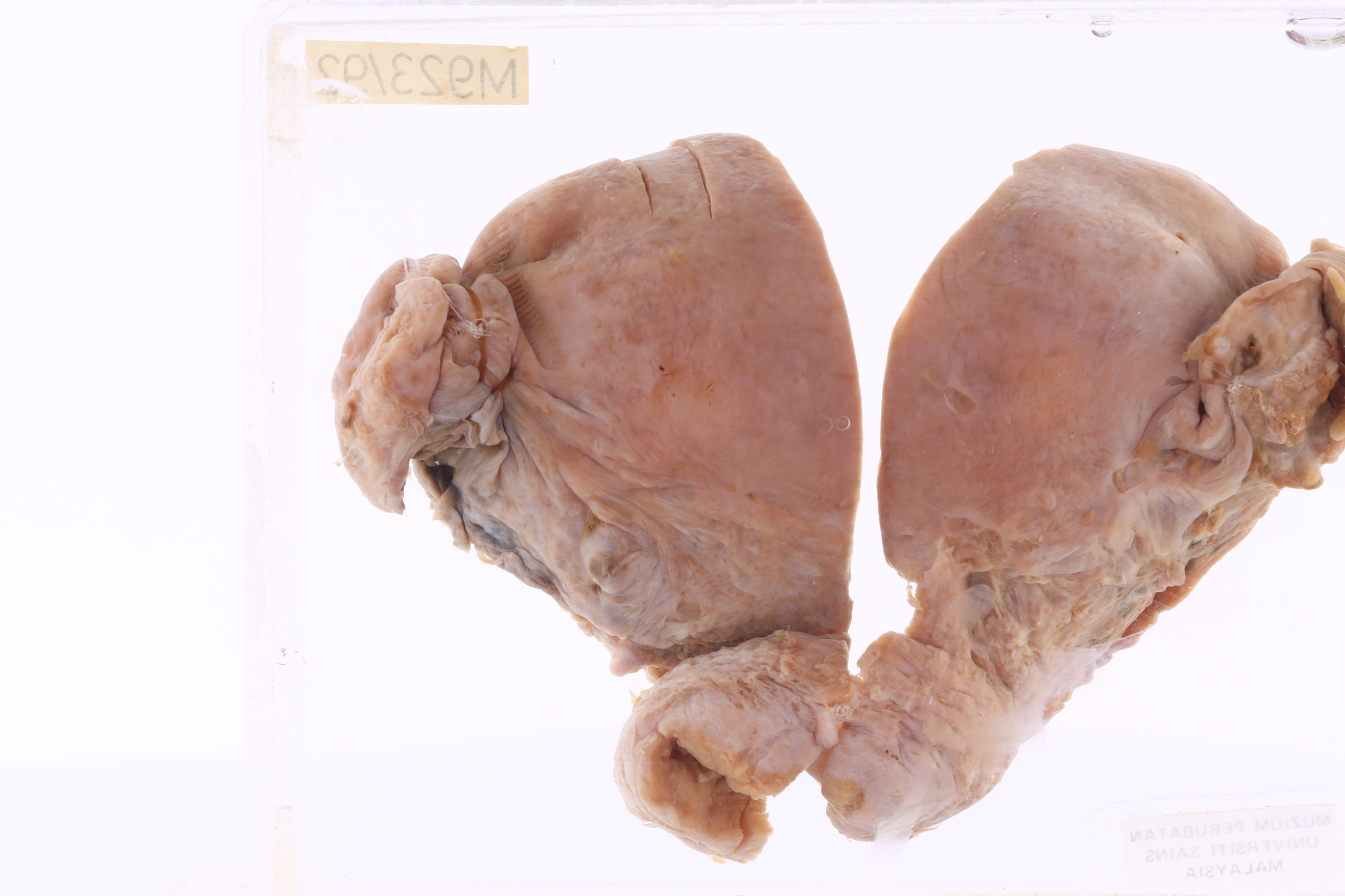
Uterus IUCD
UTERUS with IUCD
SPICES No.

-
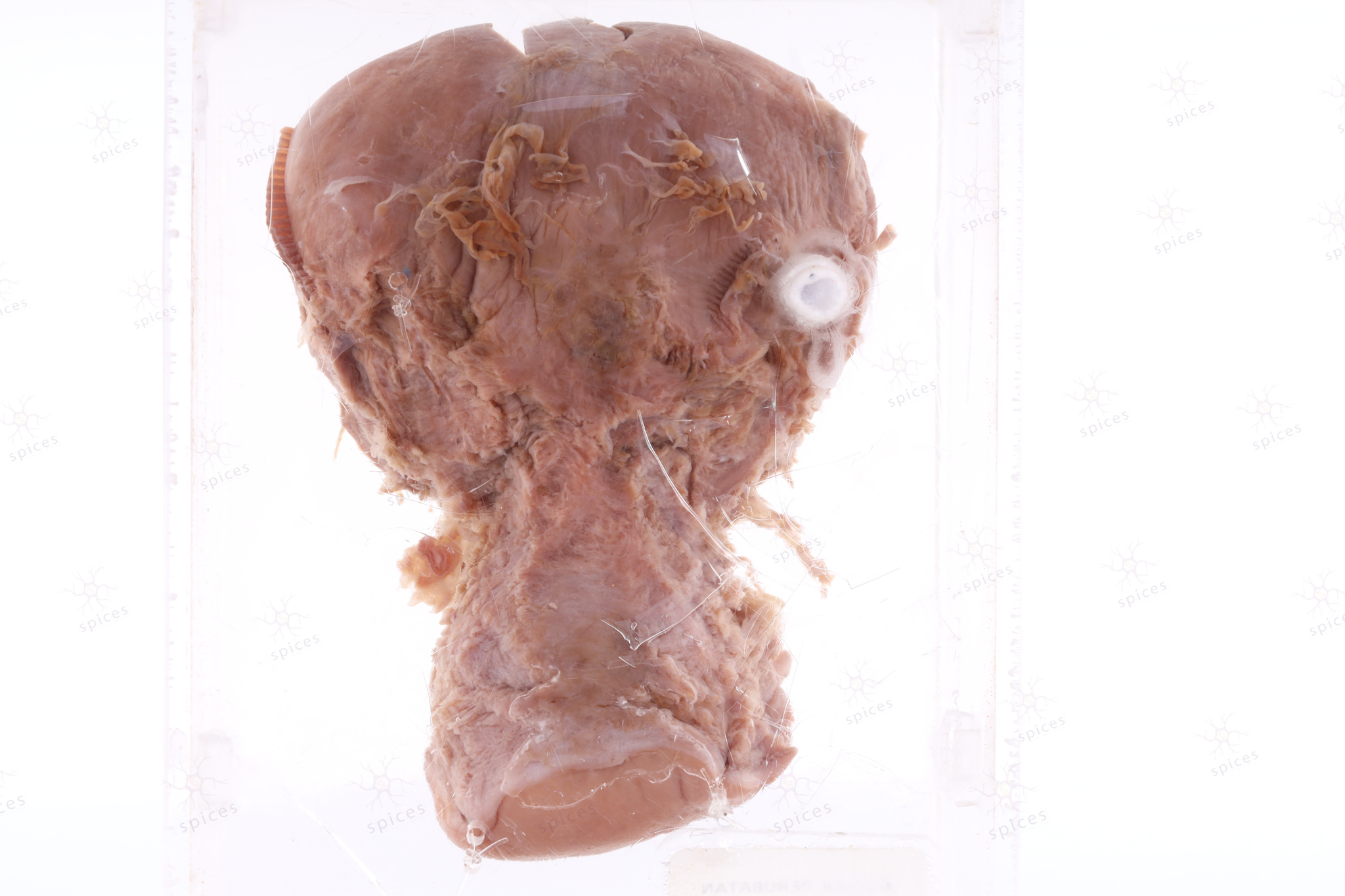
GROSS DESCRIPTION
The exhibit displays uterus with presence of intrauterine contraceptive device.
-
DESCRIPTION
Intrauterine contraceptive device are a safe and cost-effective contraceptive method for women for preventing unintended pregnancies. The effectiveness of the IUCD as a contraceptive method is approximately 99.2% to 99.8% within the first year of use, which is higher than other shorter-term reversible contraceptive methods, such as the oral contraceptive pill, within the same timeframe of use. Advantages of IUCDs include long-term effectiveness, easily reversible, and safe for use in post-abortion patients.
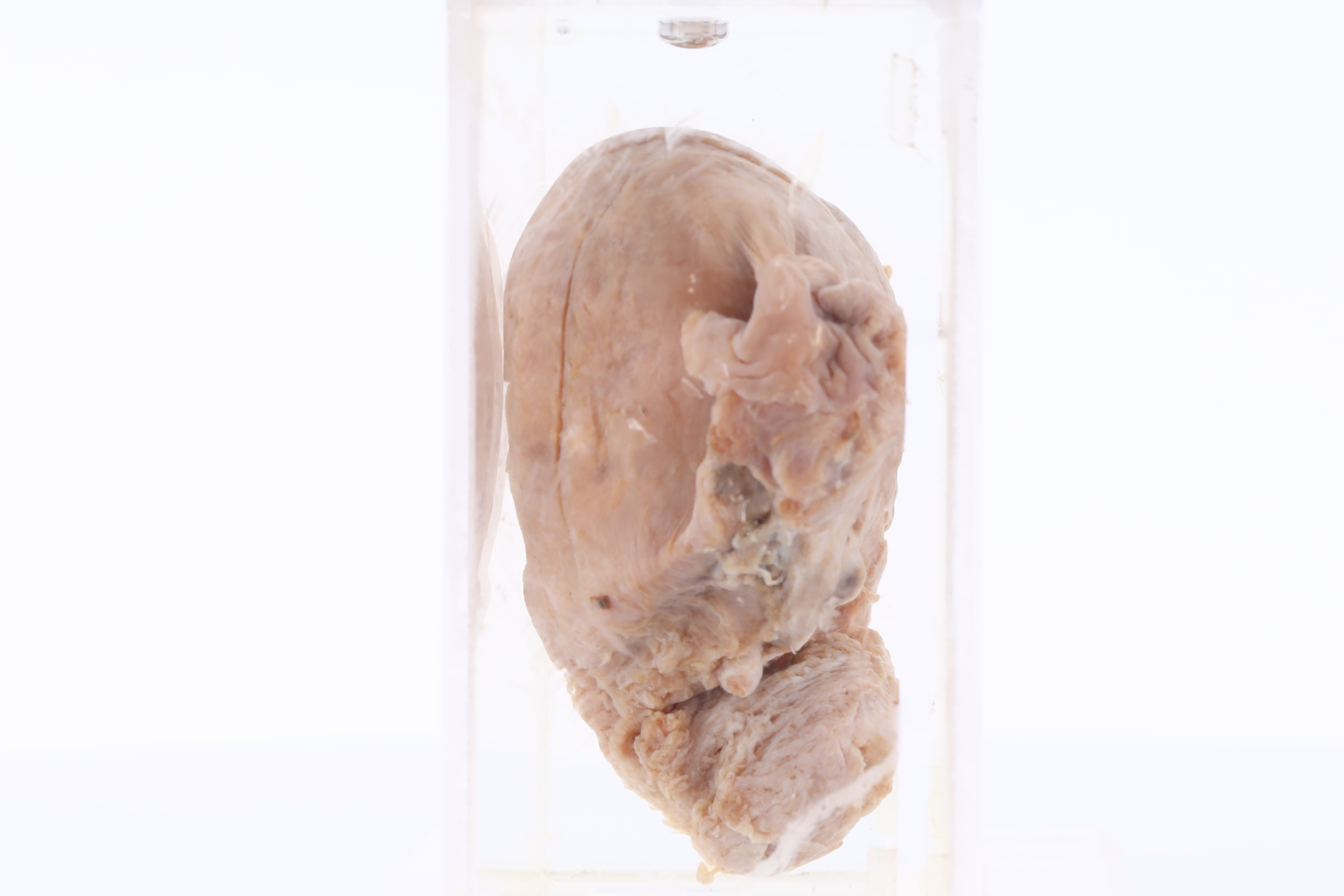
Uterus
UTERUS
SPICES No. M923/92

-
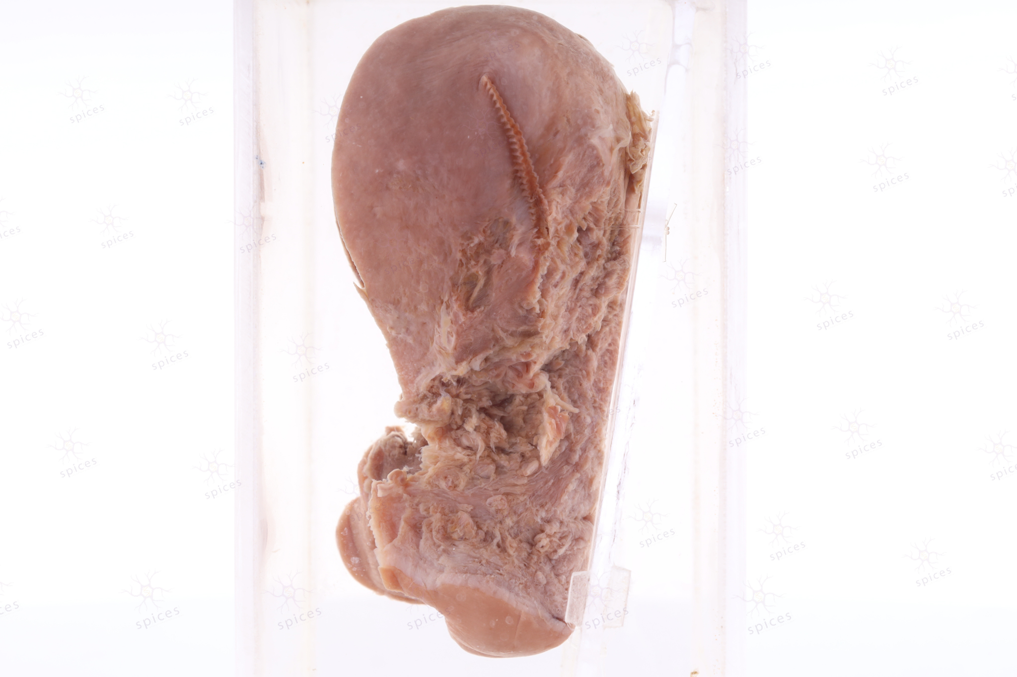
GROSS DESCRIPTION
The exhibit displays a thicken endometrium.
-
HISTOPATHOLOGY DESCRIPTION
Endometrial hyperplasia shows increase in the ratio of endometrial glands to stroma. It is futher categorized into atypia or without atypia depends on the cytology features.
-
DIAGNOSIS
Endometrial hyperplasia
-
QUIZ
Precusor of the endometrial carcinoma
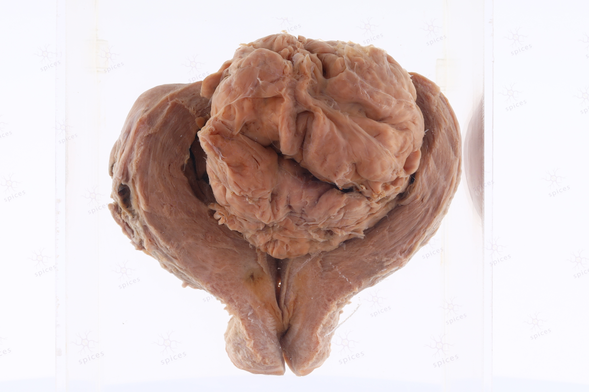
Uterus
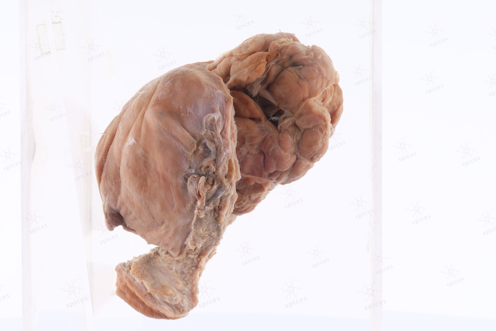
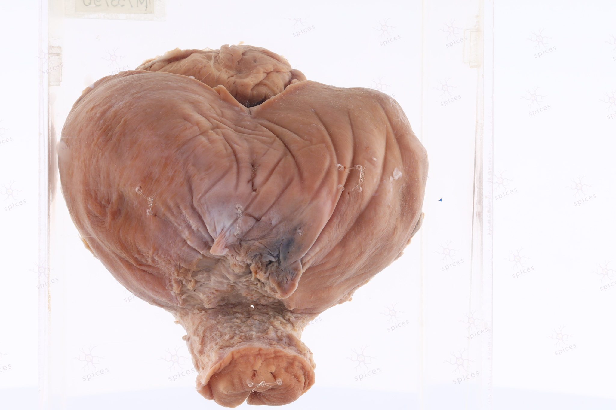
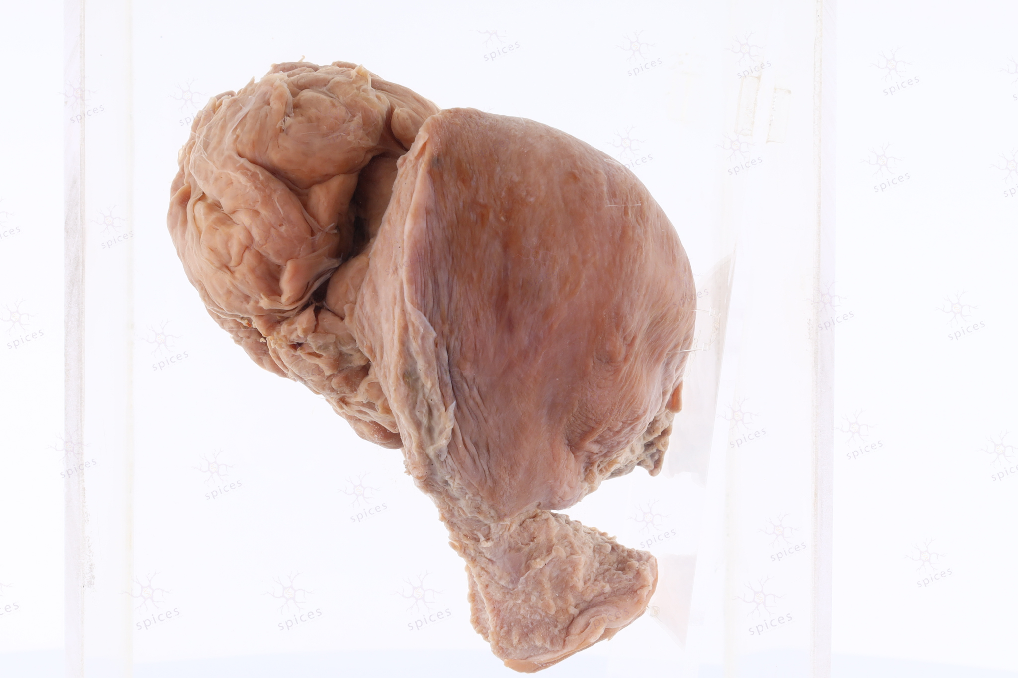
UTERUS
SPICES No. M753/90
-
GROSS DESCRIPTION
The exhibit diplay large submucosal fibroid bulging into the endometrial cavity. It show well circumscribed margin, with lobulated appearance. The surface is homogenous. Trabeculation can be observed.
-
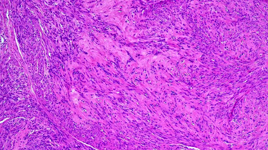
HISTOPATHOLOGY DESCRIPTION
Leimyoma is well circumscribed and unencapsulated. It is composed of spindle cells arranged in intersecting fascicles. The cells have cigar-shaped nuclei, indistinct cytoplasmic borders and eosinophilic cytoplasm.
-
DIAGNOSIS
Leiomyoma