
SPICES, School of Medical Sciences  |  Health Campus, USM

Retroperitoneal tumour - M350/85

RETROPERITONEAL TUMOUR

Spices No: M350/85

  • Front

  • Left

  • BACK

  • RIGHT

  • GROSS DESCRIPTION

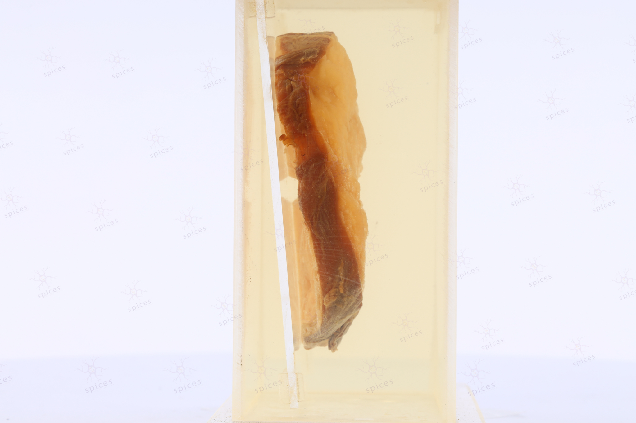
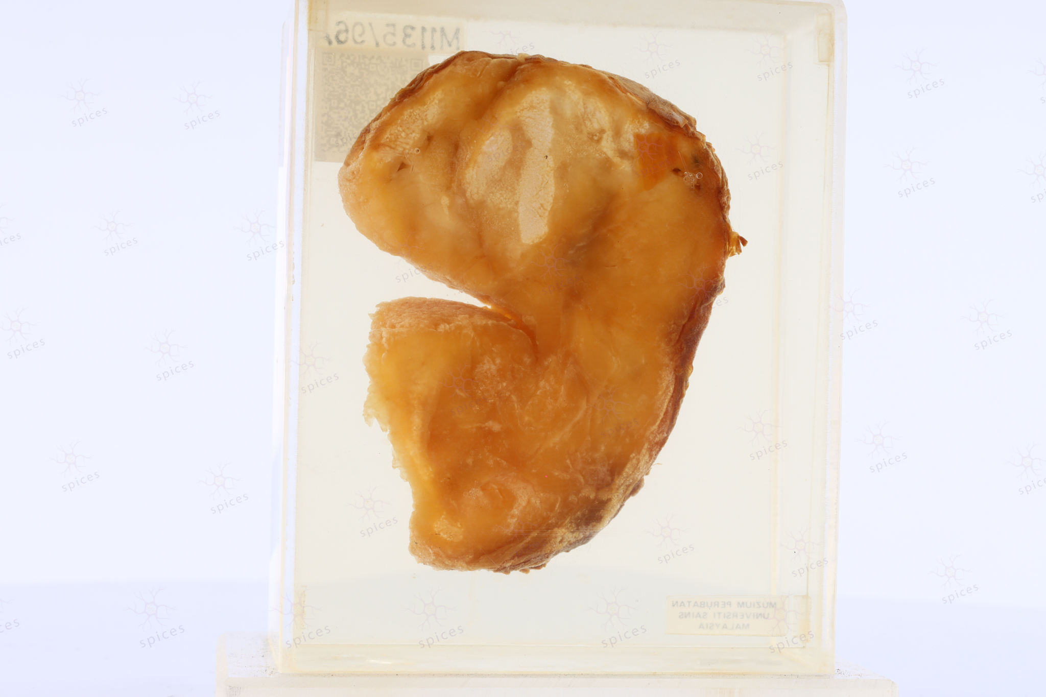
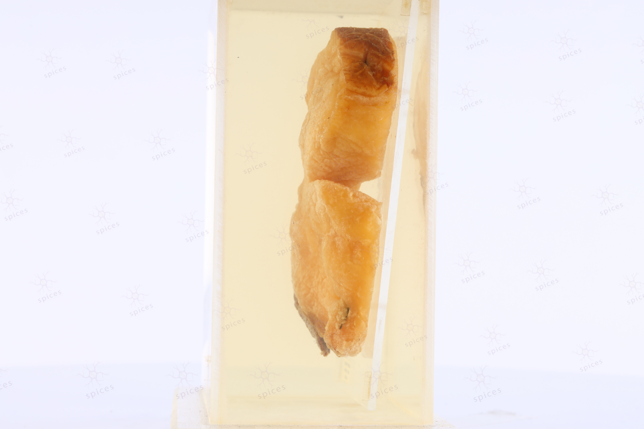
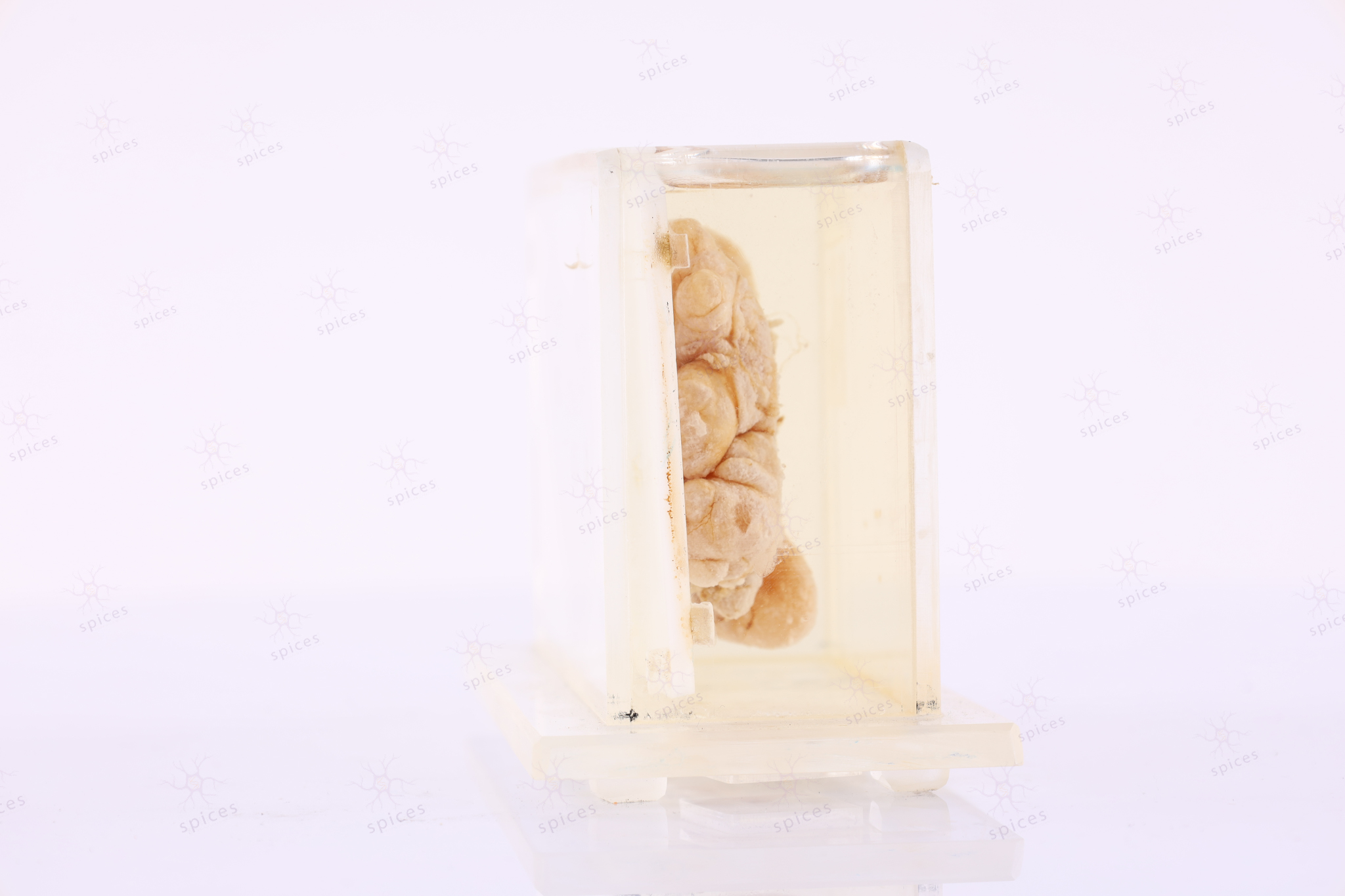
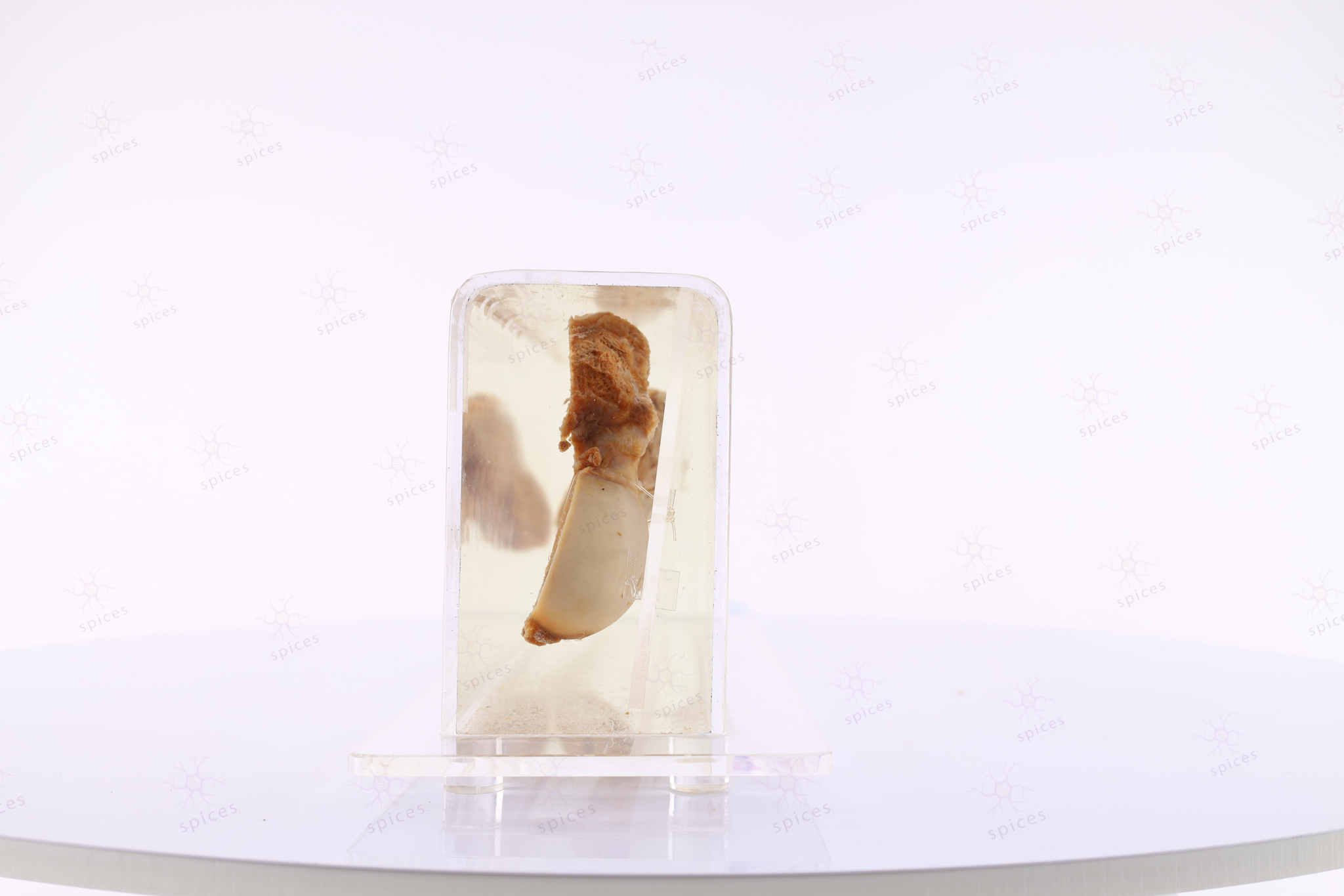
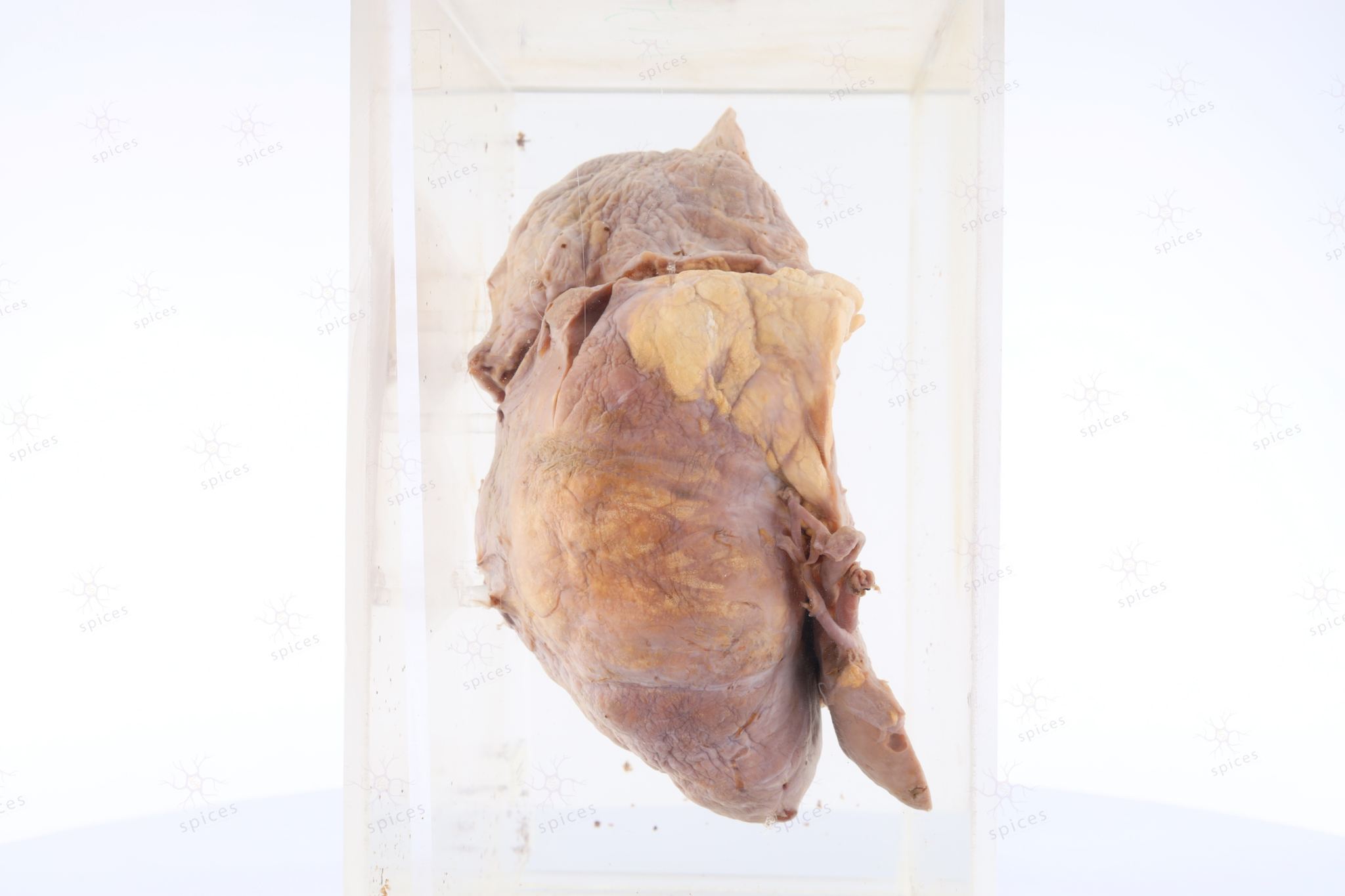
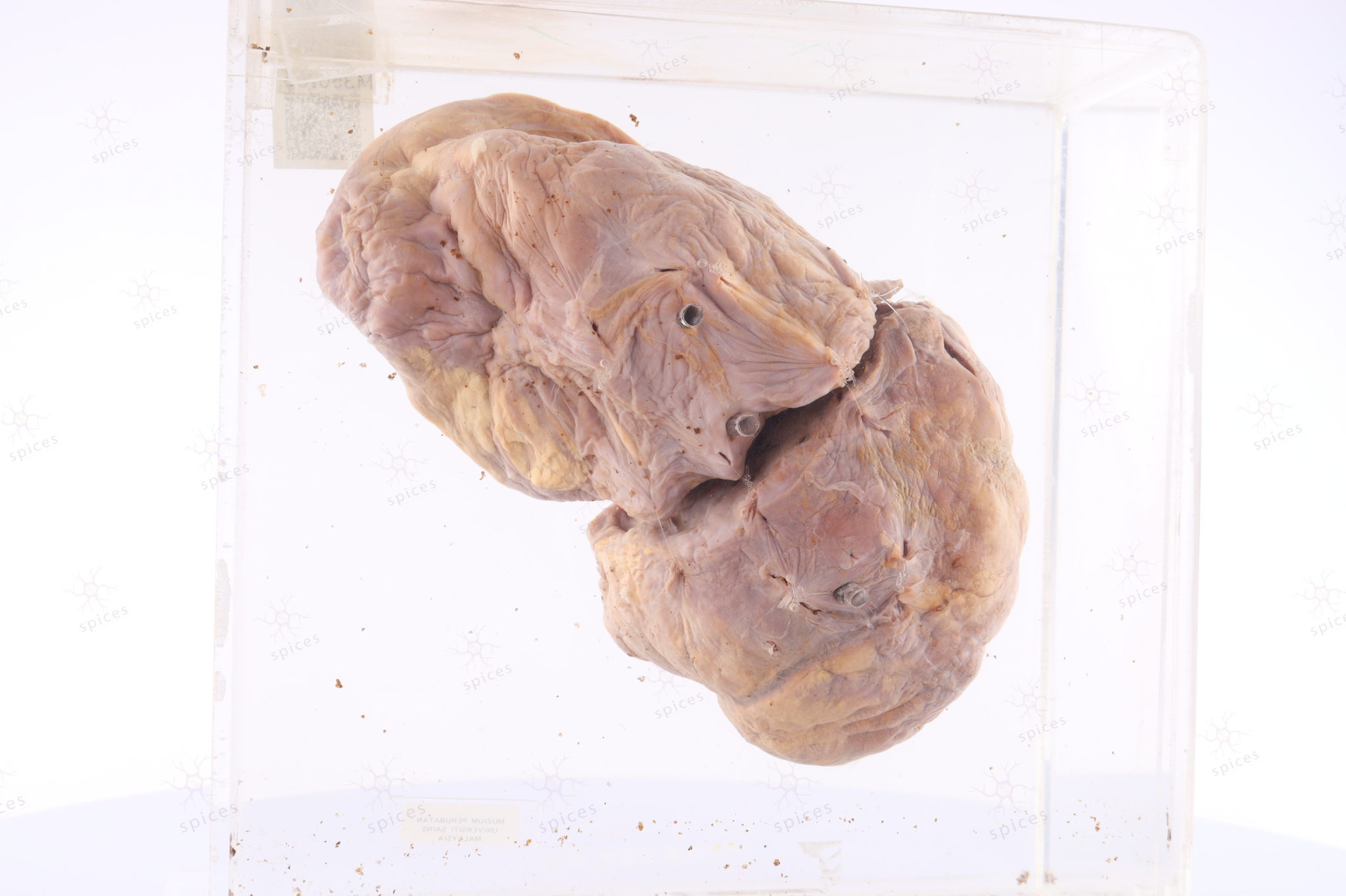
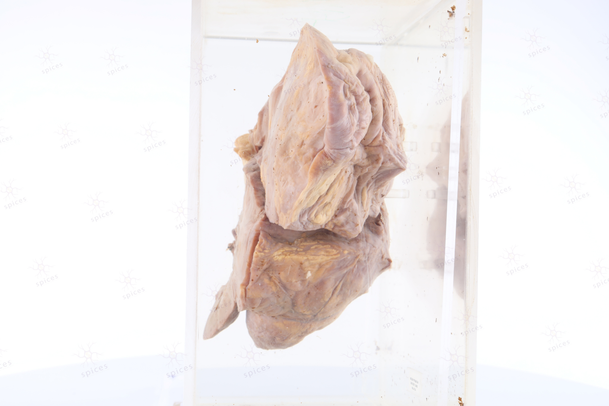
    It is a soft tissue mass, fairly circumscribed, covered by thin fibrous capsule. Cut section shows solid brownish to yellowish cut-surface.Take note that the brownish area is the dedifferentiated area with an abrupt transition to a well differentiated area (yellow). The tumour abutting the kidney (left bottom). Margins are involved by the tumour.

  • HISTOPATHOLOGY DESCRIPTION

    m350 85   microscopic

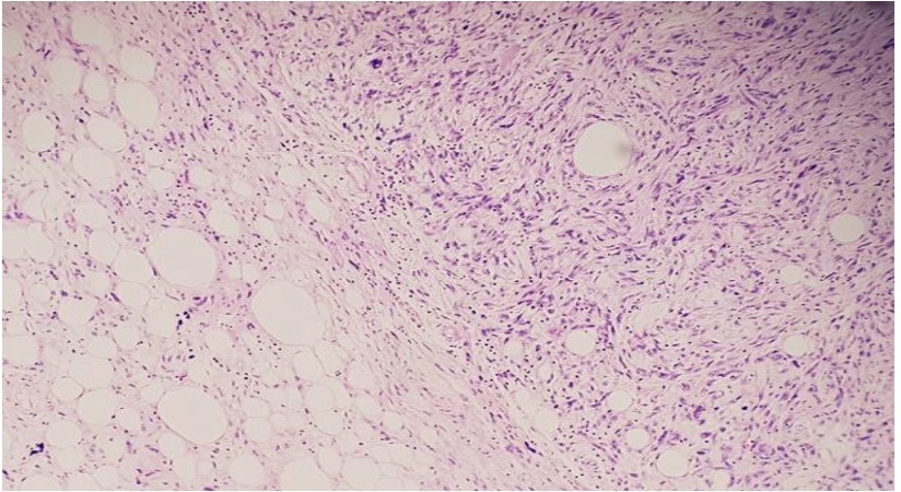
    Section shows well differentiated liposarcoma (left) with an abrupt transition to the dedifferentiated component, exhibiting pleomorphic spindle shape cells arranged in vague storiform pattern (non-lipogenic component) on the right side.


    Fluorescence In Situ Hybridisation (FISH) analysis shows amplifications of MDM2.

    m350 85    comb18a

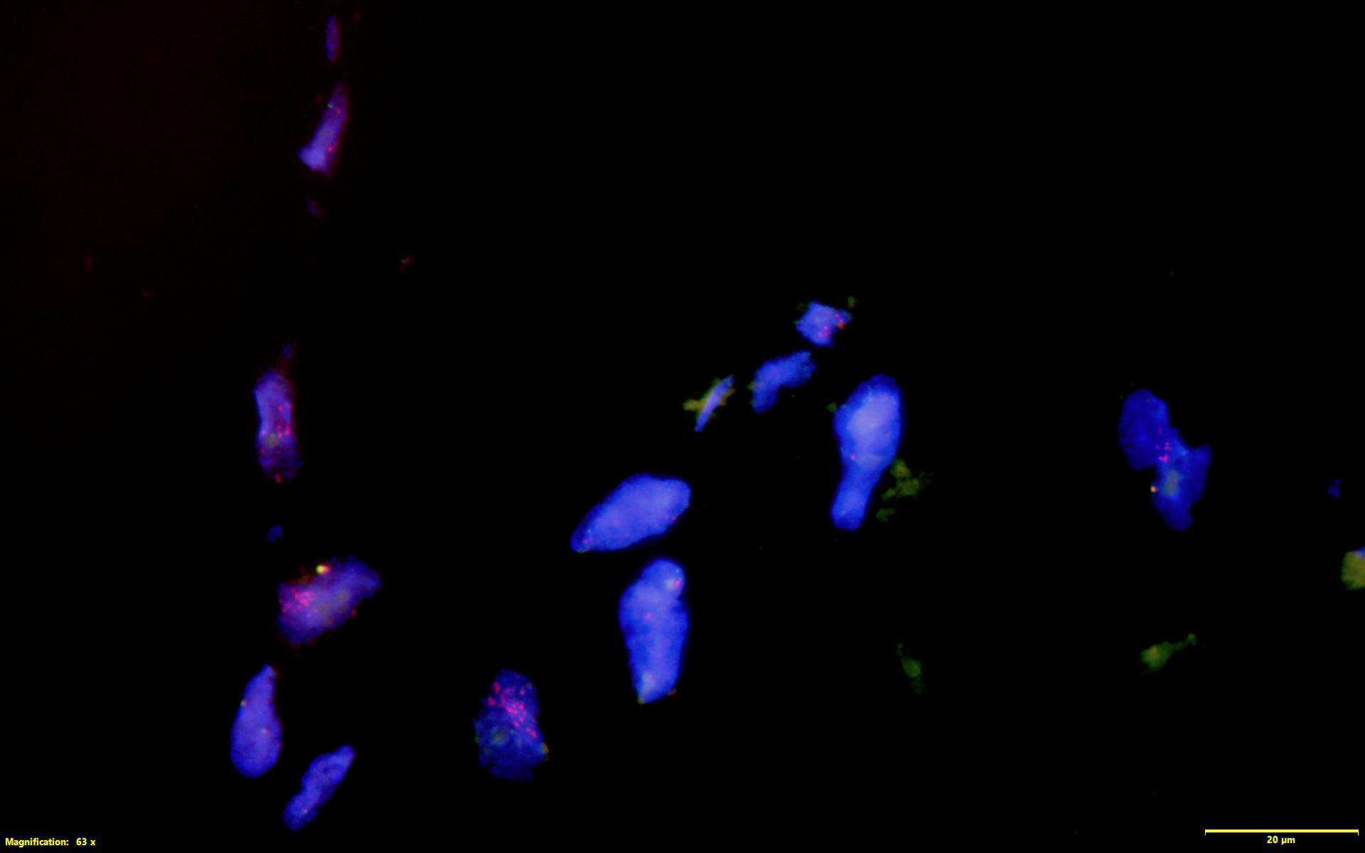
    Image shows MDM2 amplification by FISH technique

  • DIAGNOSIS

    Dedifferentiated liposarcoma

Soft tissue mass : M1135/96A

SOFT TISSUE MASS

Spices No: M1135/96A

  • Front

  • Left

  • BACK

  • RIGHT

  • GROSS DESCRIPTION

    The exhibit displays well circumscribed and thinly encapsulated lobulated mass. It shows homogenous yellowish cut surface.

  • HISTOPATHOLOGY DESCRIPTION

    Lipoma is composed of lobules of mature univacuolated adipocytes. Minimal fibrous septae can be observed. Atypical hyperchromatic stromal cell should not be present.

  • DIAGNOSIS

    Lipoma

  • BAHASA MELAYU

    Penerangan kasar: Spesimen menunjukkan ketumbuhan yang mempunyai kapsul nipis dibahagian paling luar. Permukaan ketumbuhan ini adalah berwarna kekuningan.

    Penerangan histopatologi: Di bawah mikroskop, lipoma terdiri daripada sel lemak yang matang dimana hanya terdapat univacuolated sitoplasma dengan nukleus kecil terletak di tepi sel.

    Diagnosis: Lipoma

Foot : M1166/99

FOOT

Spices No: M1166/99

  • GROSS DESCRIPTION

    The exhibit displays a third, fourth and fifth toe with presence of exophytic fungating malignant lesion arising from the skin. The mass shows irregular hyperkeratotic scaly plaque surface.

  • HISTOPATHOLOGY DESCRIPTION

    Squamous cell carcinoma show malignant epithelium with evidence of squamous differentiation. These include the presence of individual keratinization, intercellular bridges, and keratin pearl.

  • DIAGNOSIS

    Squamous cell carcinoma

Soft tissue mass : M198/83

SOFT TISSUE MASS

Spices No: M198/83

  • Front

  • Left

  • RIGHT

  • GROSS DESCRIPTION

    The exhibit displays well circumscribed and thinly encapsulated lobulated mass. It shows homogenous yellowish cut surface.

  • HISTOPATHOLOGY DESCRIPTION

    Lipoma is composed of lobules of mature univacuolated adipocytes. Minimal fibrous septae can be observed. Atypical hyperchromatic stromal cell should not be present.

  • DIAGNOSIS

    Lipoma

  • BAHASA MELAYU

    Penerangan kasar: Spesimen menunjukkan ketumbuhan yang mempunyai kapsul nipis dibahagian paling luar. Permukaan ketumbuhan ini adalah berwarna kekuningan.

    Penerangan histopatologi: Di bawah mikroskop, lipoma terdiri daripada sel lemak yang matang dimana hanya terdapat univacuolated sitoplasma dengan nukleus kecil terletak di tepi sel.

    Diagnosis: Lipoma

Femur : M1162/99

FEMUR

Spices No: M1162/99

  • Front

  • Left

  • BACK

  • RIGHT

  • GROSS DESCRIPTION

    The exhibit show a tumour arising from the metaphysis. It shows densely solid, tan-white surface resembling the cortical bone. The margin is irregular and ill defined.

  • HISTOPATHOLOGY DESCRIPTION

    Osteosarcoma shows malignant osteoid deposited in a lace like pattern or as delicate trabeculae. The malignant cells are pleomorphic with irregular hyperchromatic nuclei and high mitotic activity.

  • DIAGNOSIS

    Osteosarcoma

  • QUIZ

    In long bone, osteosarcoma commonly develops in

    A. Diaphysis [TRUE/FALSE]

    B. Metaphysis [TRUE/FALSE]

  • BAHASA MELAYU

    Penerangan kasar: Spesimen menunjukkan barah tulang di bahagian metaphysis iaitu bahagian sebelum hujung tulang panjang berdekatan dengan bahagian pertumbuhan tulang atau physis.

    Penerangan histopatologi: Osteosarcoma atau kanser tulang yang agresif mempunyai karakter menghasilkan bahan atau material tulang yang dipanggil osteoid. Sel kanser ini sangat pleomorphic/ mempunyai rupa, bentuk dan saiz yang pelbagai serta aktiviti mitosis dan pertumbuhan yang tinggi.

    Diagnosis: Osteosarcoma