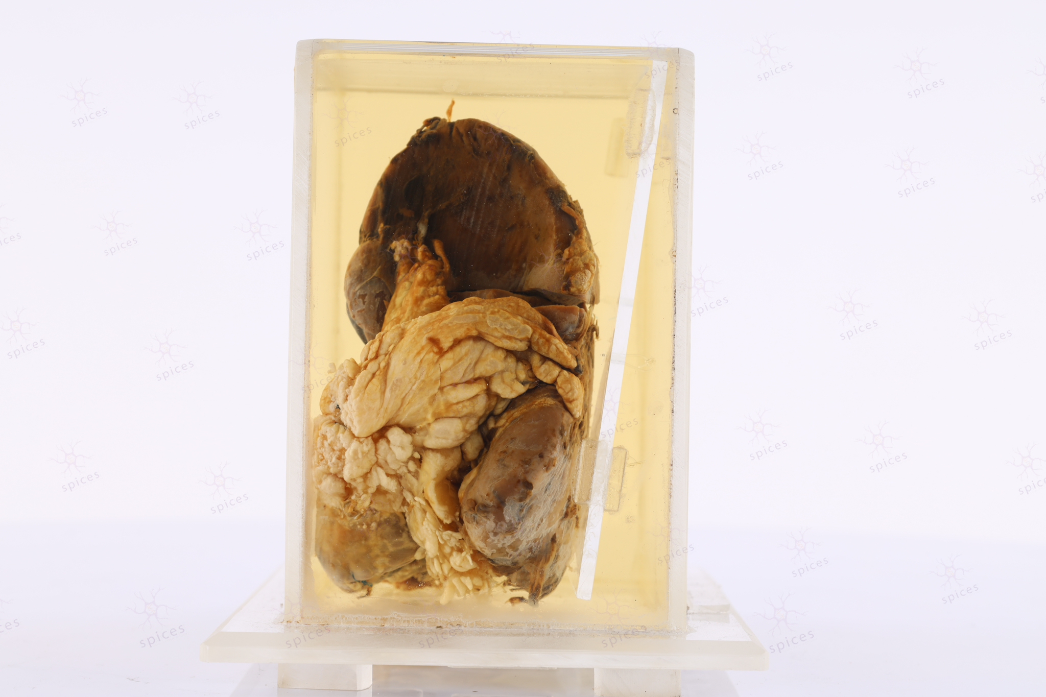
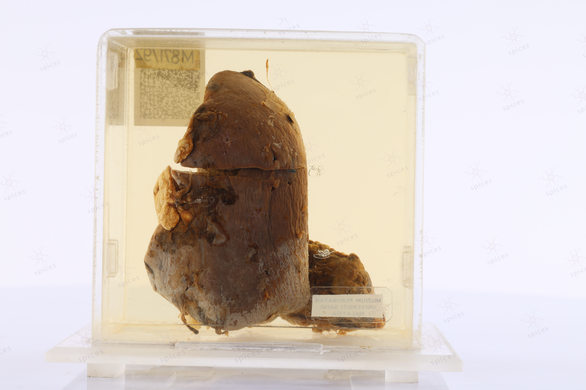
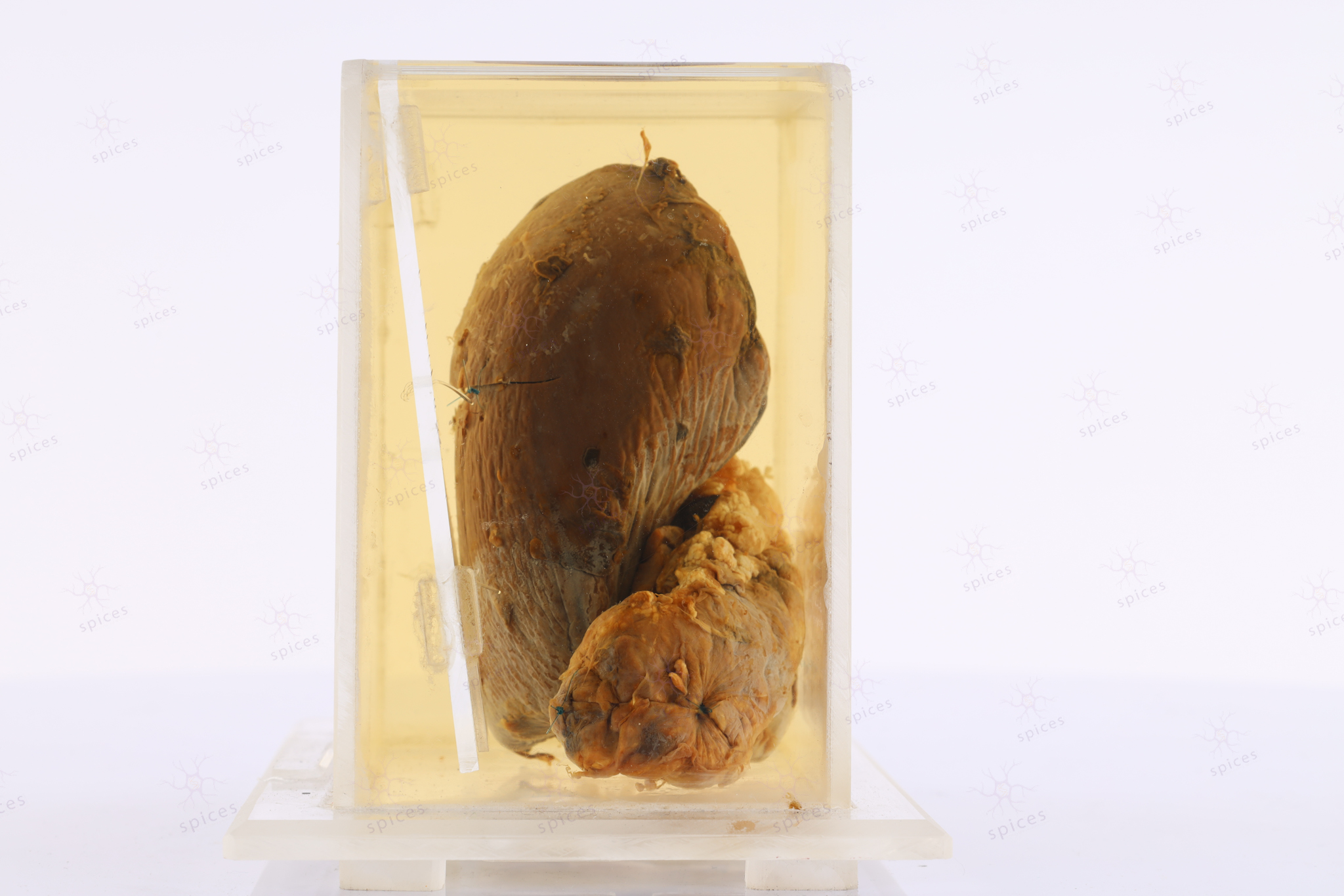
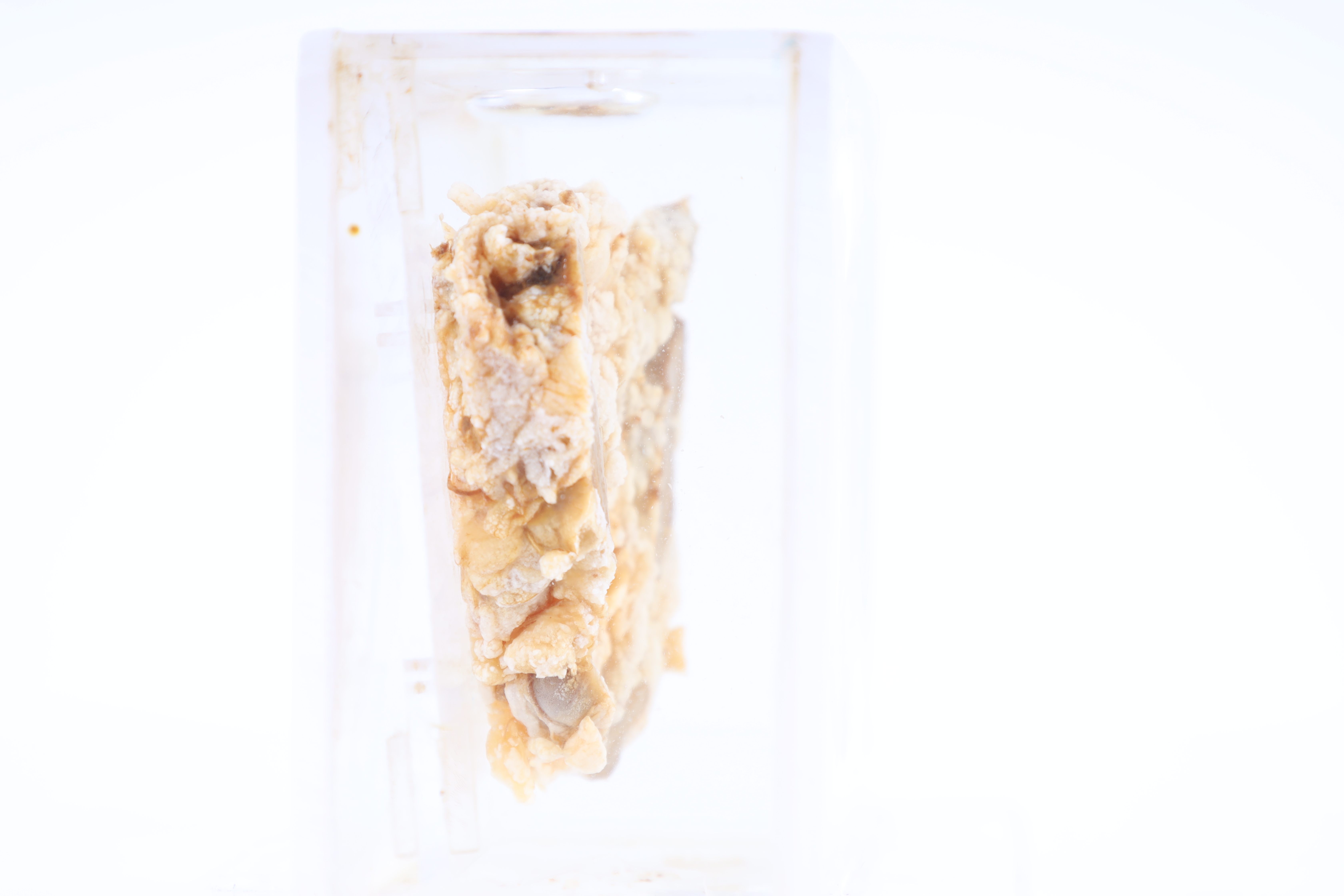
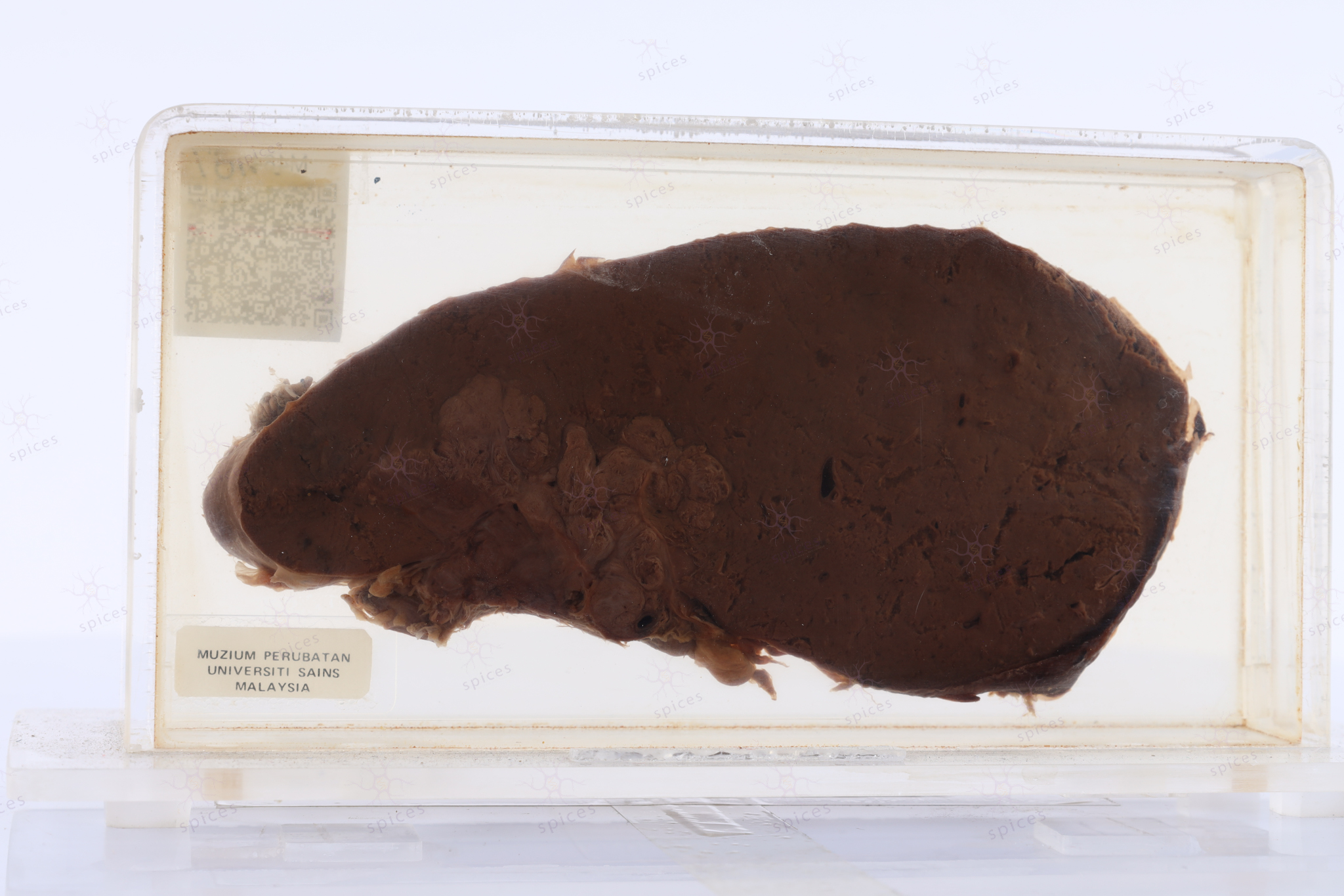
SPICES, School of Medical Sciences  |  Health Campus, USM

Spleen : M484/87

SPLEEN

Spices No: M484/87

  • Front

  • Left

  • BACK

  • RIGHT

  • GROSS DESCRIPTION

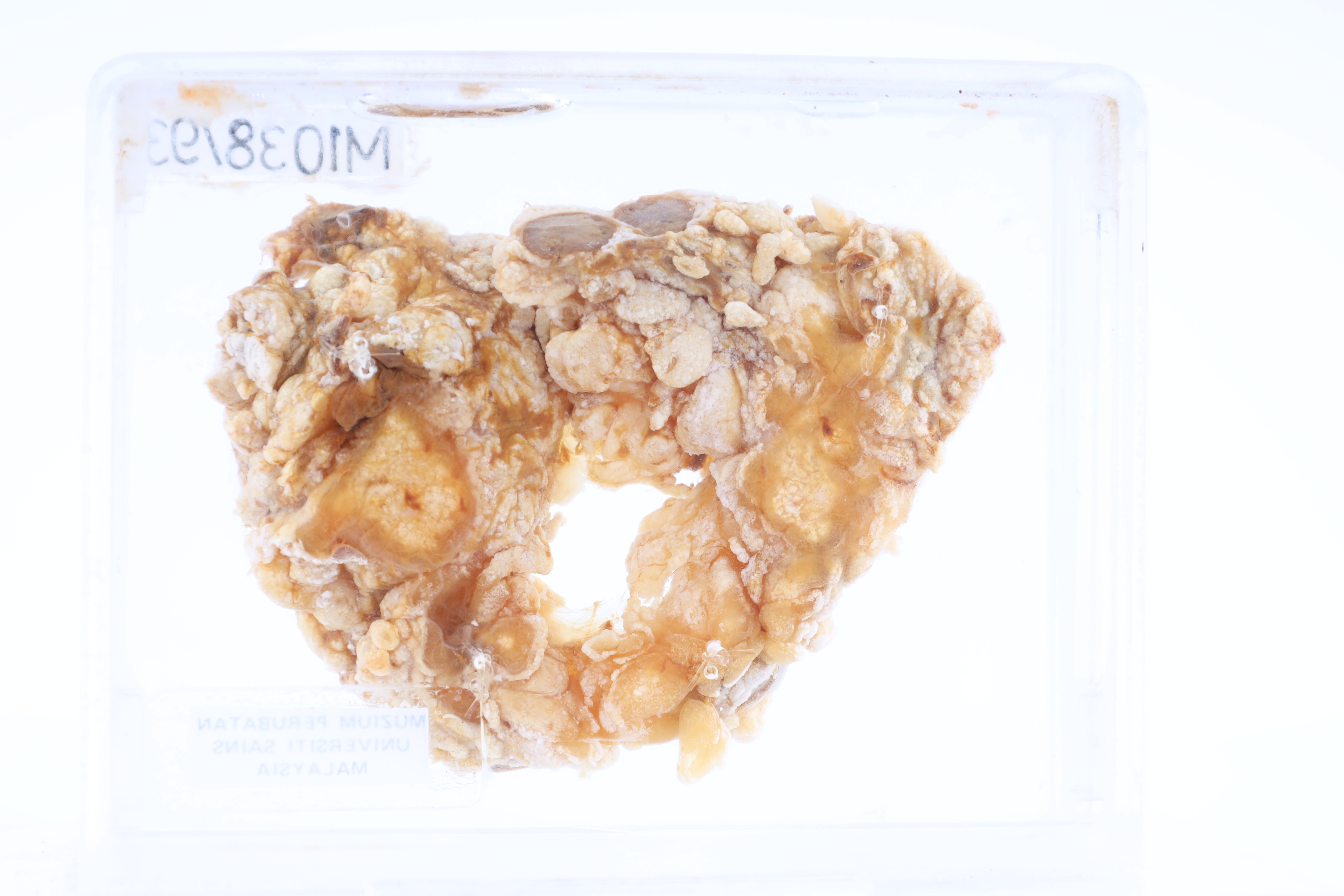
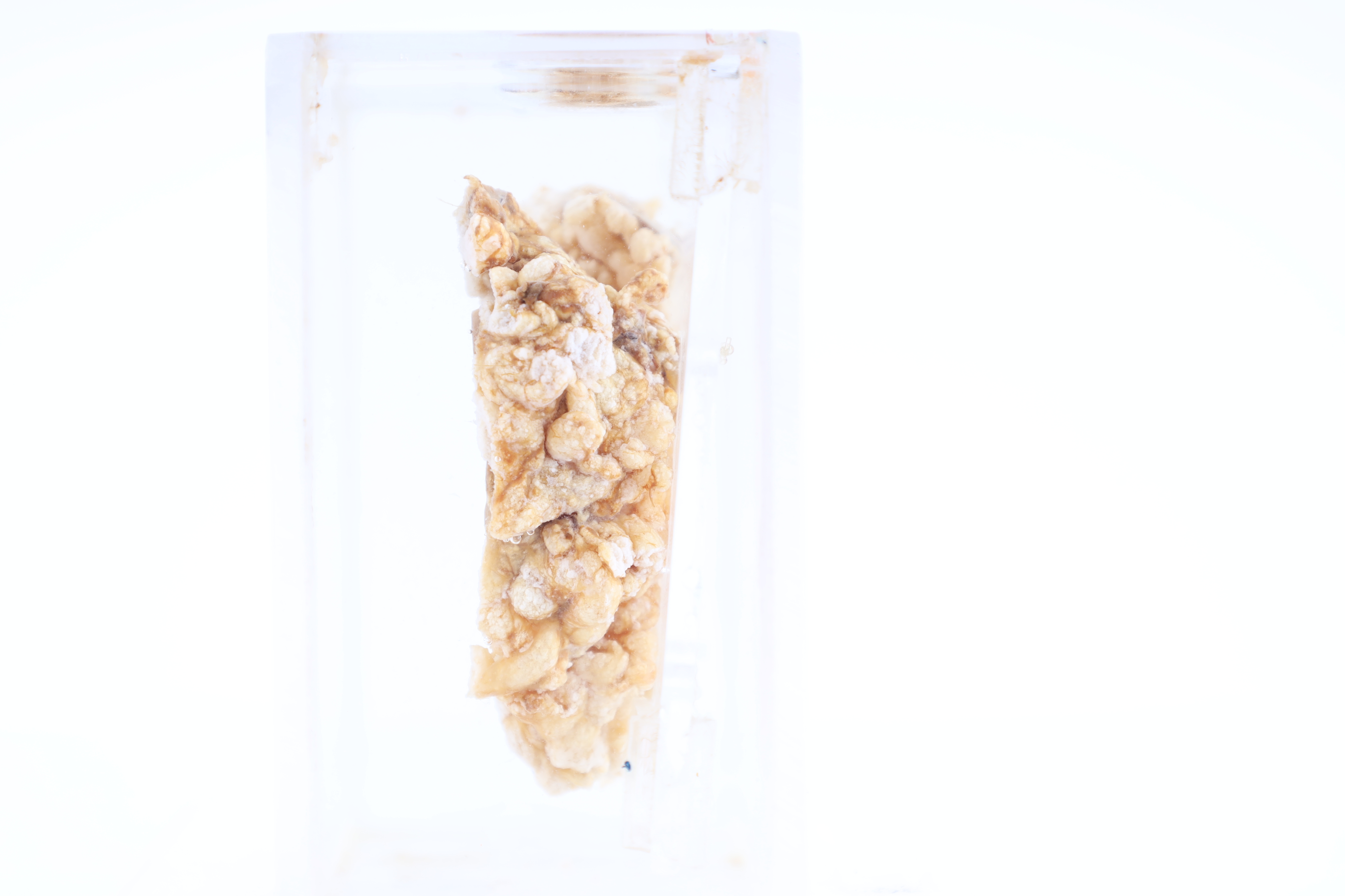
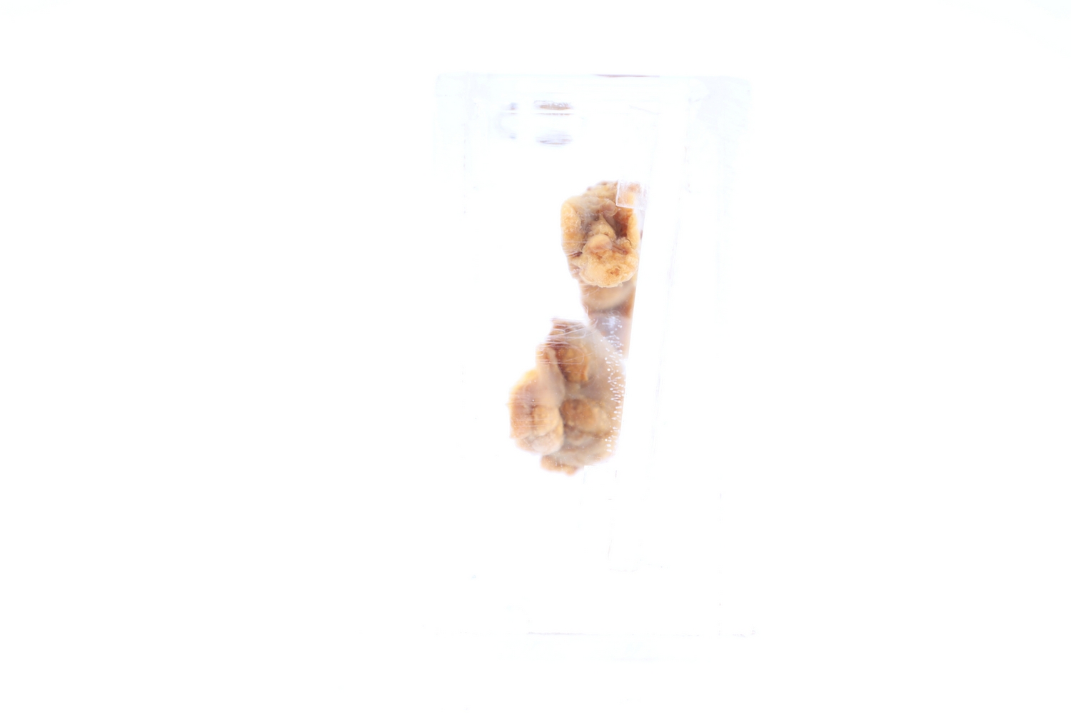
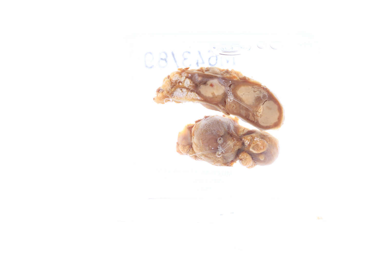
    The exhibti displays multiple small minute nodules distributed in the spleenic parenchyma

  • HISTOPATHOLOGY DESCRIPTION

    Metastatic carcinoma shows malignant epithelial cell in glandular and tubular pattern. It consist of malignant epithelium displaying pleomorphic vesicular nuclei in eosinophilic cytoplasm. Immunohistochemistry is important ancillary test to defined the primary organ.

  • DIAGNOSIS

    Metastatic carcinoma

Spleen : M871/92

SPLEEN

Spices No: M871/92

  • Front

  • Left

  • BACK

  • RIGHT

  • GROSS DESCRIPTION

    The exhibit displays enlarged spleen.

  • HISTOPATHOLOGY DESCRIPTION

    Splenomegaly

  • DIAGNOSIS

    Splenomegaly

  • QUIZ

    Causes of splenomegaly

Lymph node : M736/90

LYMPH NODE

Spices No: M736/90

  • GROSS DESCRIPTION

    The exhibit displays enlarged lymph node with tan homogenous surface.

  • HISTOPATHOLOGY DESCRIPTION

    The lymph node is effaced by the neoplastic lymphoid cell. The lymphoid cell exhibit large cell with vesicular nuclei and prominent nucleoli. They are positive for B-cell marker.

  • DIAGNOSIS

    Diffuse large B cell lymphoma

Lymph Node

LYMPH NODE

SPICES No. M1038/93
  • Front

  • Left

  • BACK

  • RIGHT

  • GROSS DESCRIPTION

    The exhibit displays lymph nodes with metastatic carcinoma
  • HISTOPATHOLOGY DESCRIPTION

    Metastatic carcinoma shows malignant epithelial cell in glandular and tubular pattern. It consist of malignant epithelium displaying pleomorphic vesicular nuclei in eosinophilic cytoplasm. Immunohistochemistry is important ancillary test to defined the primary organ.

  • DIAGNOSIS

    Metastatic adenocarcinoma

Lymph Node

LYMPH NODE

SPICES No. M643/89
  • Front

  • Left

  • BACK

  • RIGHT

  • GROSS DESCRIPTION

    The exhibit displays enlarged lymph node with presence of whitish nodule.

  • HISTOPATHOLOGY DESCRIPTION

    Section from lymphoid tissue composed of reactive lymphoid follicles with some area presence of granuloma. Granuloma is composed of aggregates of epithelioid macrophages. Multinucleated giant cell Lamgerhans type is seen. Central necrosis also appreciated.

  • DIAGNOSIS

    Caseating tuberculous lymphadenitis