RETROPERITONEAL TUMOUR
Spices No: M350/85

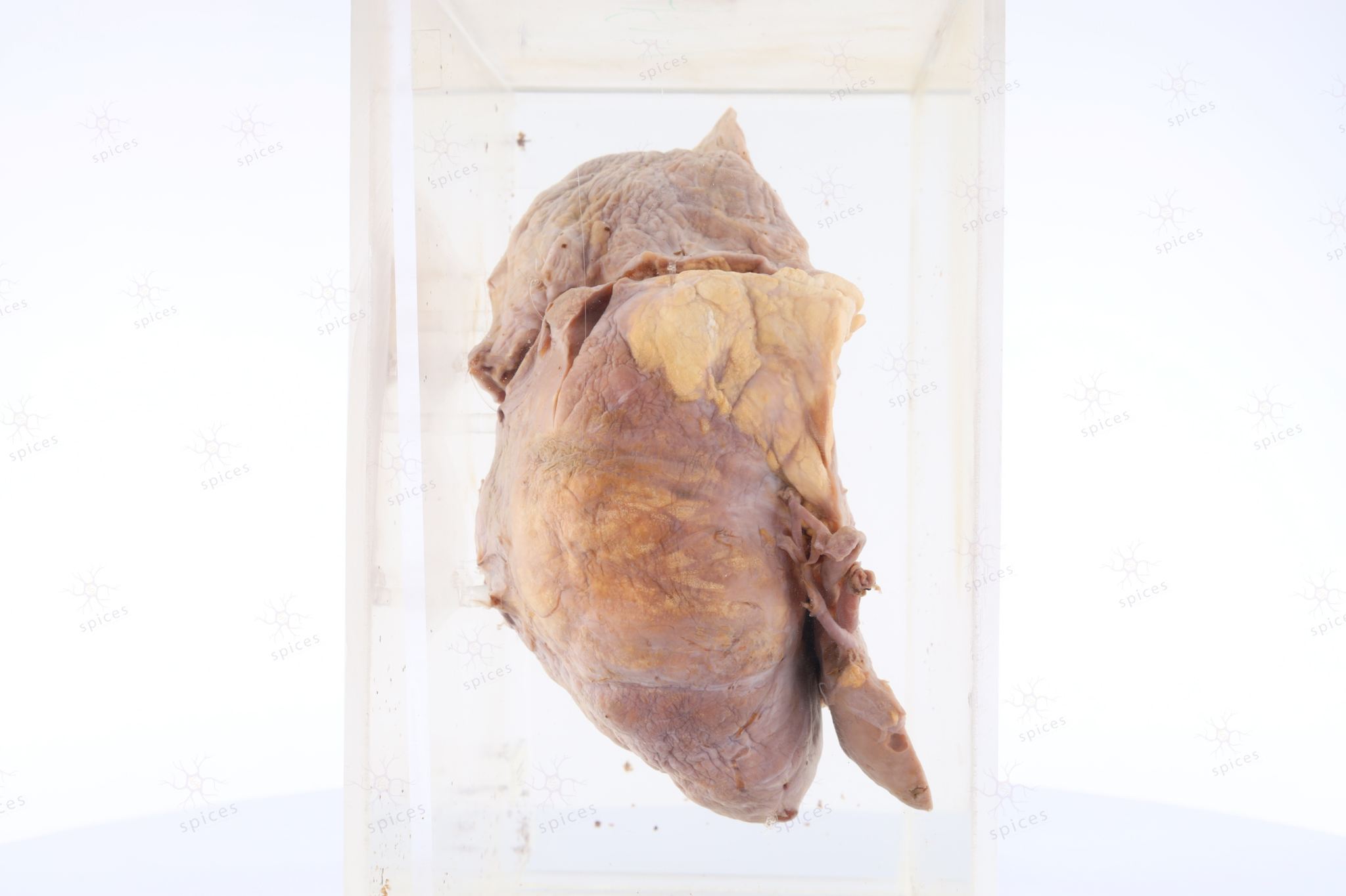
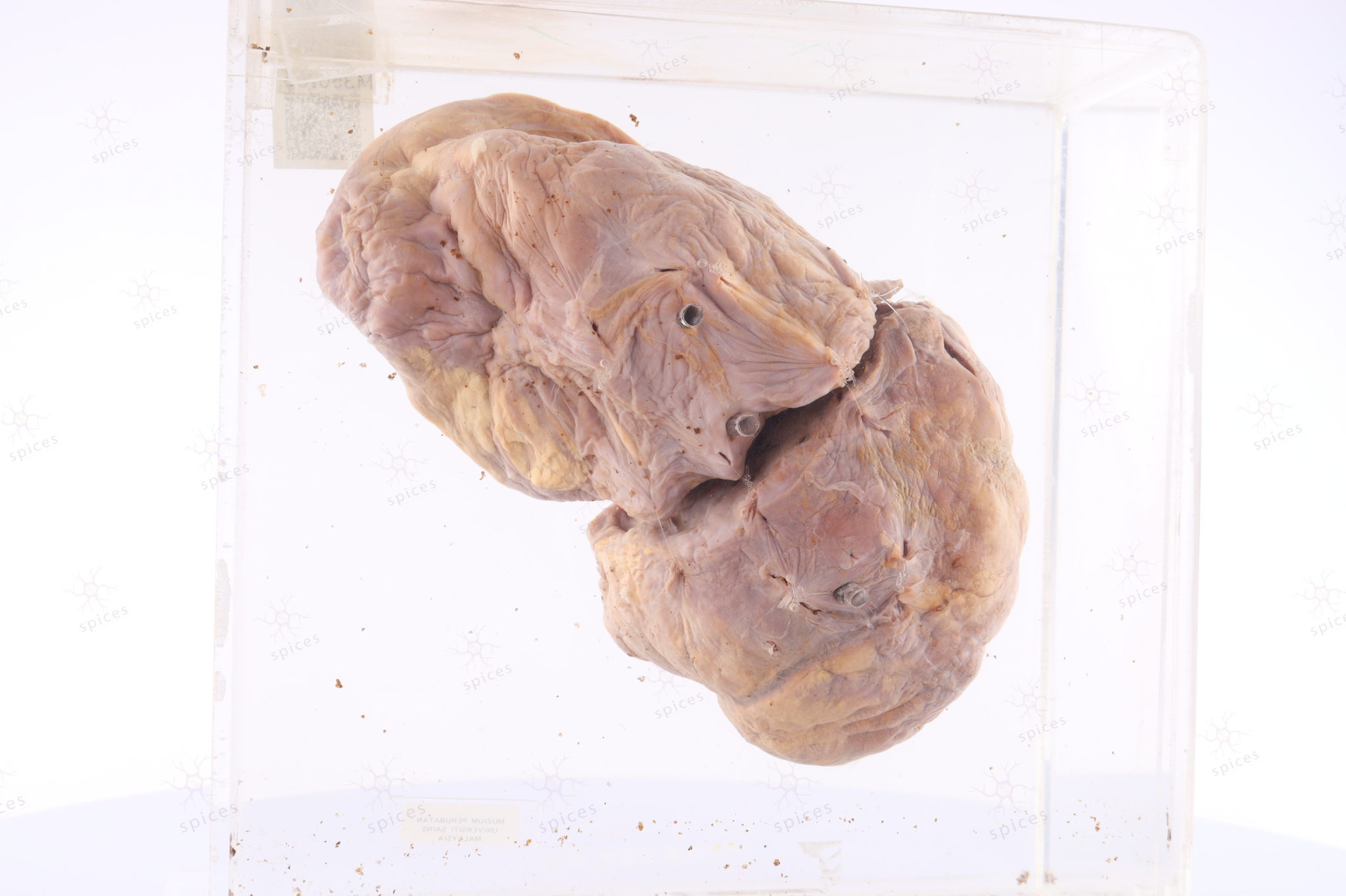
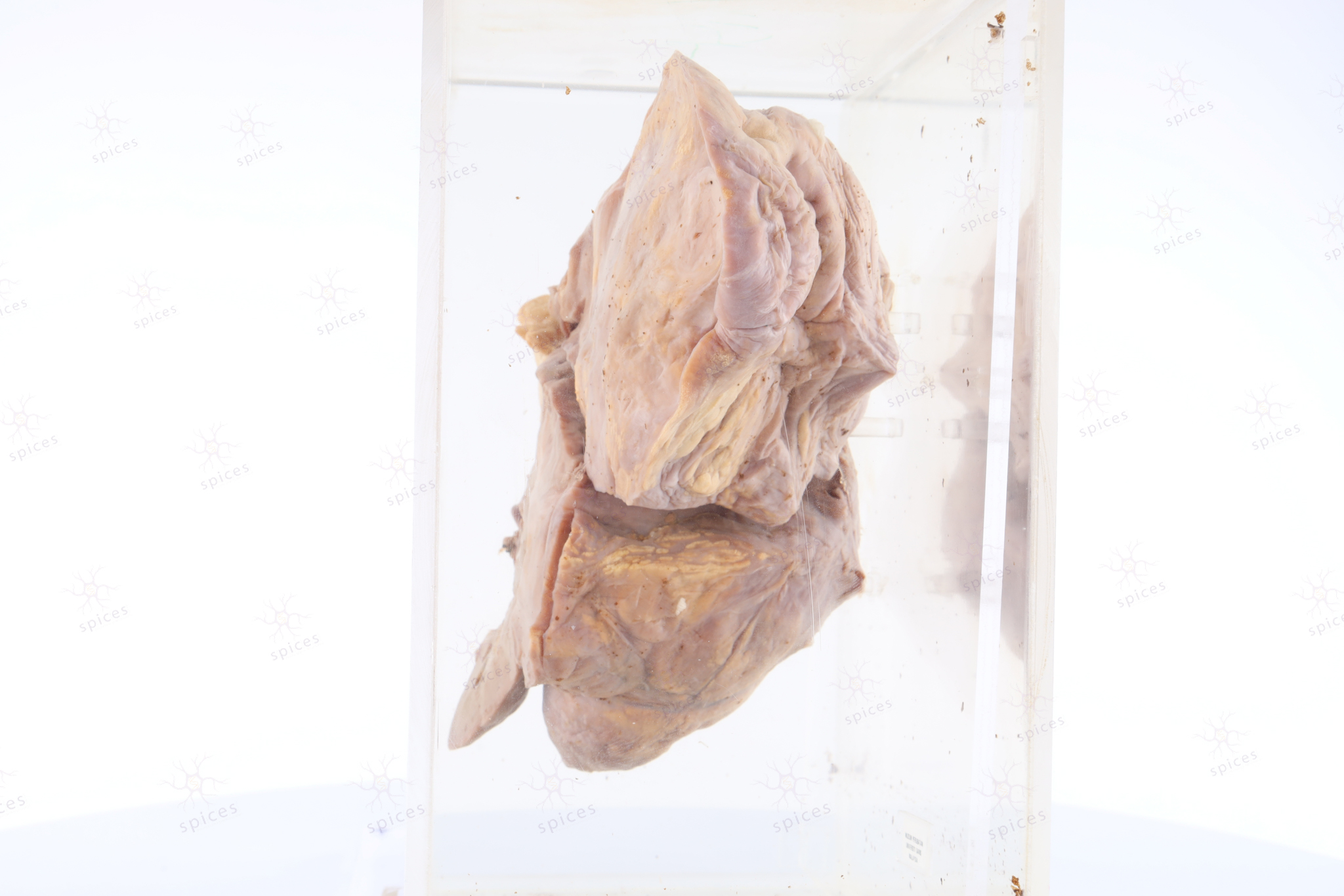
It is a soft tissue mass, fairly circumscribed, covered by thin fibrous capsule. Cut section shows solid brownish to yellowish cut-surface.Take note that the brownish area is the dedifferentiated area with an abrupt transition to a well differentiated area (yellow). The tumour abutting the kidney (left bottom). Margins are involved by the tumour.
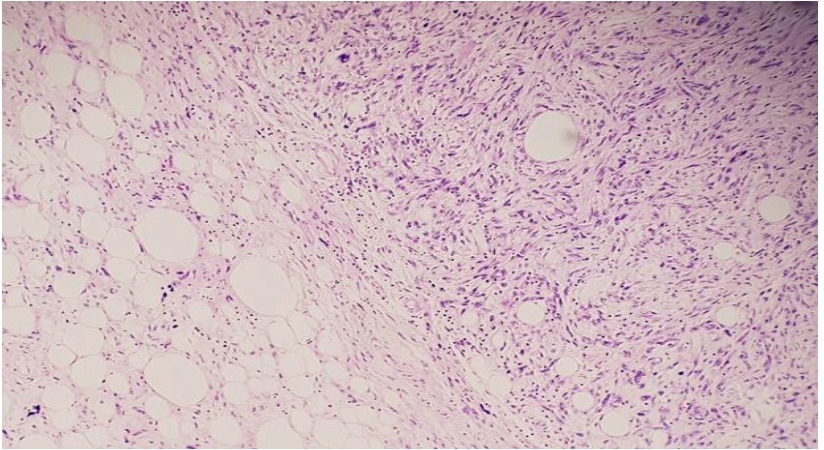
Section shows well differentiated liposarcoma (left) with an abrupt transition to the dedifferentiated component, exhibiting pleomorphic spindle shape cells arranged in vague storiform pattern (non-lipogenic component) on the right side.
Fluorescence In Situ Hybridisation (FISH) analysis shows amplifications of MDM2.
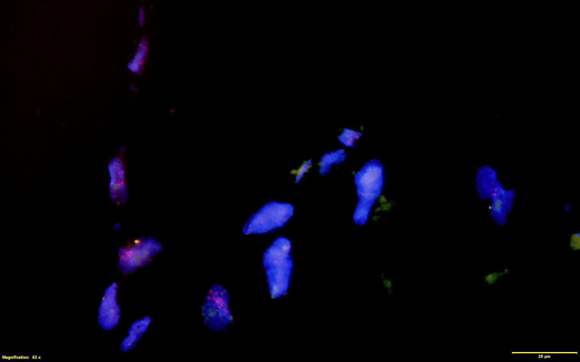
Image shows MDM2 amplification by FISH technique
Dedifferentiated liposarcoma