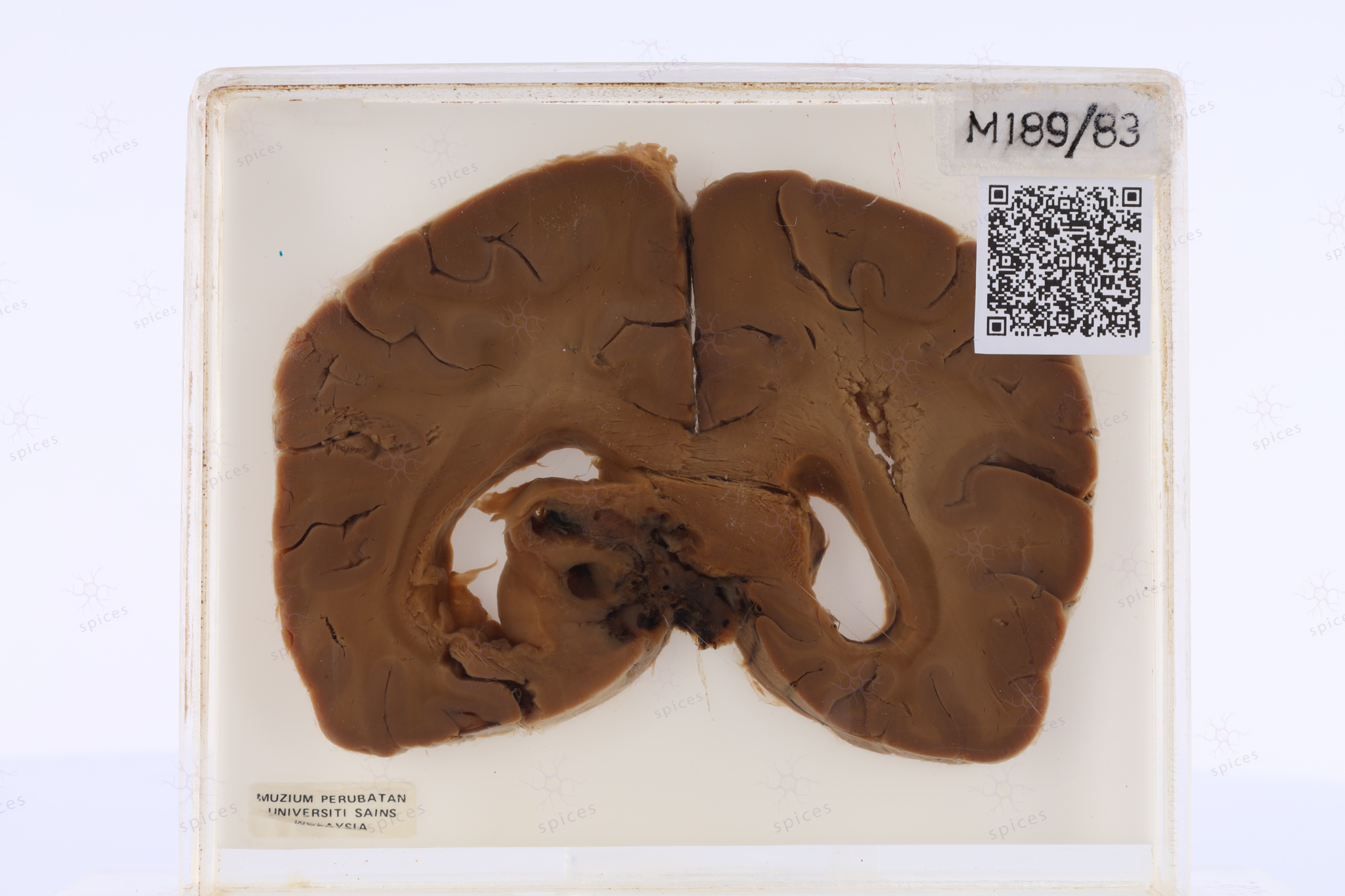
The exhibit displays ill defined lesion over the corpus callosum.
Section shows infiltrative tumour cells with sheets pattern in the fibrillary background. The cells are moderate to markedly pleomorphic, round to oval nuclei with some bizarre cells, hyperchromatic chromatin pattern, and eosinophilic cytoplasm. Geographical necrosis and proliferation of blood vessels is also noted. Mitosis easily seen.
High grade glioma
Common location for high grade glioma