
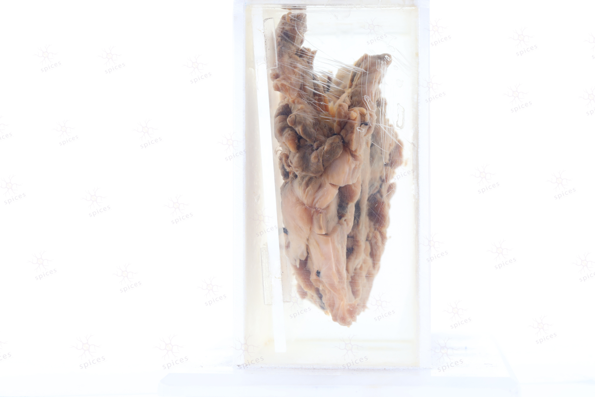
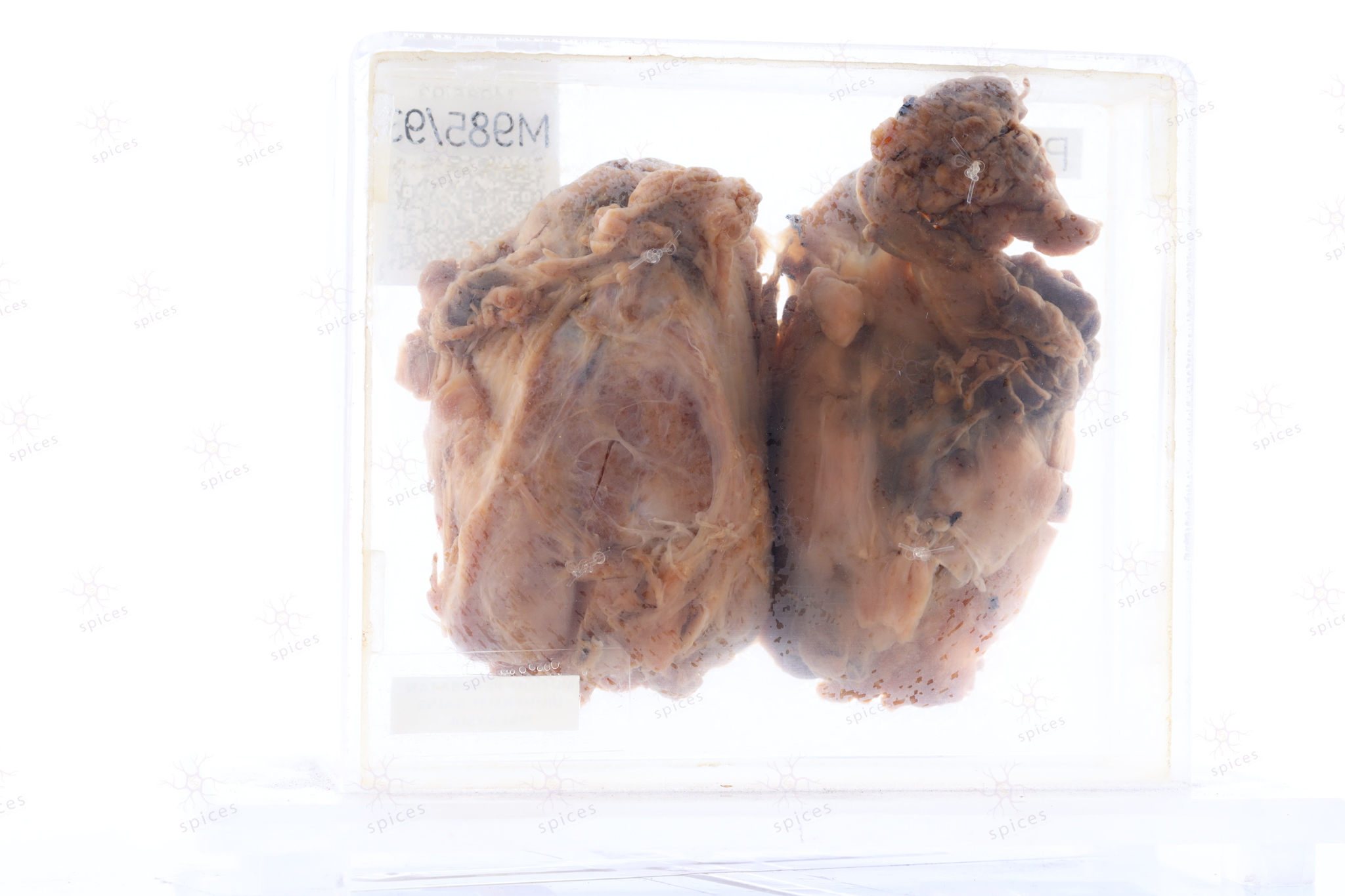
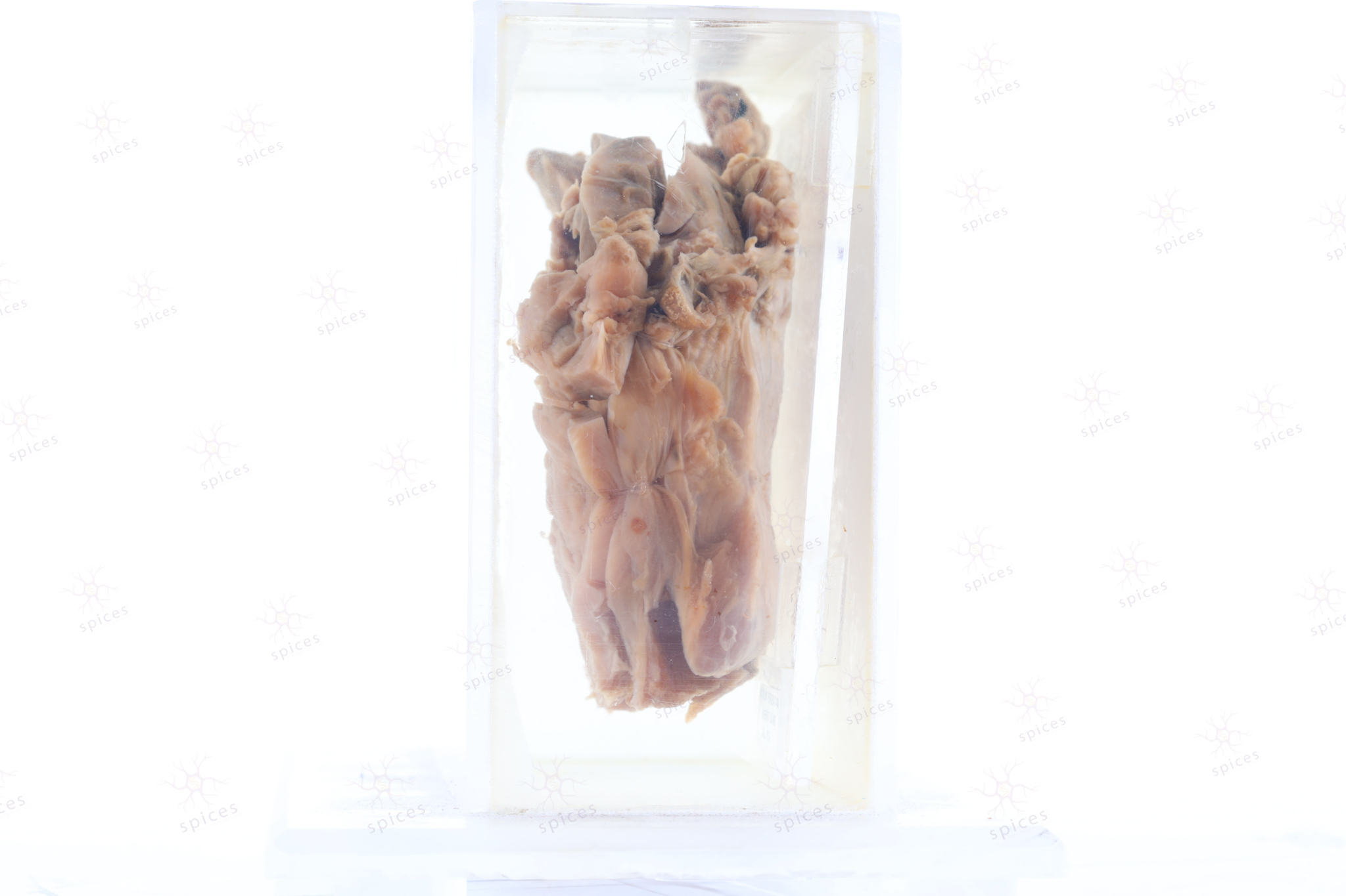
The exhibit is an enlarged thyroid which has been replaced by the tumour. The tumour is solid, gray-white with papillary projection on the cut surface. The tumour invaded the capsule and extended to the extracapsular tissue.
Sections of the thyroid show papillary thyroid carcinoma characterised by true papillary structure lined by enlarged oval follicular cells, exhibiting grooved nuclei and intra-nuclear cytoplasmic inclusion. In areas, anaplastic features are also seen with large pleomorphic and hyperchromatic nuclei with sarcomatoid changes.
Papillary thyroid carcinoma