
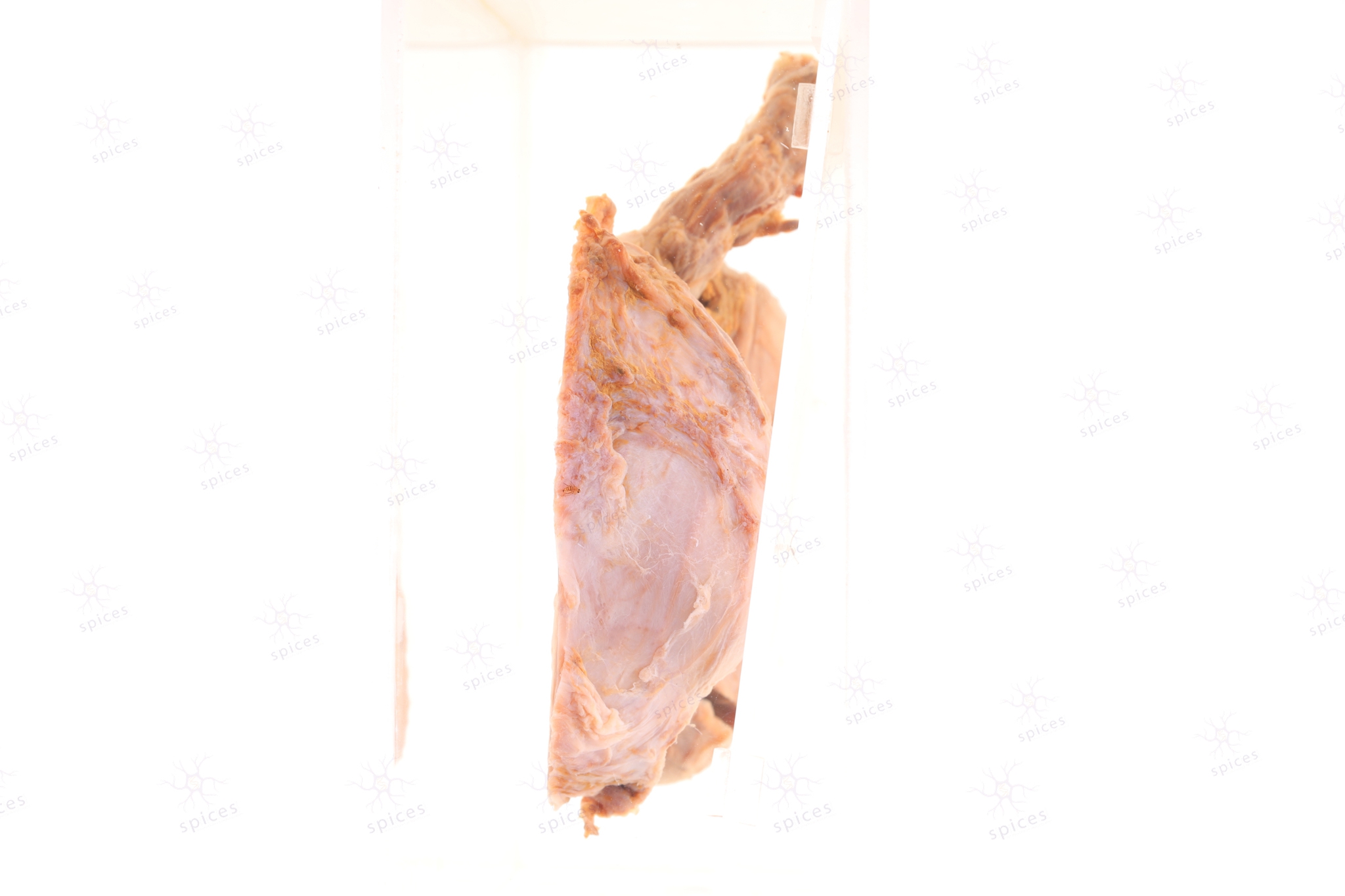
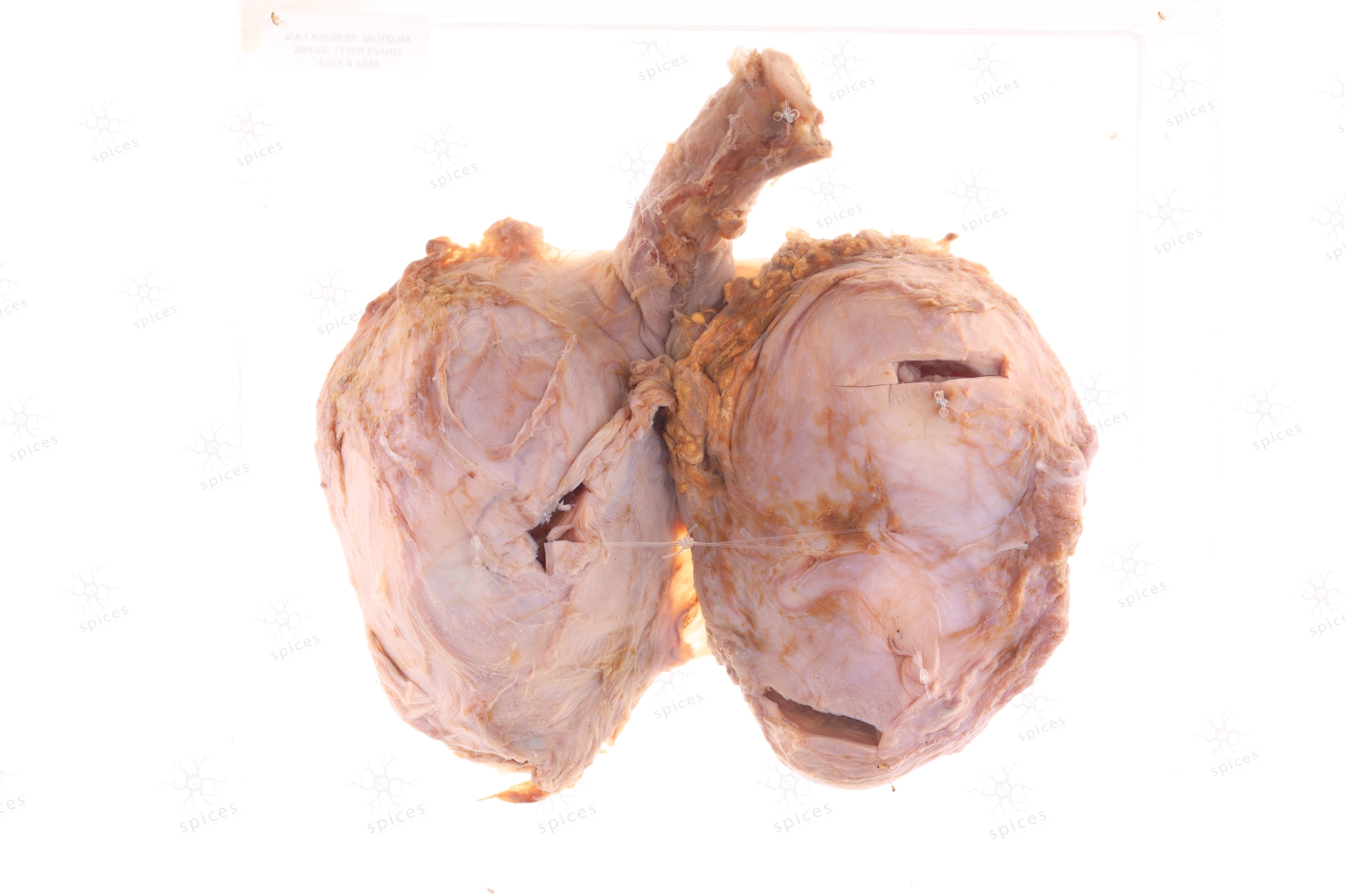
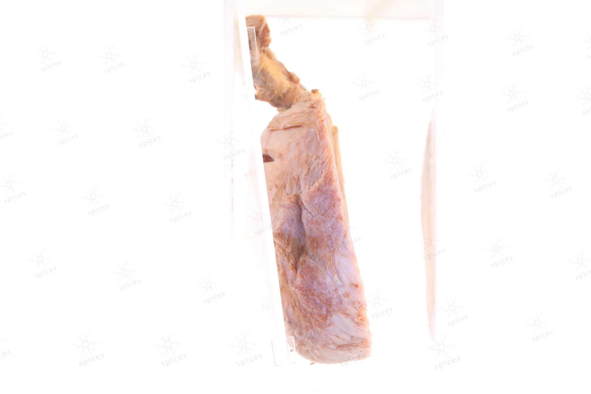
The exhibit displays a solid tumour occupying the whole testis with. It show tan- brownish cut surface replacing almost the whole testicular parenchyma. Cystic structures are noted.
Seminoma show nests of tumour tissue surrouned by thin fibrous septe with sprinkles of lymphocytes within the septae. The cells are polygonal in shape, with clear cytoplasm and centrally located nuclei. Cystoplasm is moderate in amount and clear. Mitosis is seen. No aother germ cell tumour component noted.
Testicular tumour - Seminoma