Musculoskeletal
Bone Proximal Tibia : S016/2024
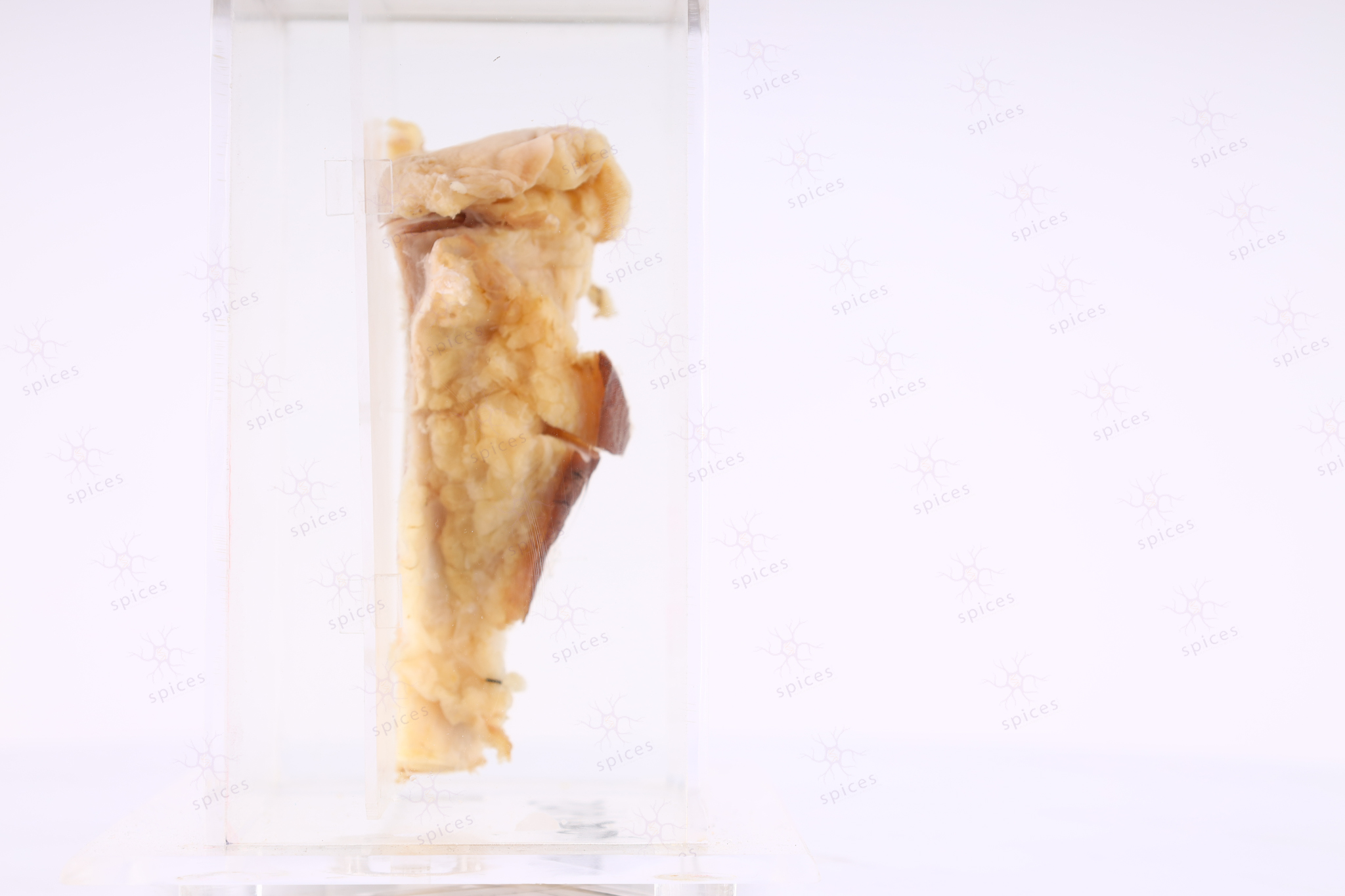
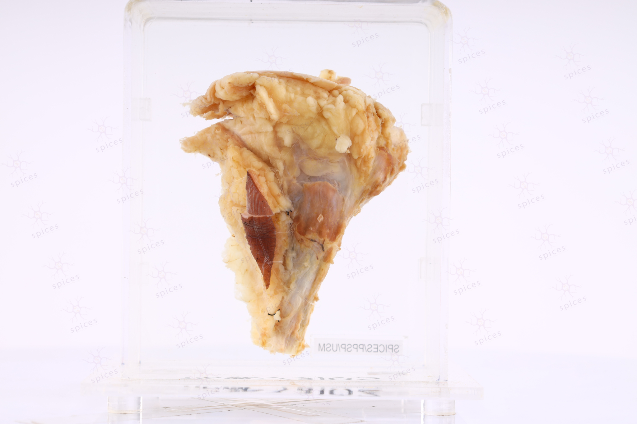
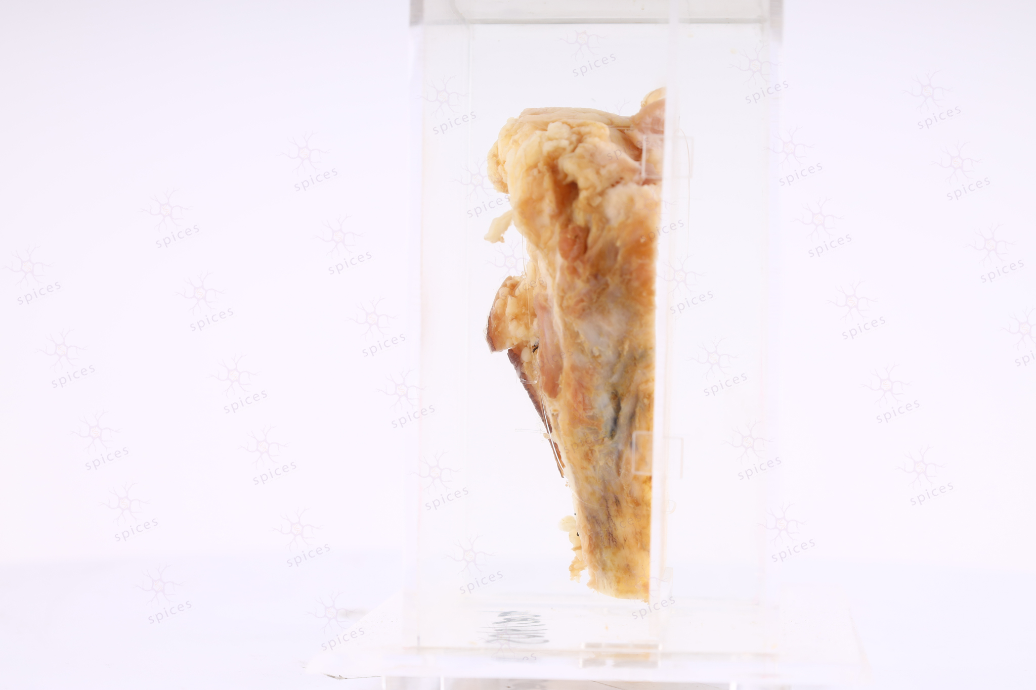
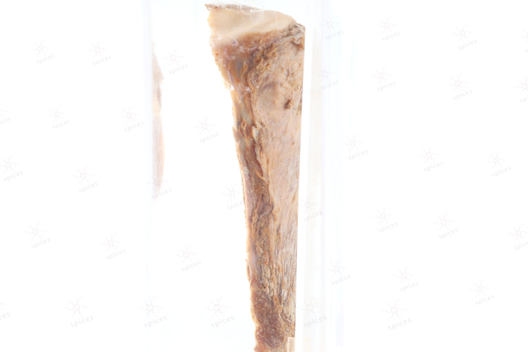
Bone Proximal Tibia
Spices No: S016/2024

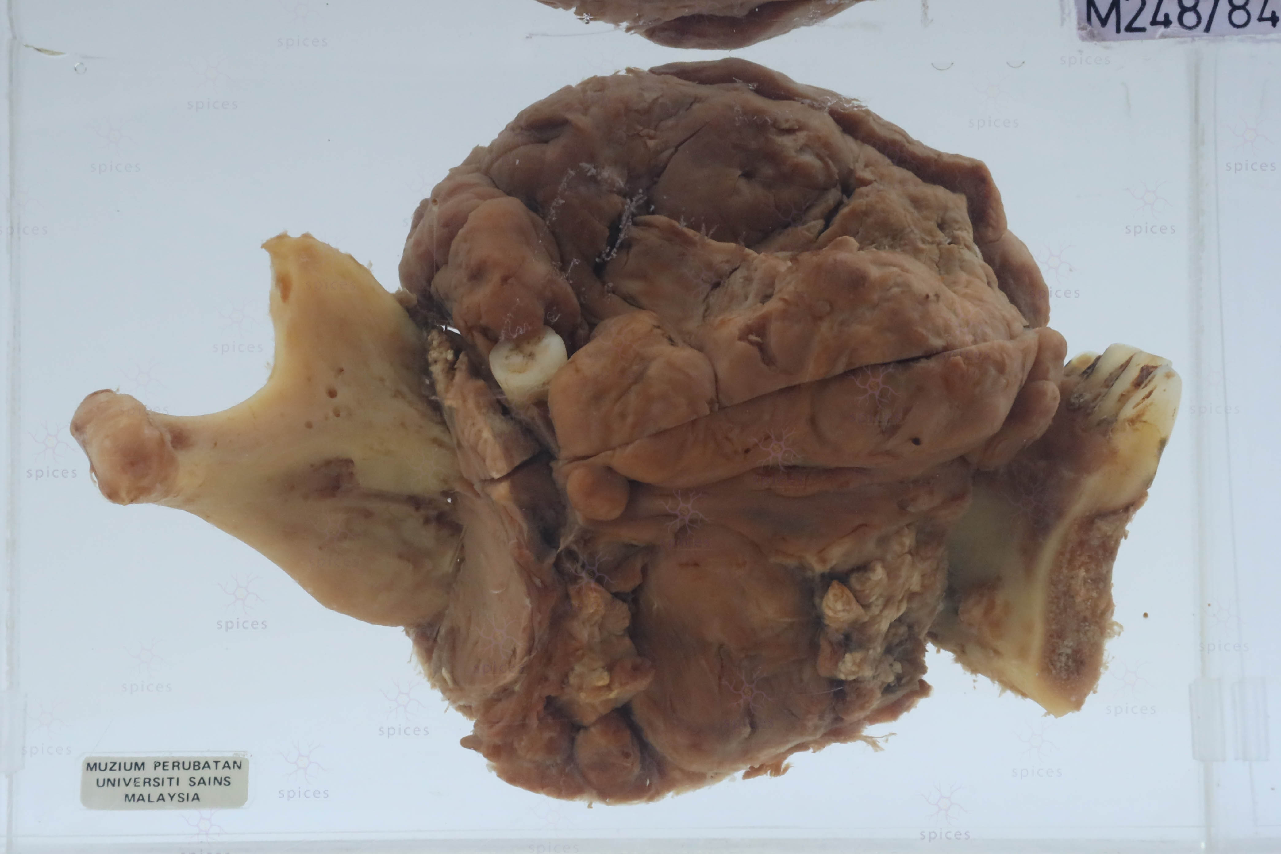
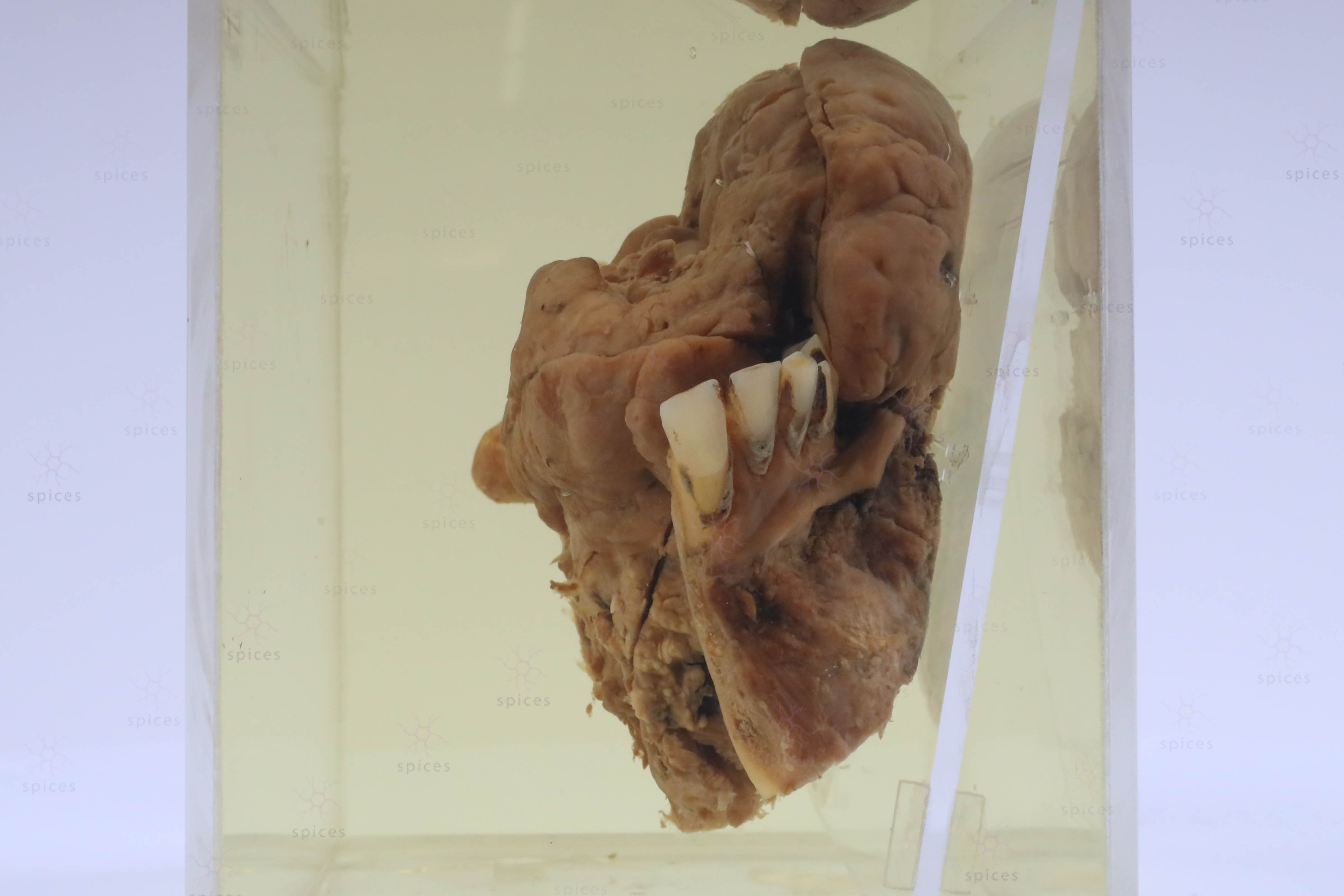
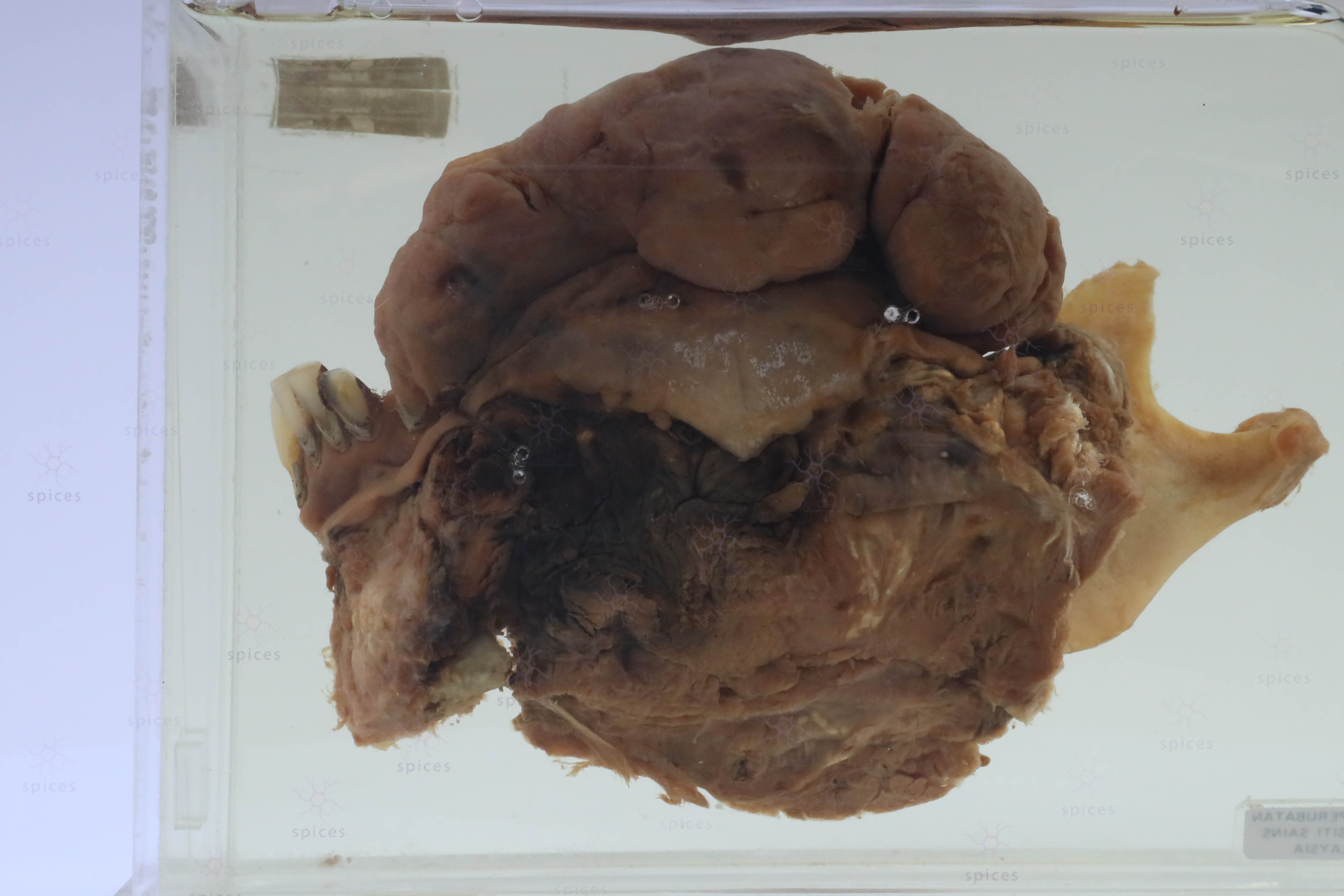
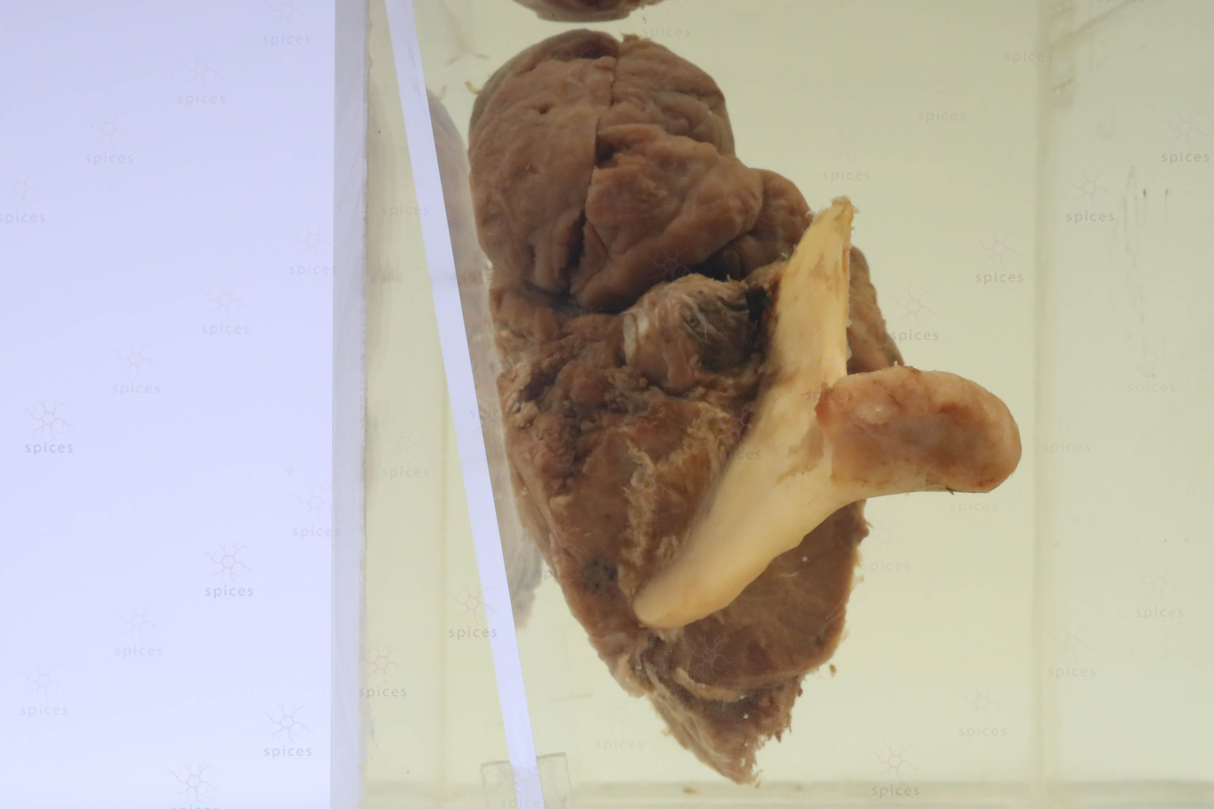
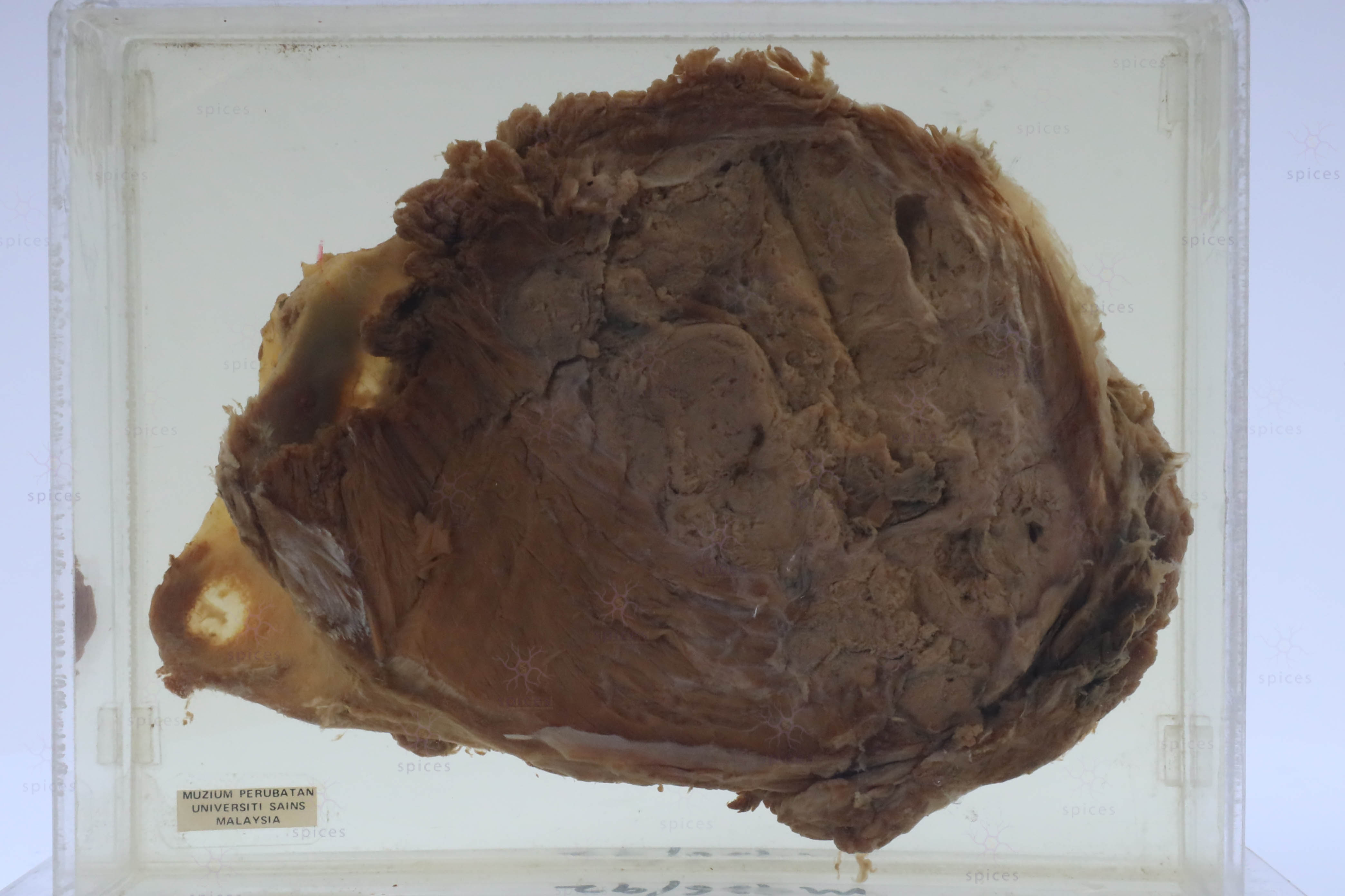
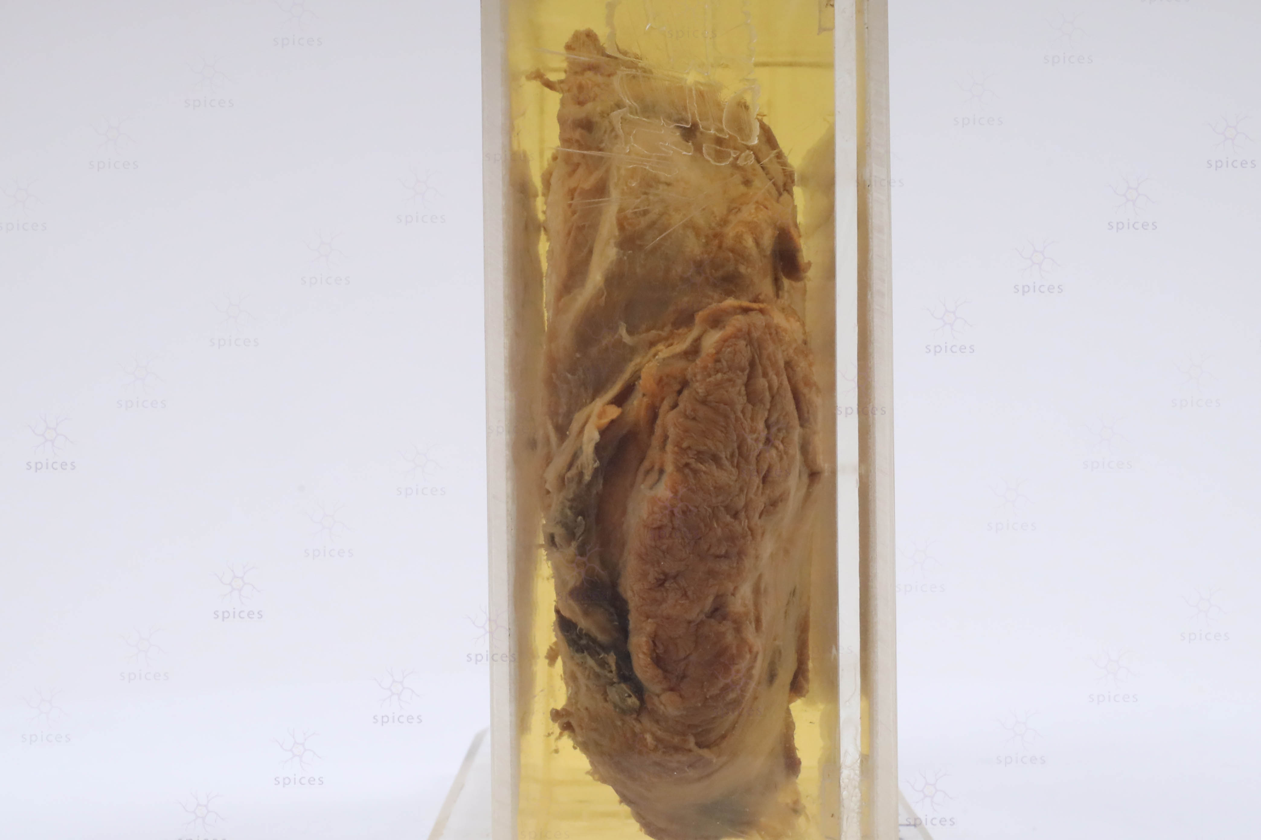
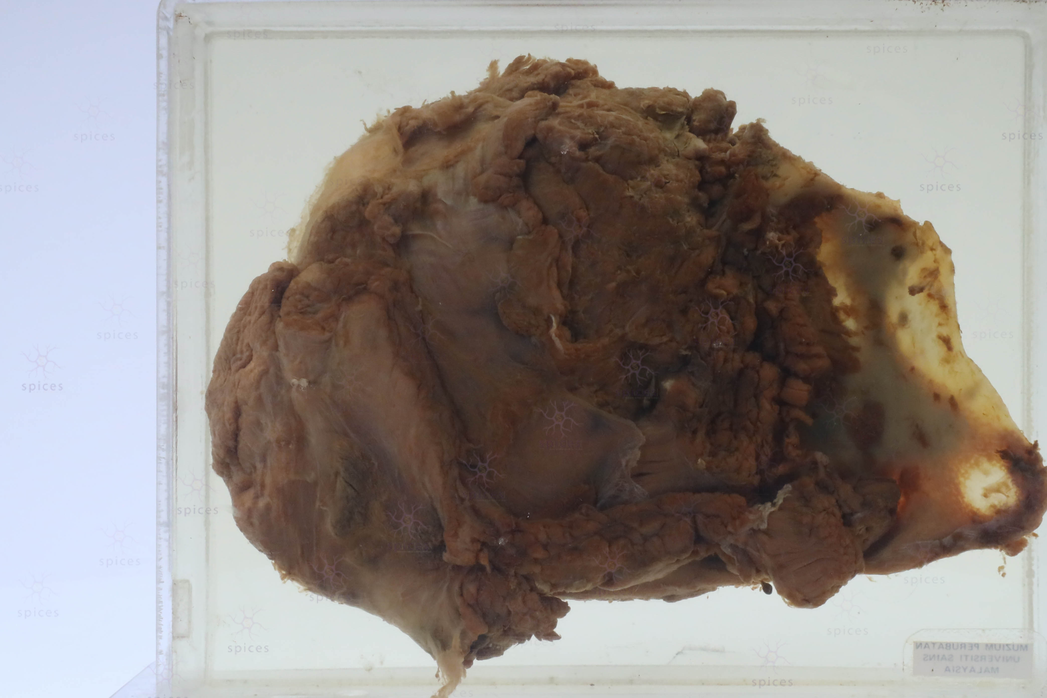
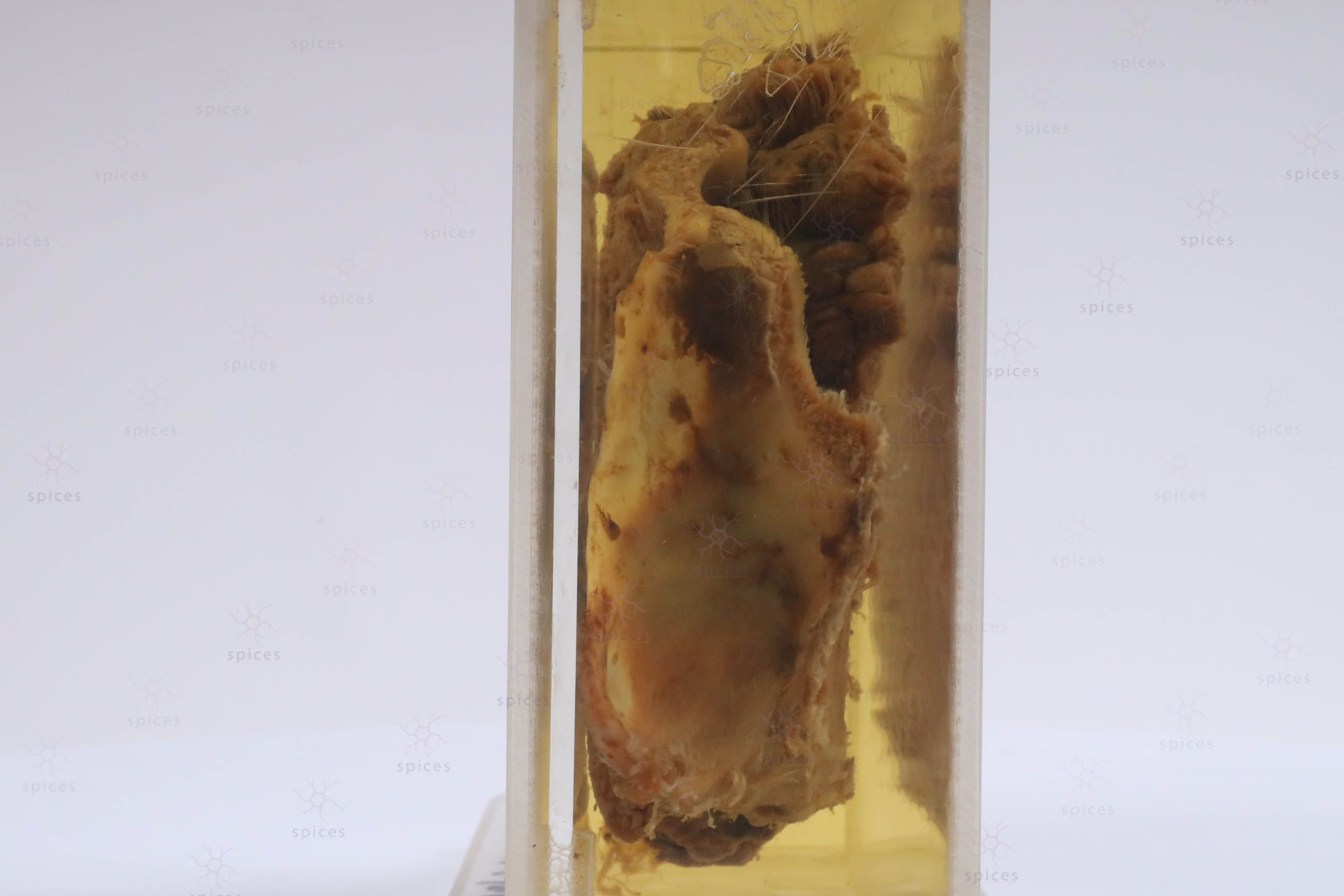
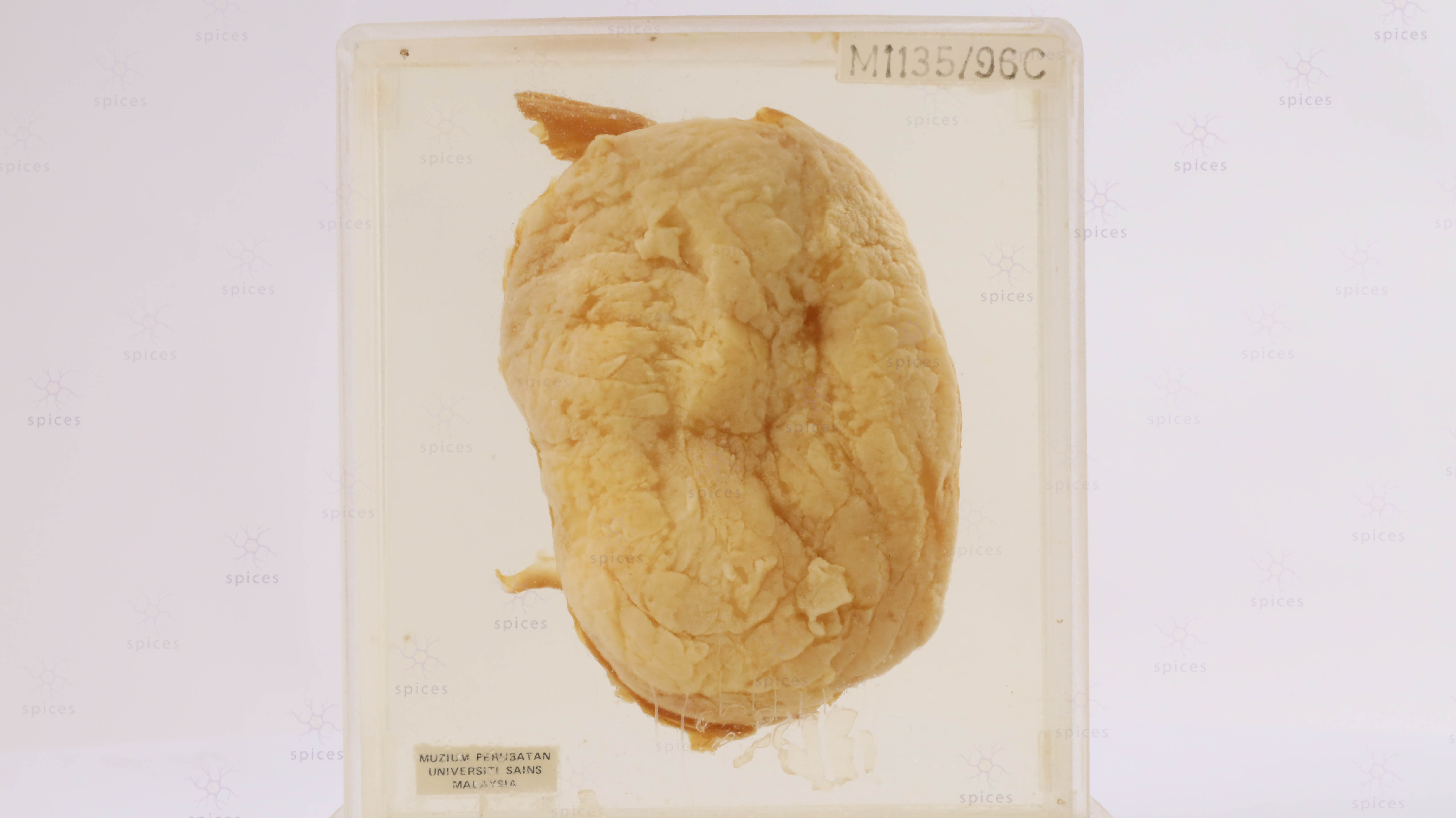
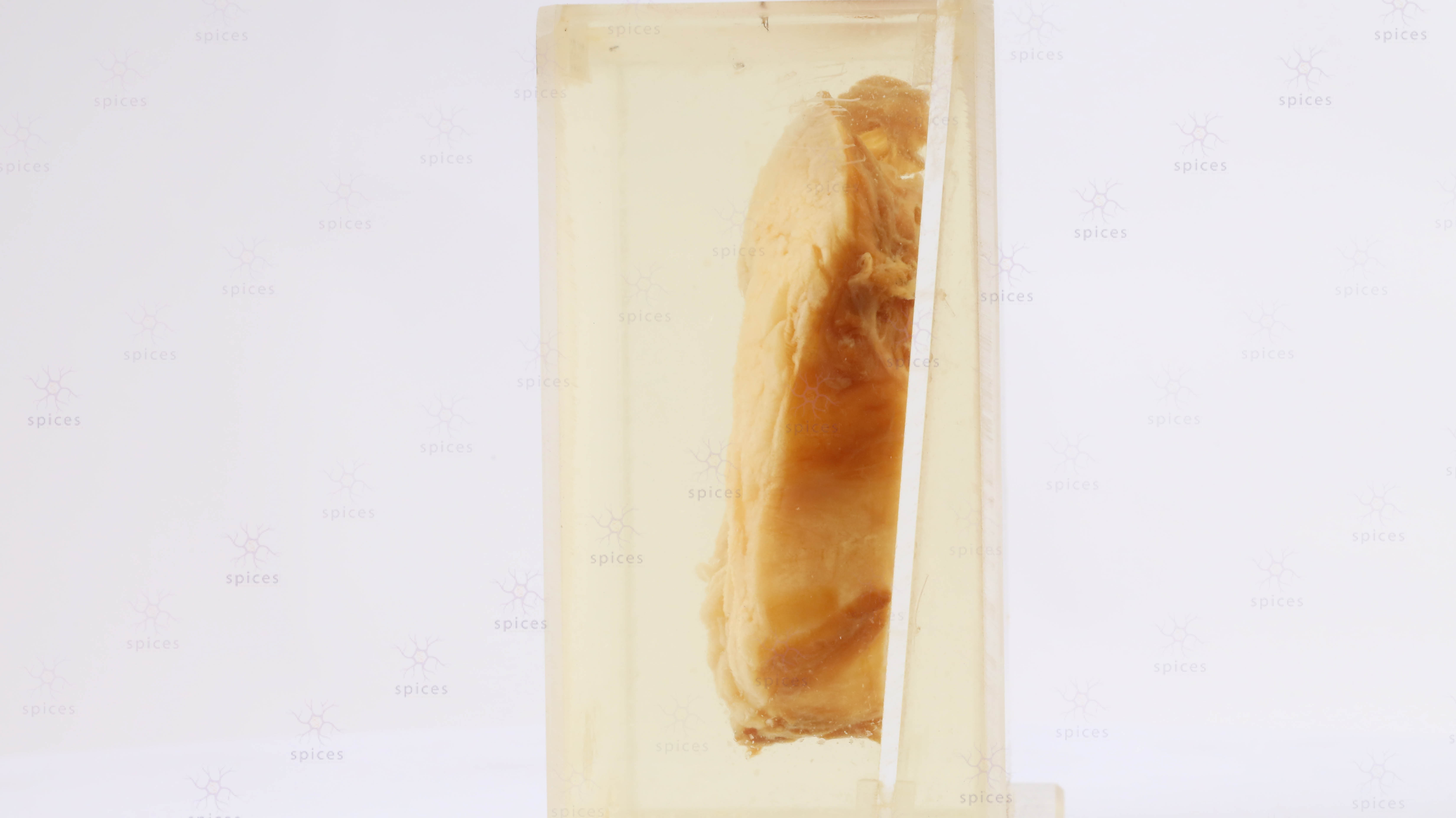
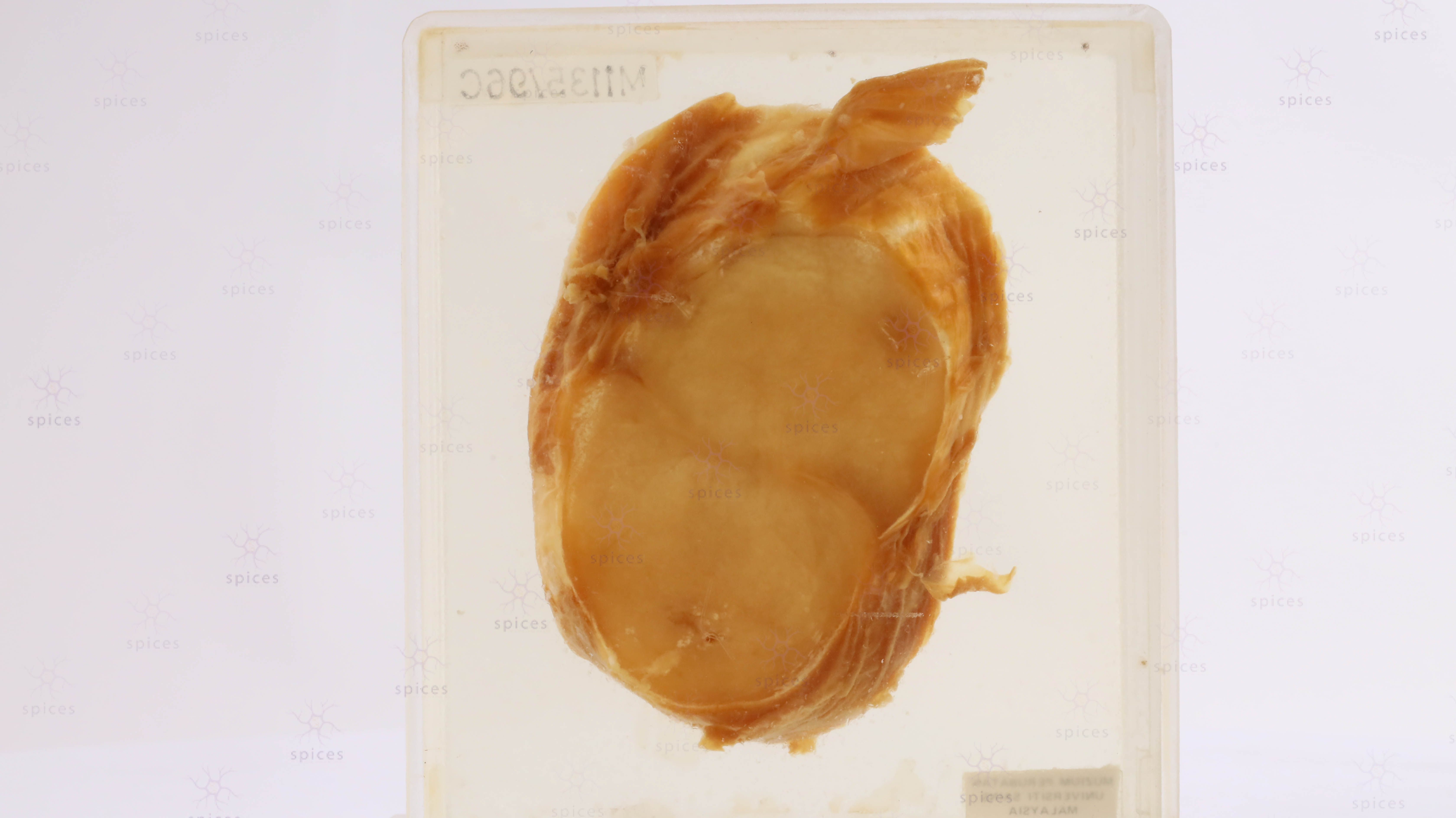
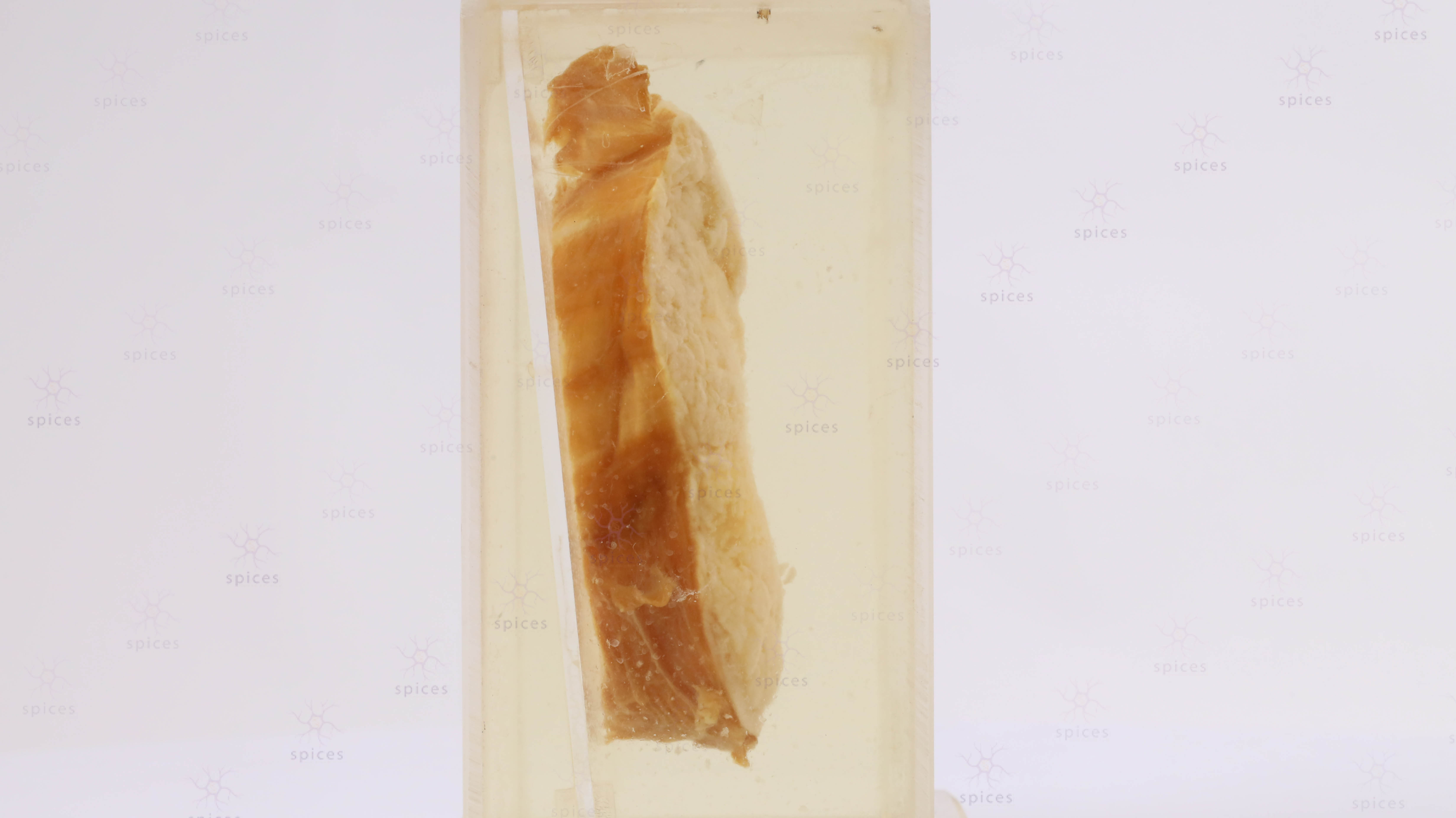
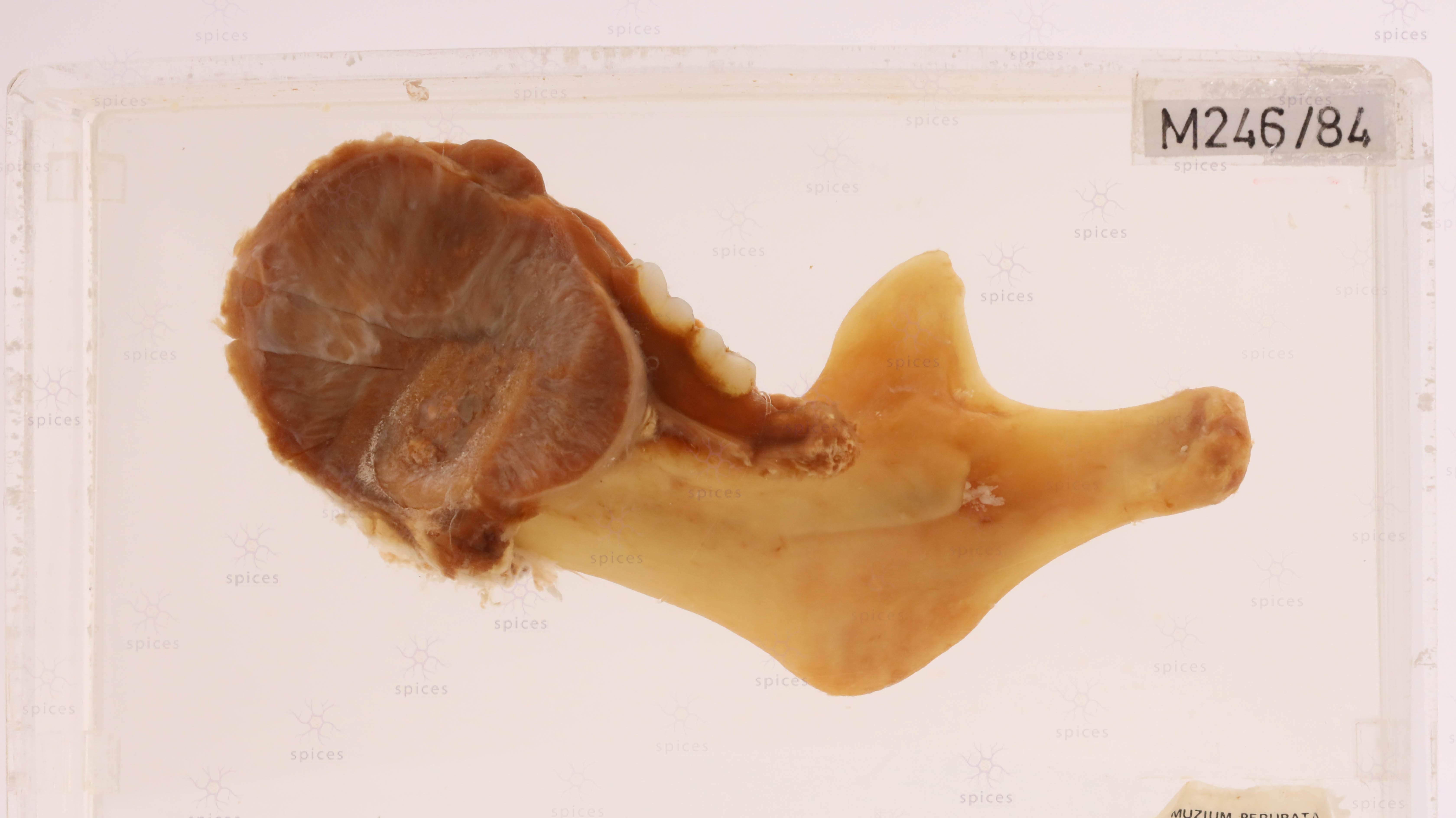
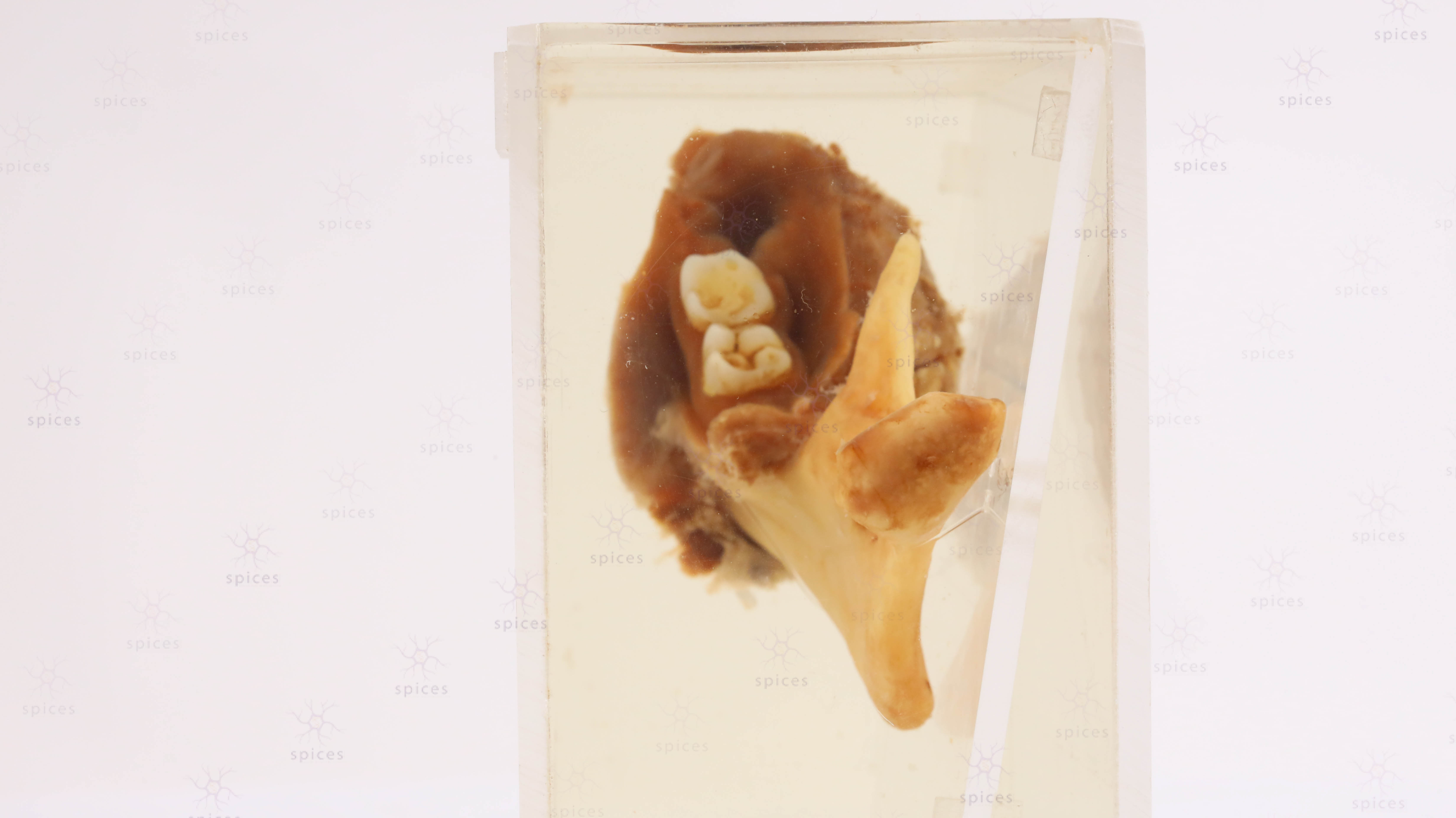
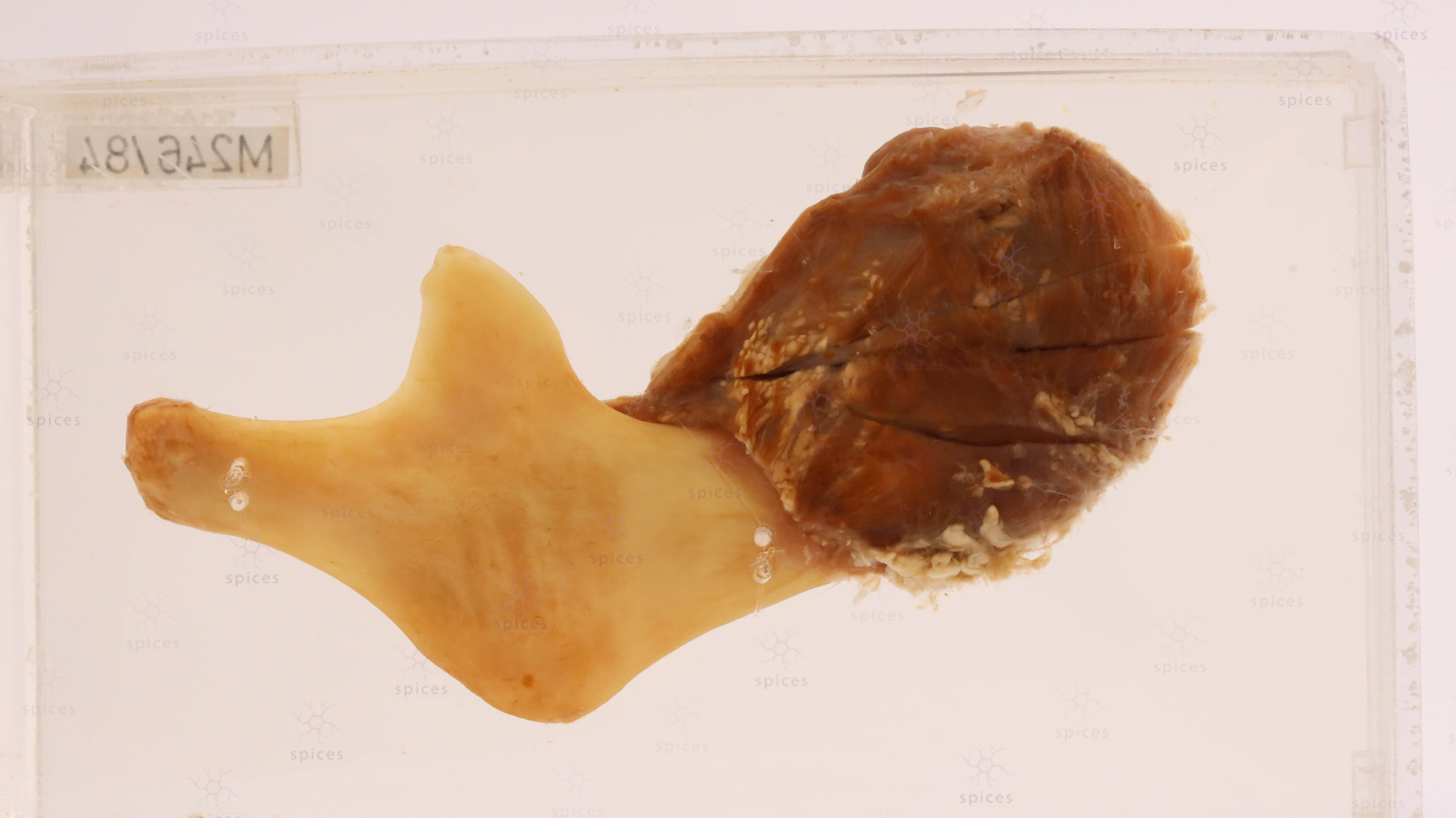
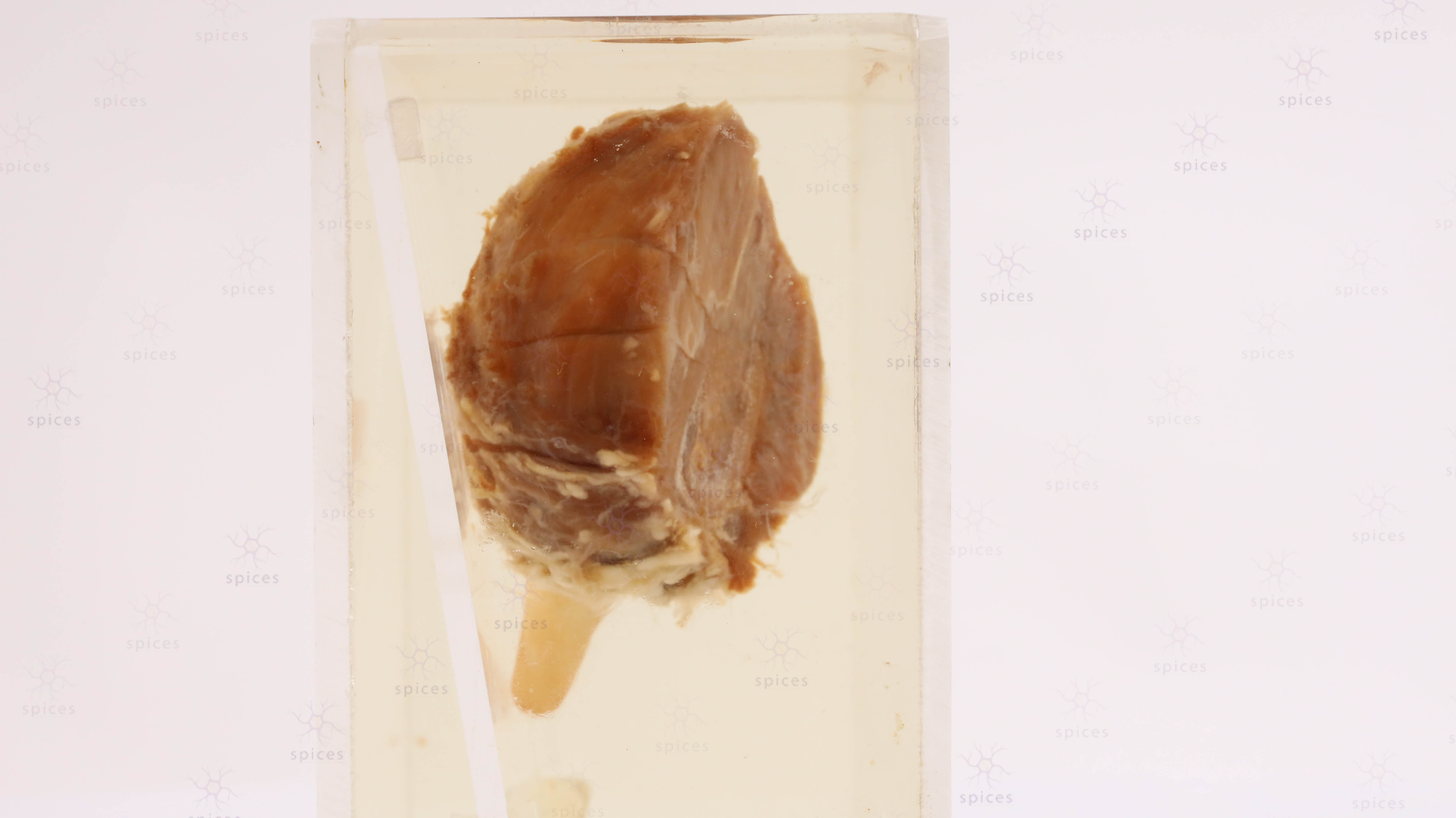
GROSS DESCRIPTION
• Expansile lytic lesion involving epiphysis extending to metaphysis• Well-defined but not encapsulated• Marked cortical thinning and expansion• Cut surface: soft, flesy, tan-brown to grey• Areas of hemorrhage and dark brown discoloration (hemosiderin)• Focal cystic / blood-filled spaces• Extends close to subchondral bone• No obvious bone or cartilage matrix seenHISTOPATHOLOGY DESCRIPTION
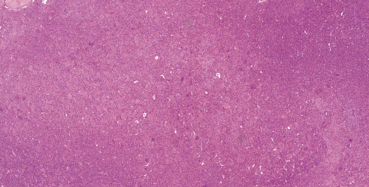 4x
4x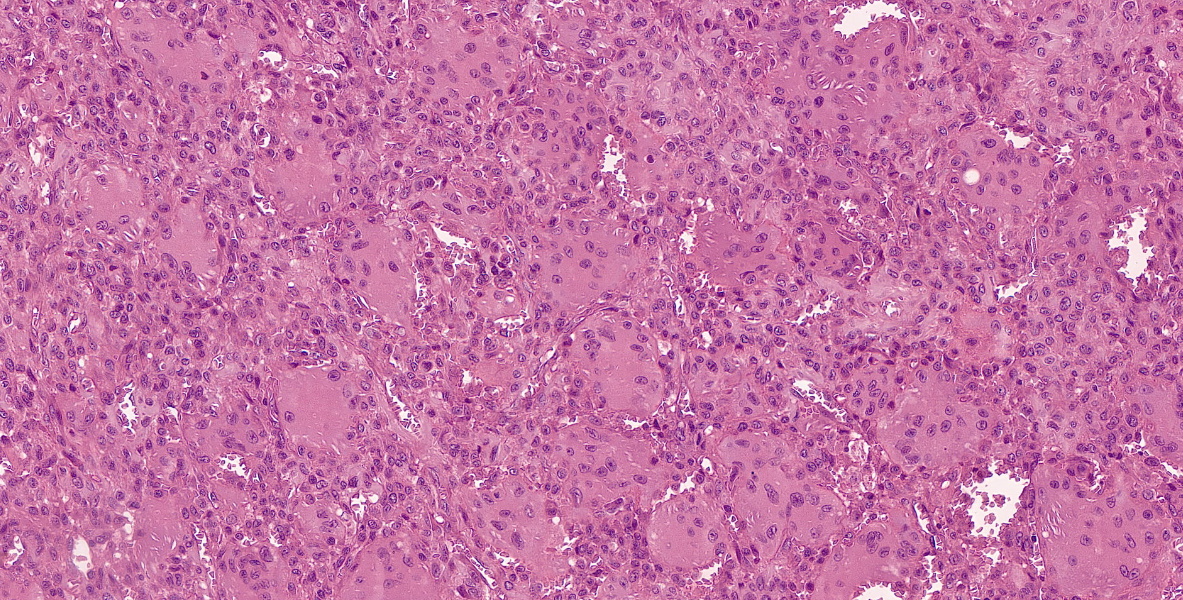 21xMicroscopy Description:Highly cellular tumor composed of mononuclear stromal cells and numerous osteoclast-type multinucleated giant cells• Giant cells are evenly distributed throughout the lesion• Mononuclear stromal cells are oval to spindle-shaped with nuclei similar to those of the giant cells• Mitotic figures may be seen but are usually typical (no atypical mitoses)• Areas of hemorrhage and hemosiderin deposition• Focal cystic change may be present• No malignant osteoid productionImmunohistochemistry:G34W shows diffuse nuclear positivity
RADIOLOGY
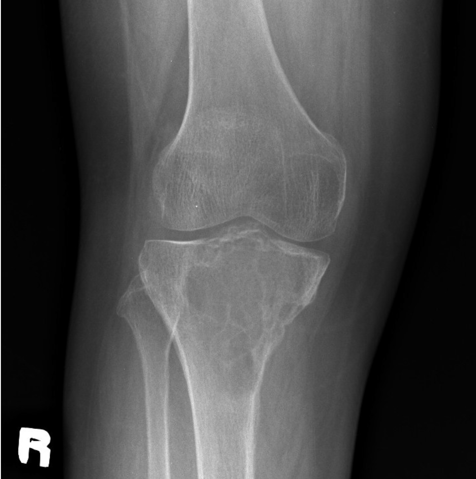
Right knee radiograph in frontal projection: A well-defined minimally expansile solitary lytic lesion in the meta-epiphysis of proximal tibia, with multiple internal trabeculations/septations (“soap-bubble” appearance). It has a narrow zone of transition with minimal marginal sclerosis. It is eccentric in location, extends to the subarticular region, with thinning of the overlying cortex. No cortical break. Minimal periosteal reaction at the inferomedial cortex of the proximal diaphysis.
*Image retrieved from PACS USM Workflow Manager
- Image description by Dr Fattah Rahiman Ghazali (Radiologist)
DIAGNOSIS
Giant Cell Tumour of Bone
QUIZ
BAHASA MELAYU