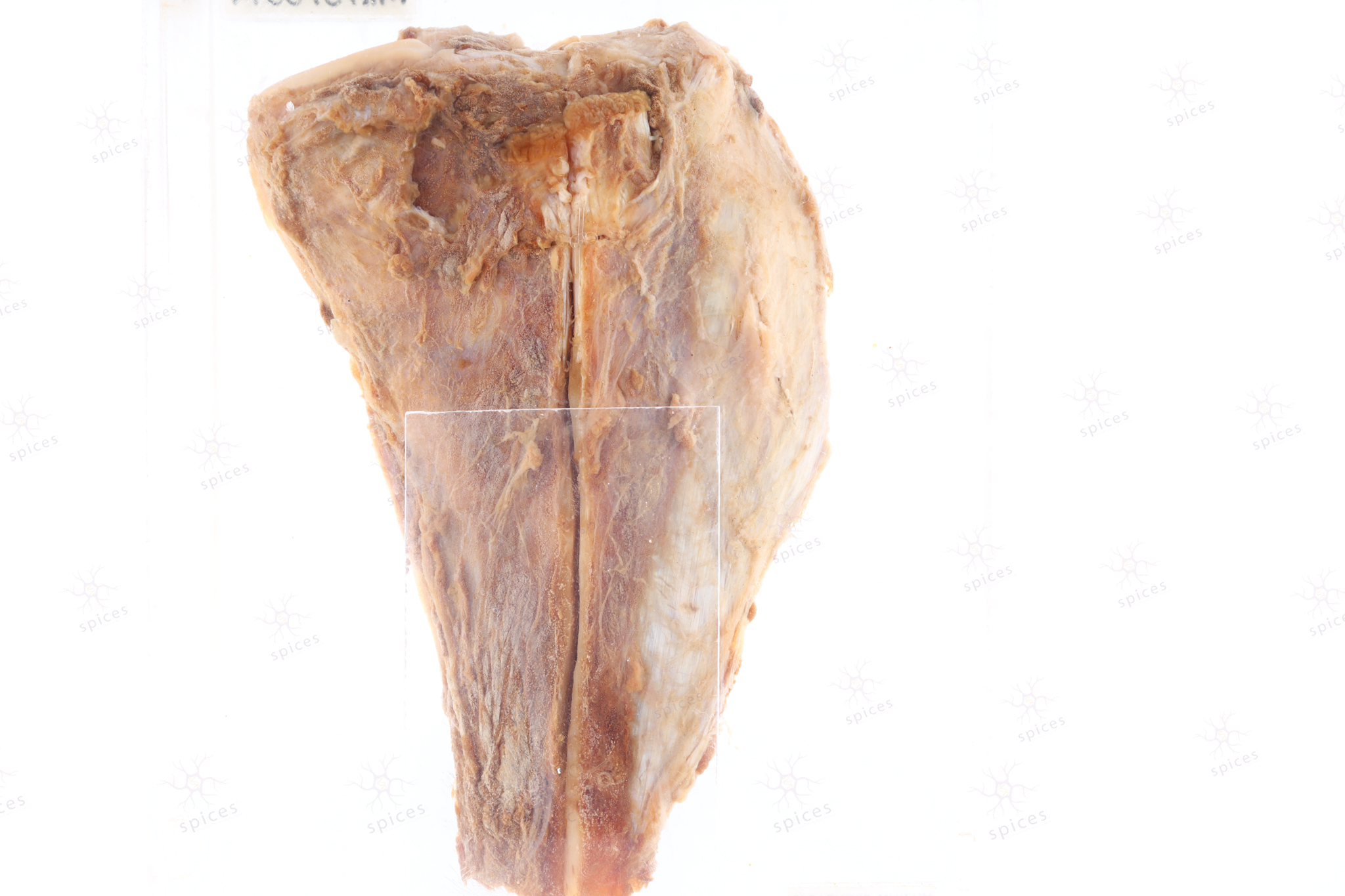
PROXIMAL TIBIA
Spices No: M1179/99A

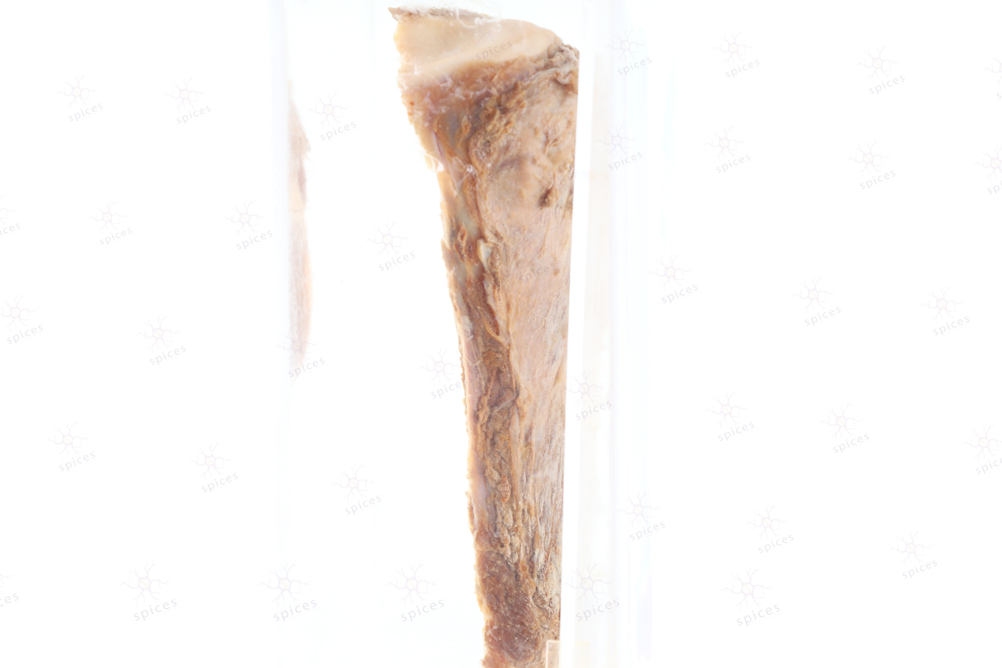
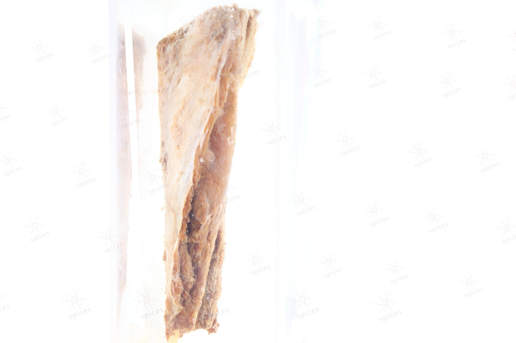
The exhibit displays ill defined large destructive tumour over proximal part of the tibia involving the epiphysis and metaphysis. The tumour is infiltrative, destroying the overlying cortex and extending into the surrounding soft tissue. It exhibit heterogenous tan to brown mass.

Osteosarcoma shows malignant osteoid deposited in a lace like pattern or as delicate trabeculae. The malignant cells are pleomorphic with irregular hyperchromatic nuclei and high mitotic activity.
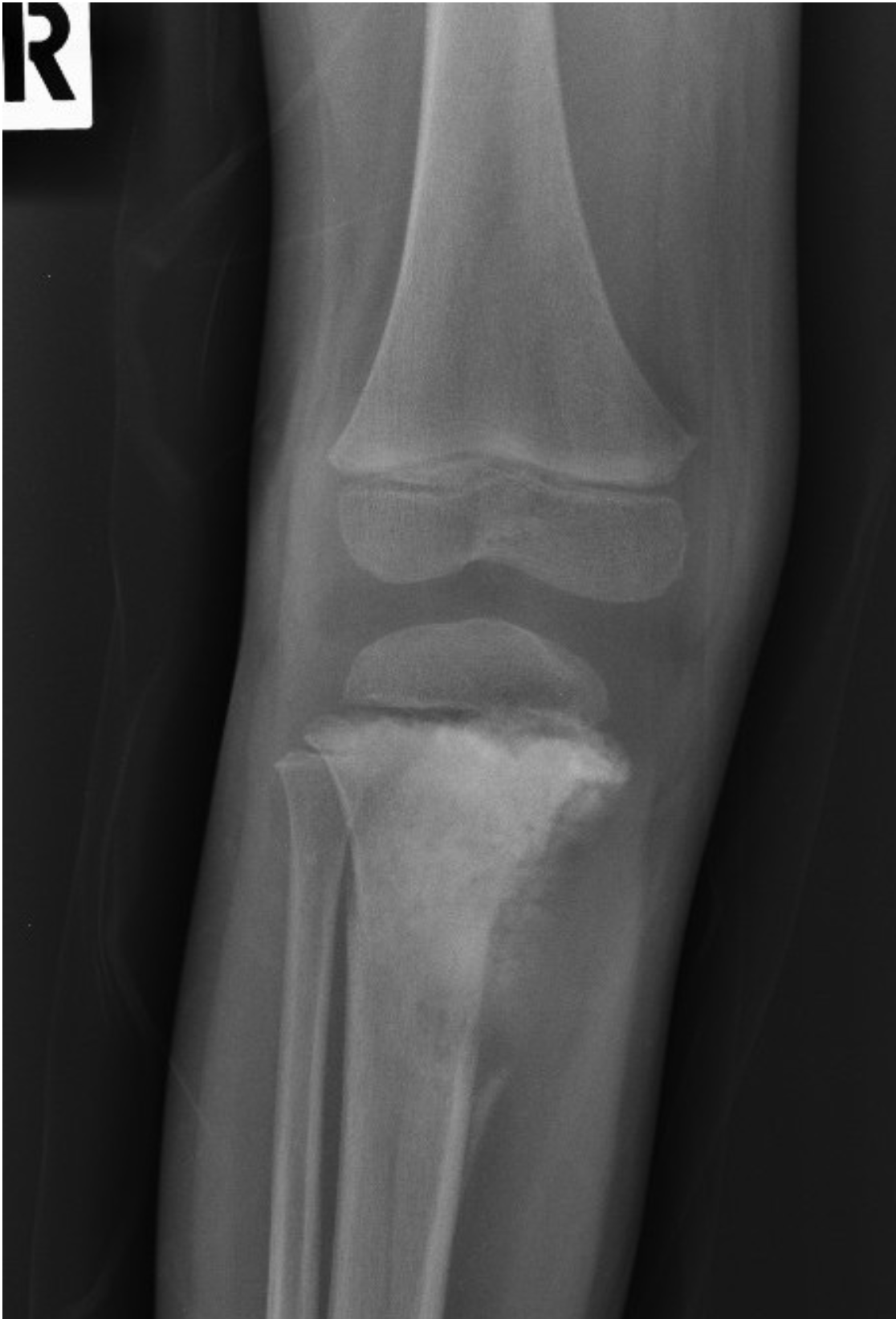
Right knee radiograph in frontal projection: An ill-defined sclerotic lesion in the dia-metaphyseal region of proximal tibia, with osteoid matrix formation. It has a wide zone of transition. Elevated periosteal reaction at the inferomedial cortex of the proximal diaphysis (Codman’s triangle). Minimal extraosseous/soft tissue extension at the medial aspect of the lesion. No intra-articular extension.
*Image retrieved from PACS USM Workflow Manager
- Image description by Dr Fattah Rahiman Ghazali (Radiologist)
Osteosarcoma
The type(s) of osteosarcoma include(s):
A. Osteoblastic [T/F]
B. Chondroblastic [T/F]
C. Fibroblastic [T/F]
D. Telangiectatic [T/F]
E. Lymphoblastic [T/F]
Answers: T T T T F