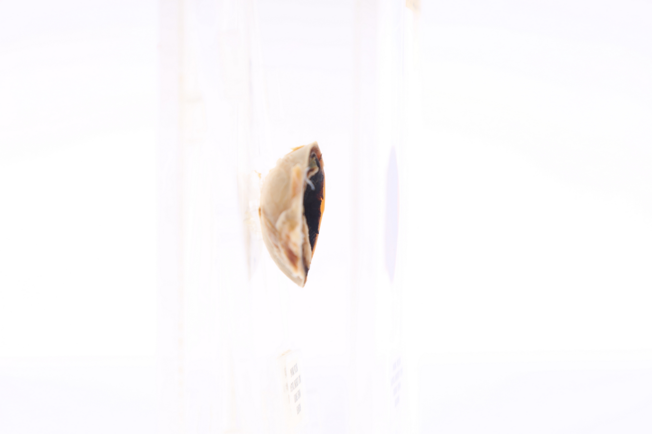
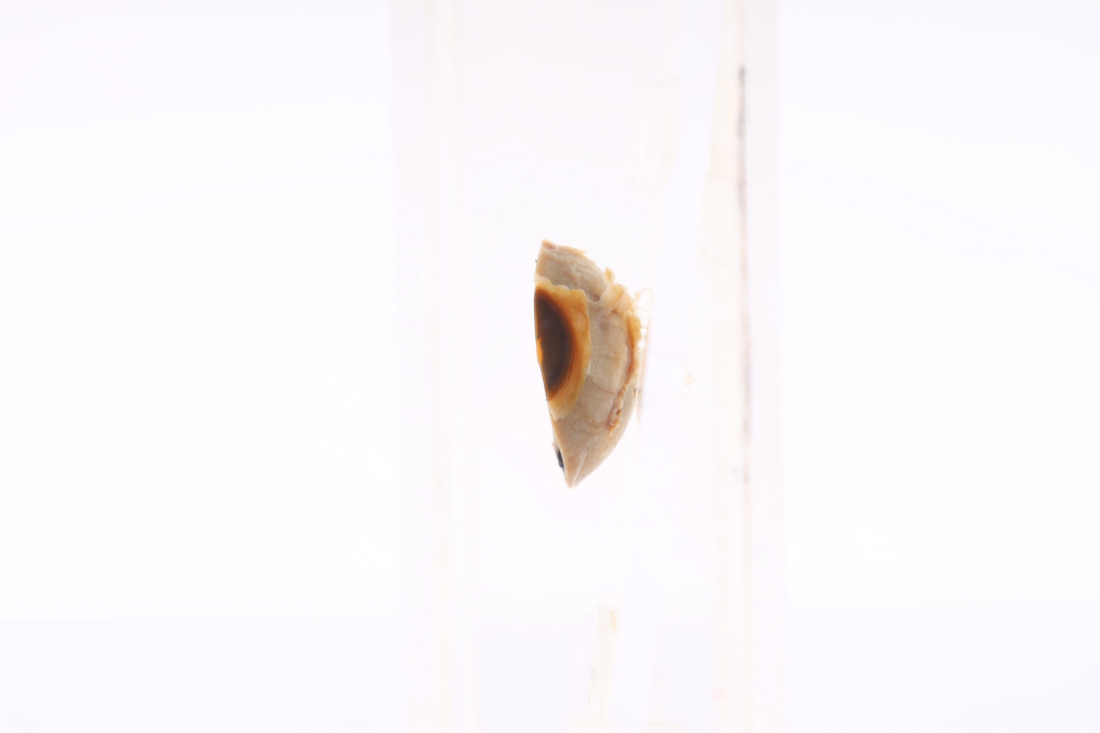
Eye: M1094/94
EYE
SPICES No. M1094/94
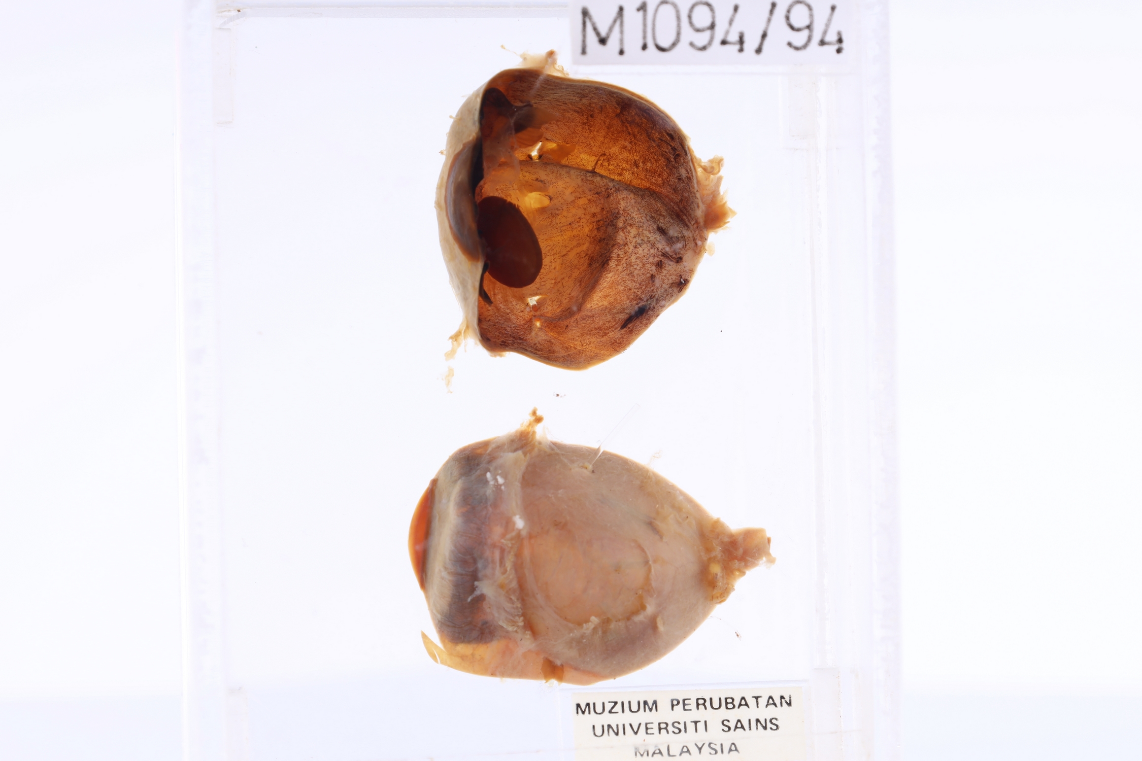
GROSS DESCRIPTION
The exhibit displays specimen of a normal eyeball. The eye is made of three basic layers but cannot be discernible grossly : the outer corneoscleral layer, uveal layer and inner retinal layer. The large cavity is a posterior compartment which is supposed to contain transparent gel known as vitreous body.
HISTOPATHOLOGY DESCRIPTION
Section shows unremarkable histology of eye balls composed of multiple layers of outer and inner structure. The sclera and cornea make up the exterior layers. The uvea is the vascular layer in the middle, subdivided into the iris, ciliary body, and choroid. The retina constitutes the innermost layer and is made up of nervous tissue.
DIAGNOSIS
Normal eyeball
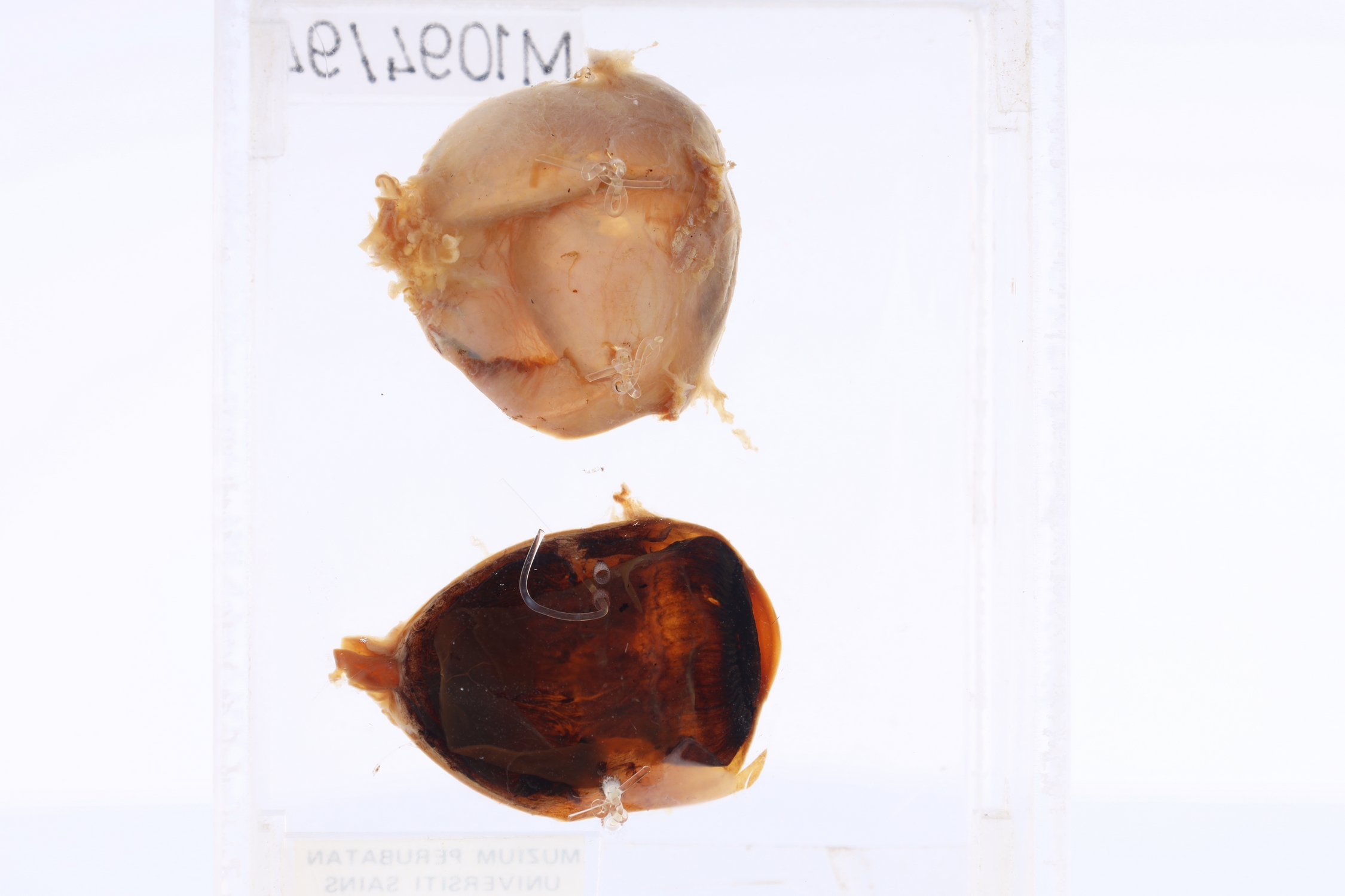
Eye: M766/90
EYE
SPICES No. M766/90
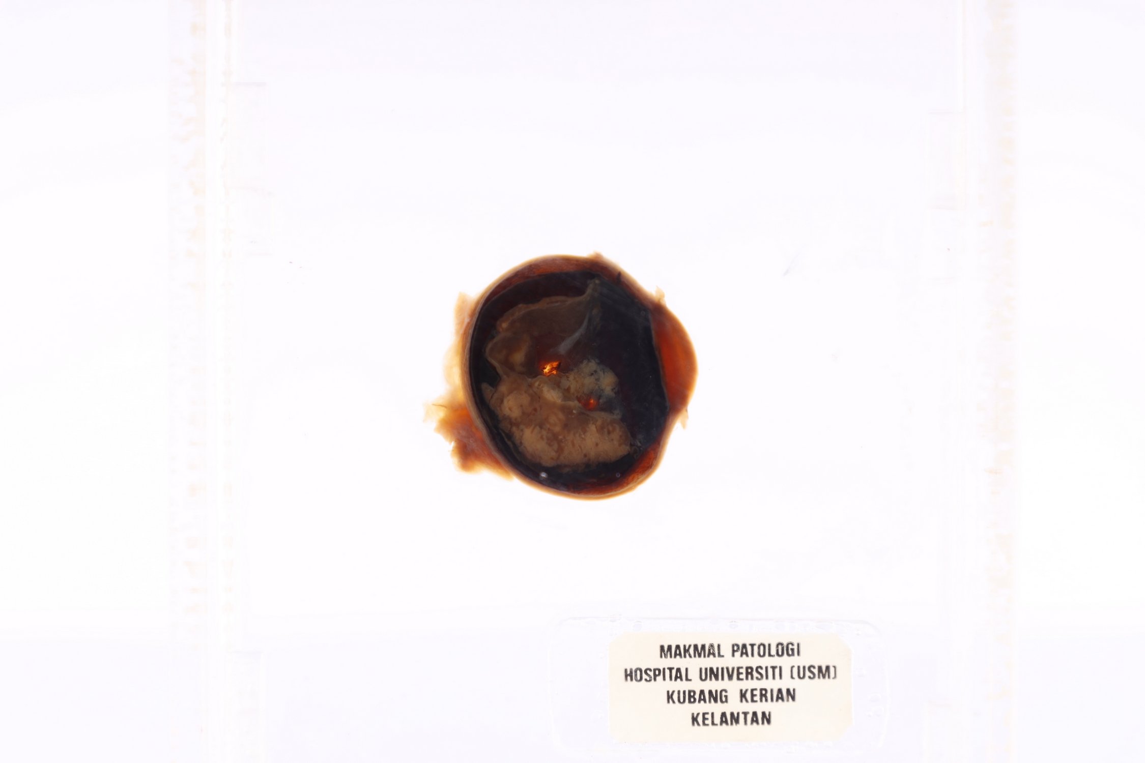
GROSS DESCRIPTION
This is exhibit displays eye with retinoblastoma. There is malignant tumor arising from the retina growing into subretinal spaces and vitreous.
HISTOPATHOLOGY DESCRIPTION
Retinoblastoma is show highly cellular tumours arranged in sheets and some in trabeculae pattern. The cells are small, round and blue with scanty cytoplasm. Flexner-Wintersteiner rosettes is seen. Calcification and perivascular necrosis also appreciated.
DIAGNOSIS
Retinoblastoma
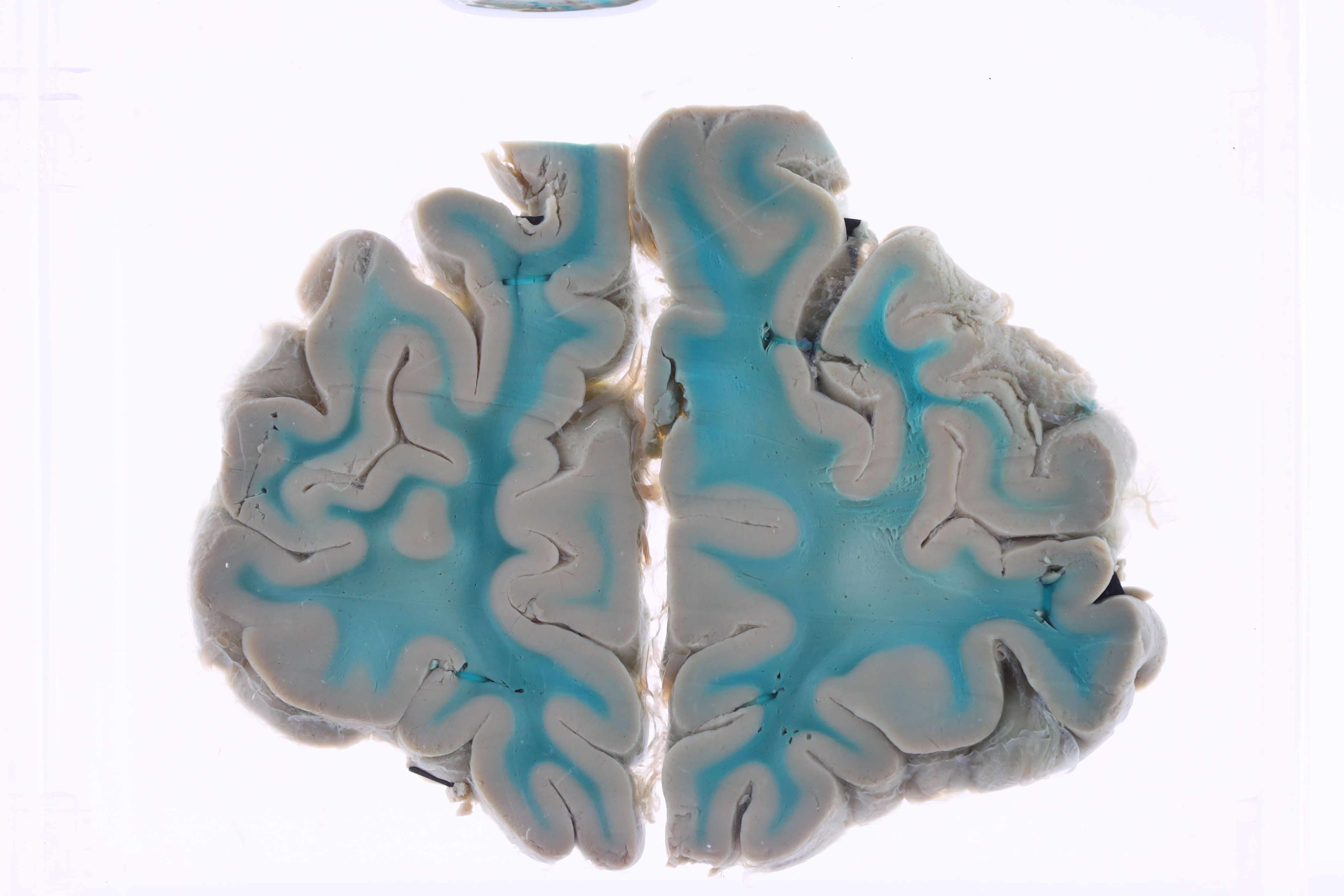
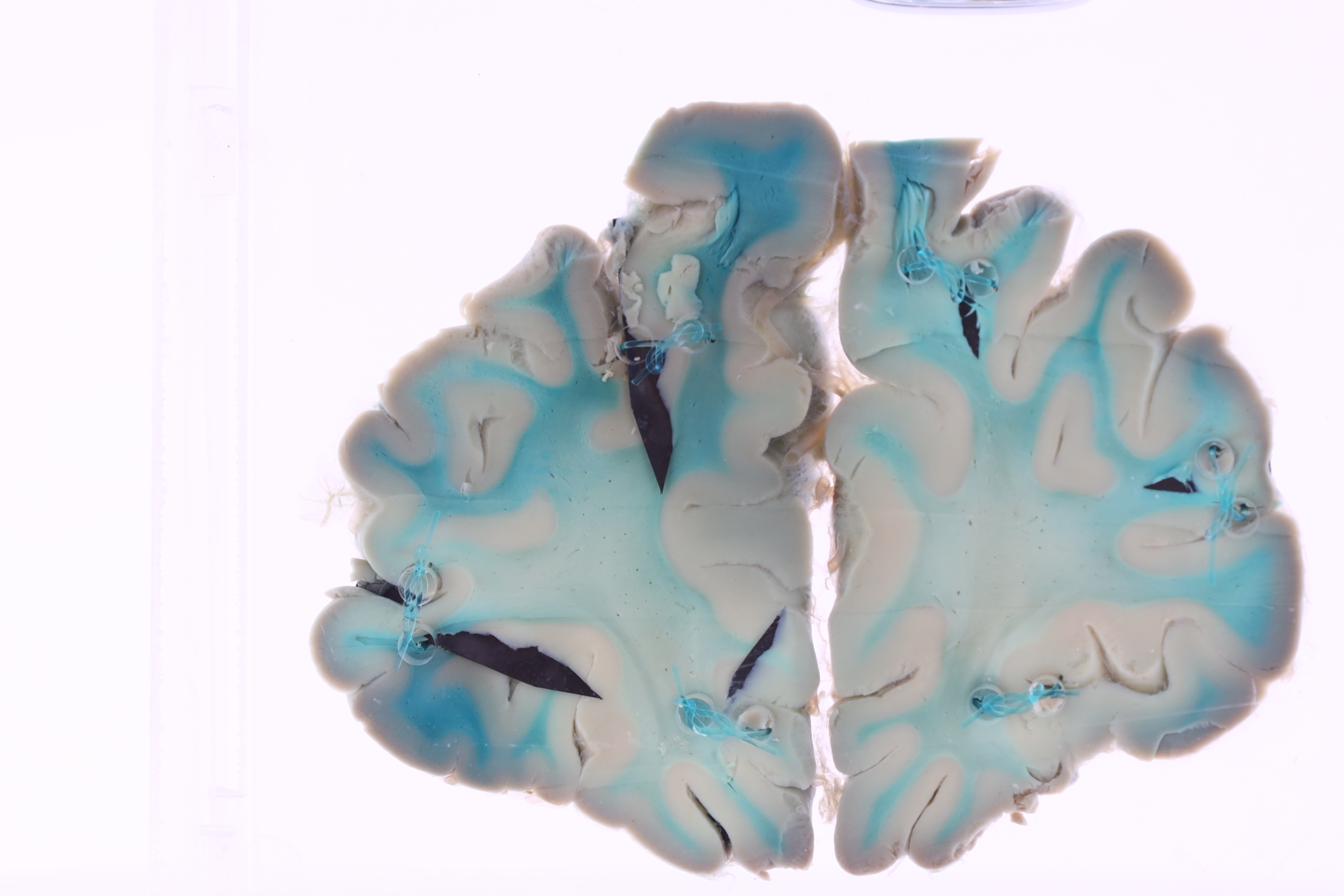
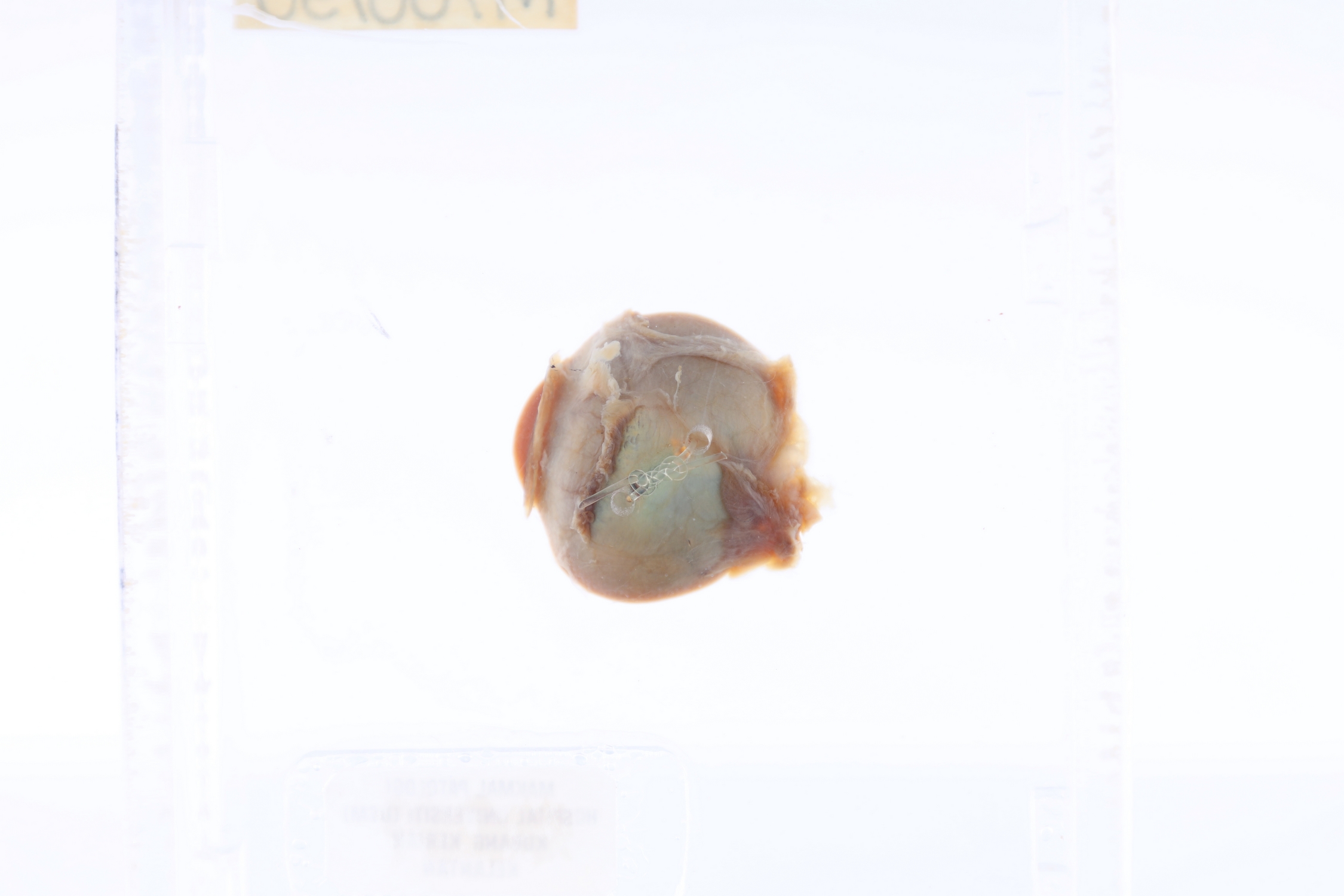
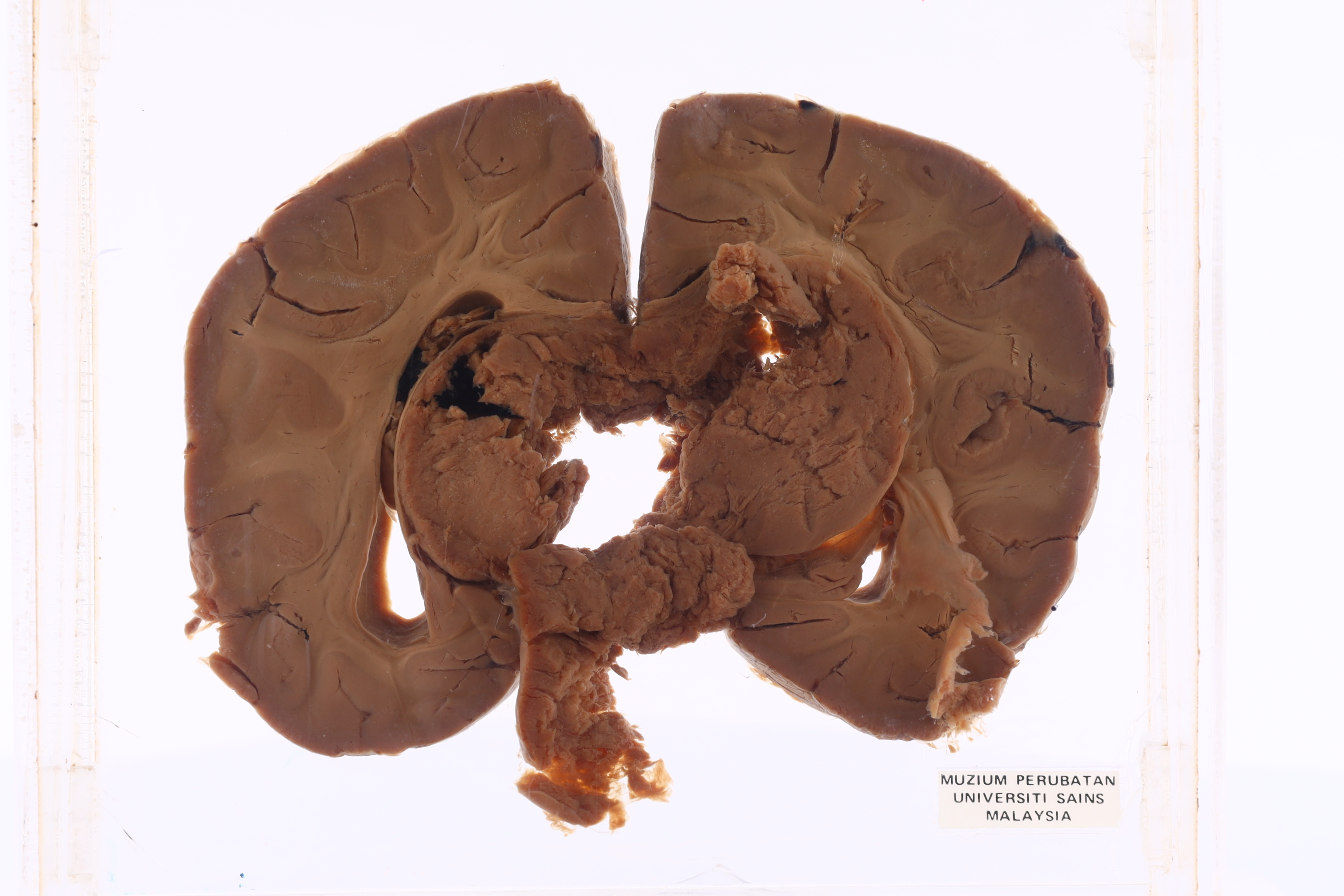
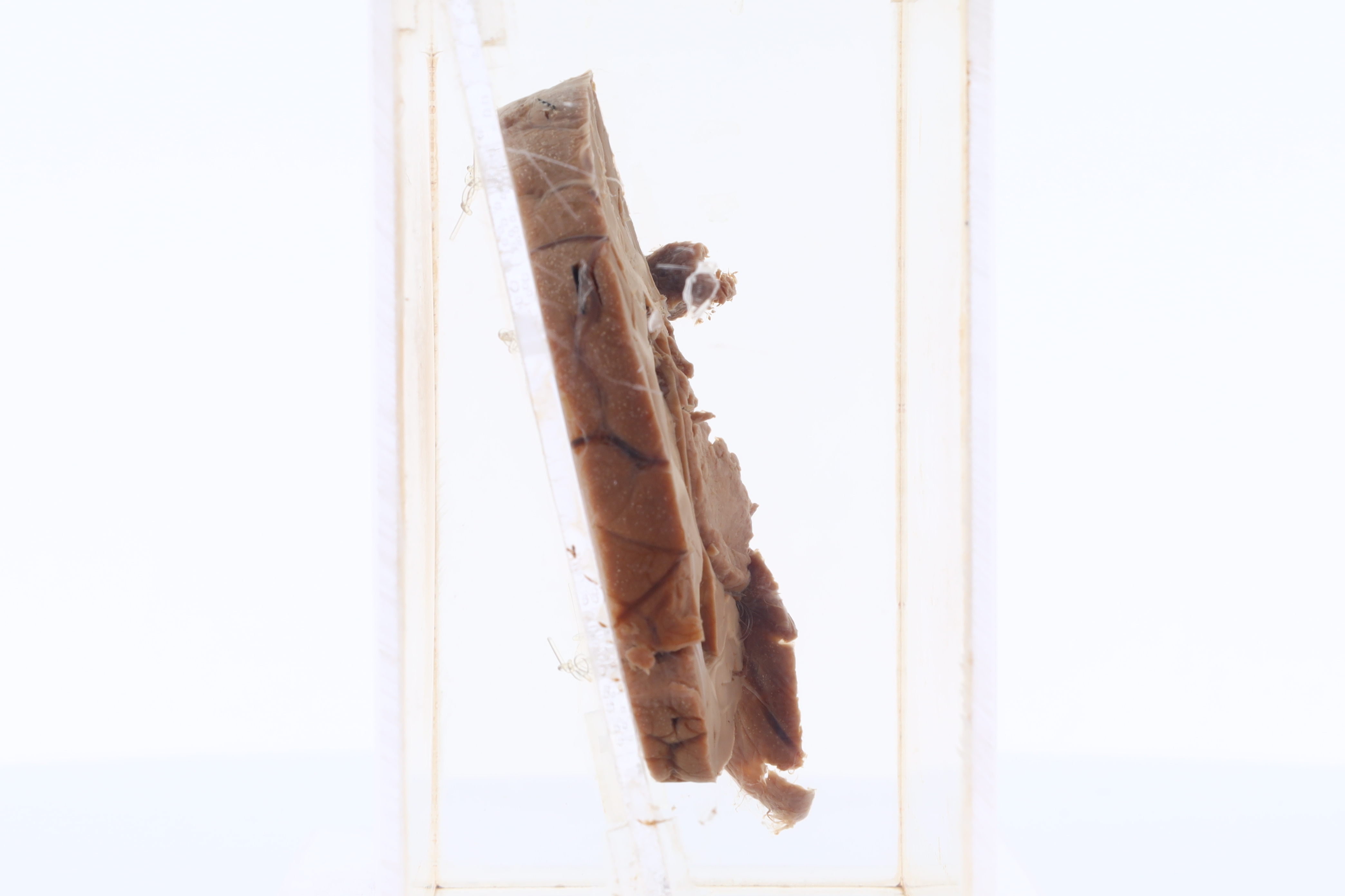
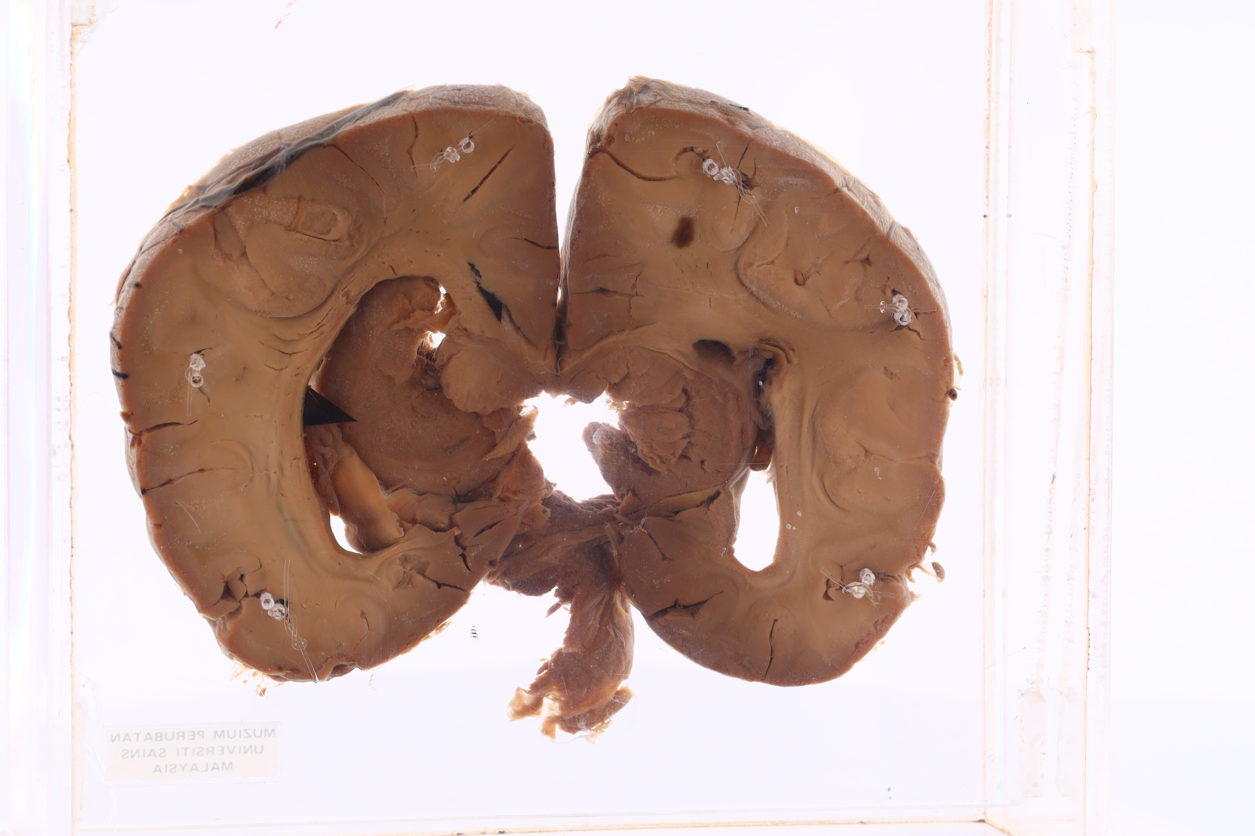
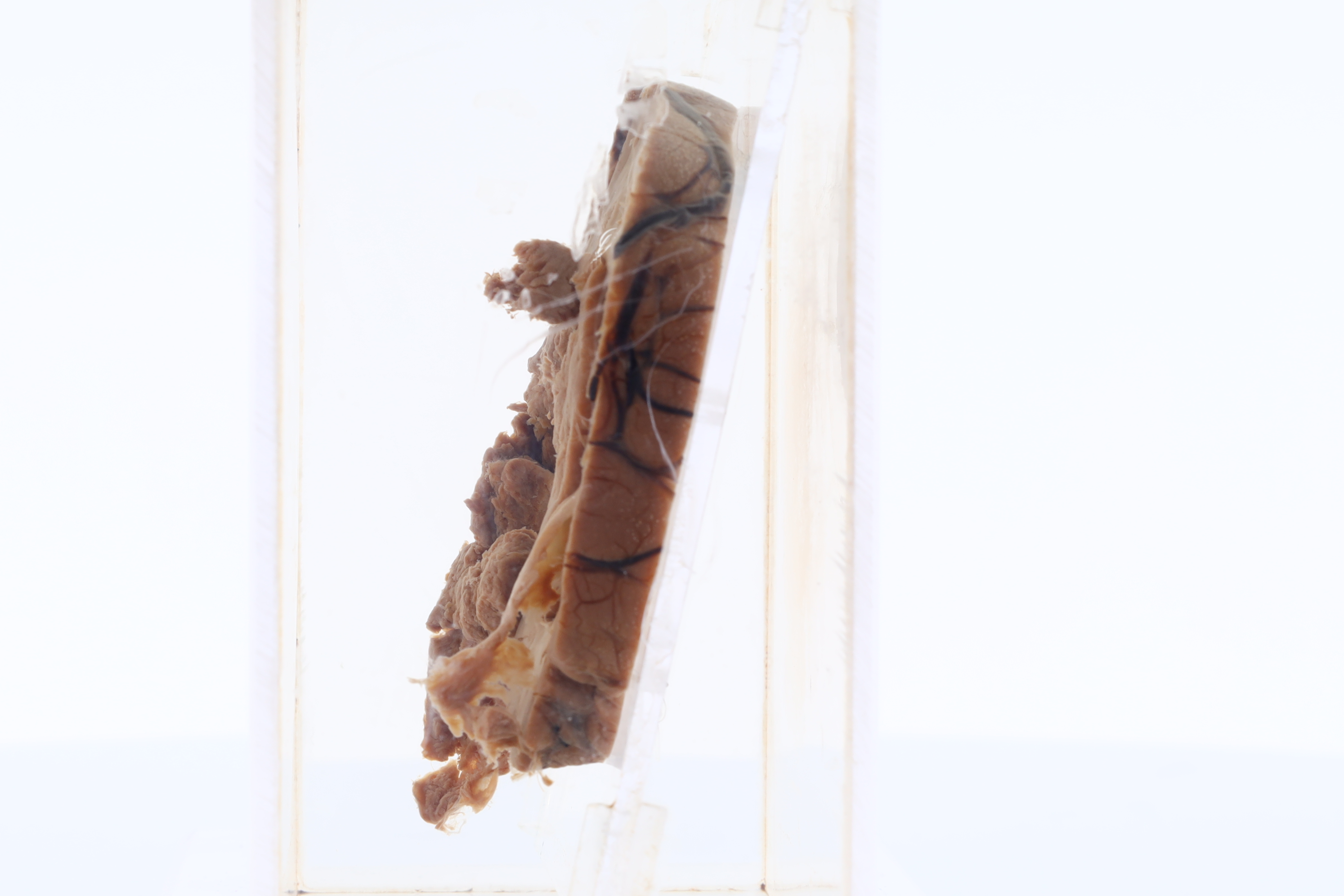
Brain
BRAIN
SPICES No. M42/82
GROSS DESCRIPTION
The exhibit displays presence of large supratentorial tumor arising in and occupying the lateral ventricle causing midline shift. Areas of central degenerated cystic changes observed.
HISTOPATHOLOGY DESCRIPTION
Section shows hypercellular tumour cells with sharply demarcated border. The cells are monomorphic with speckled chromatin pattern. Perivascular pseudorosettes and ependymal rosettes also appreciated.
DIAGNOSIS
Ependymoma