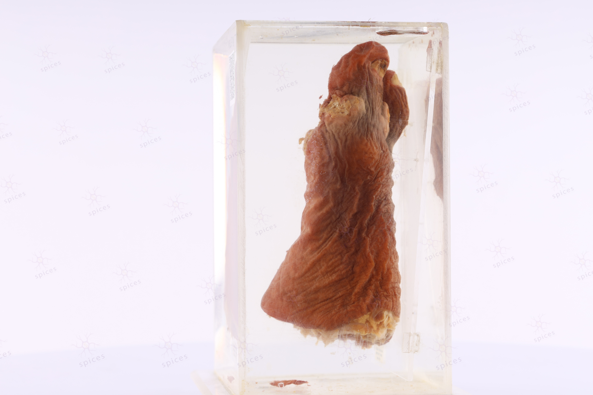
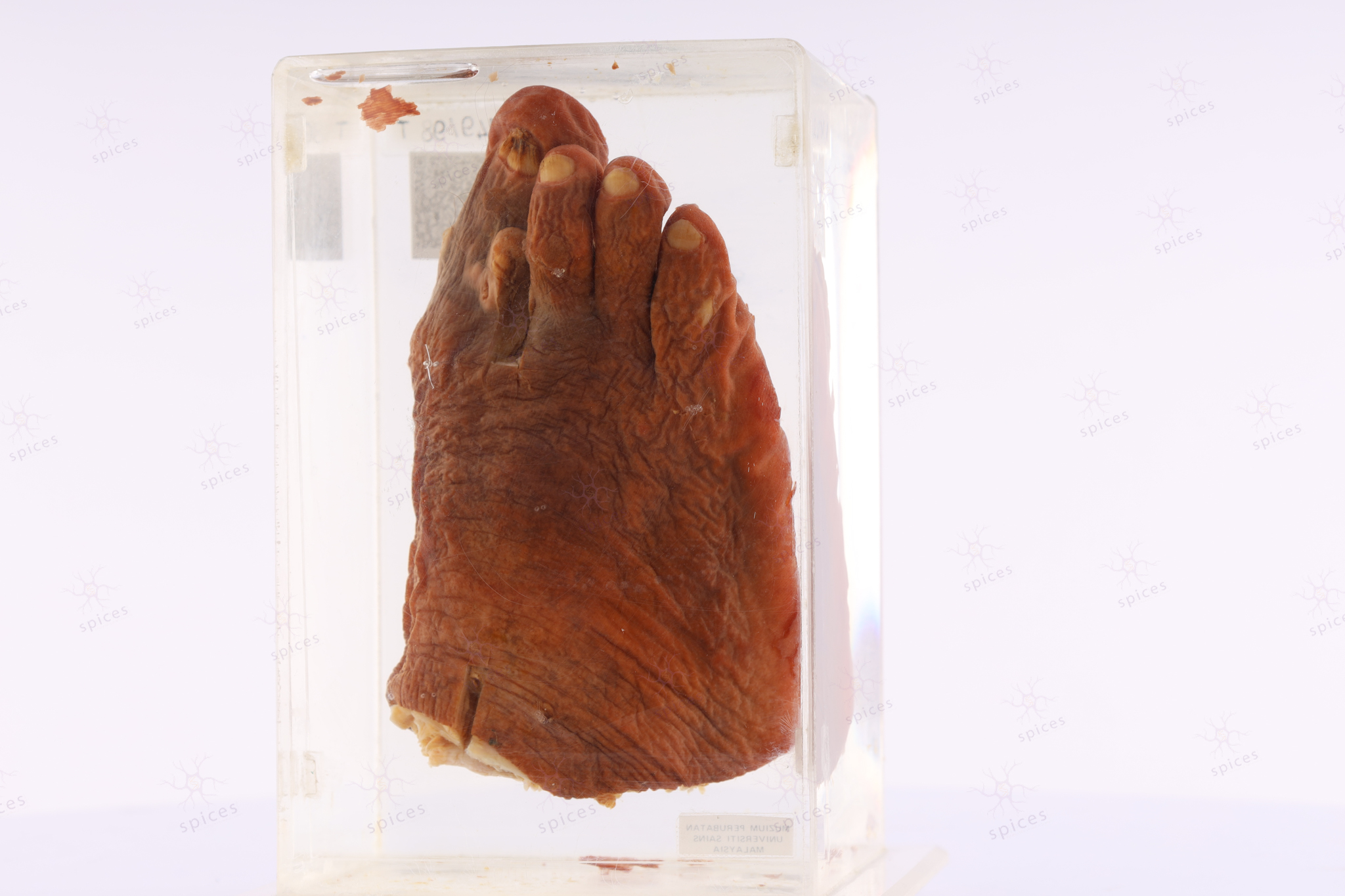
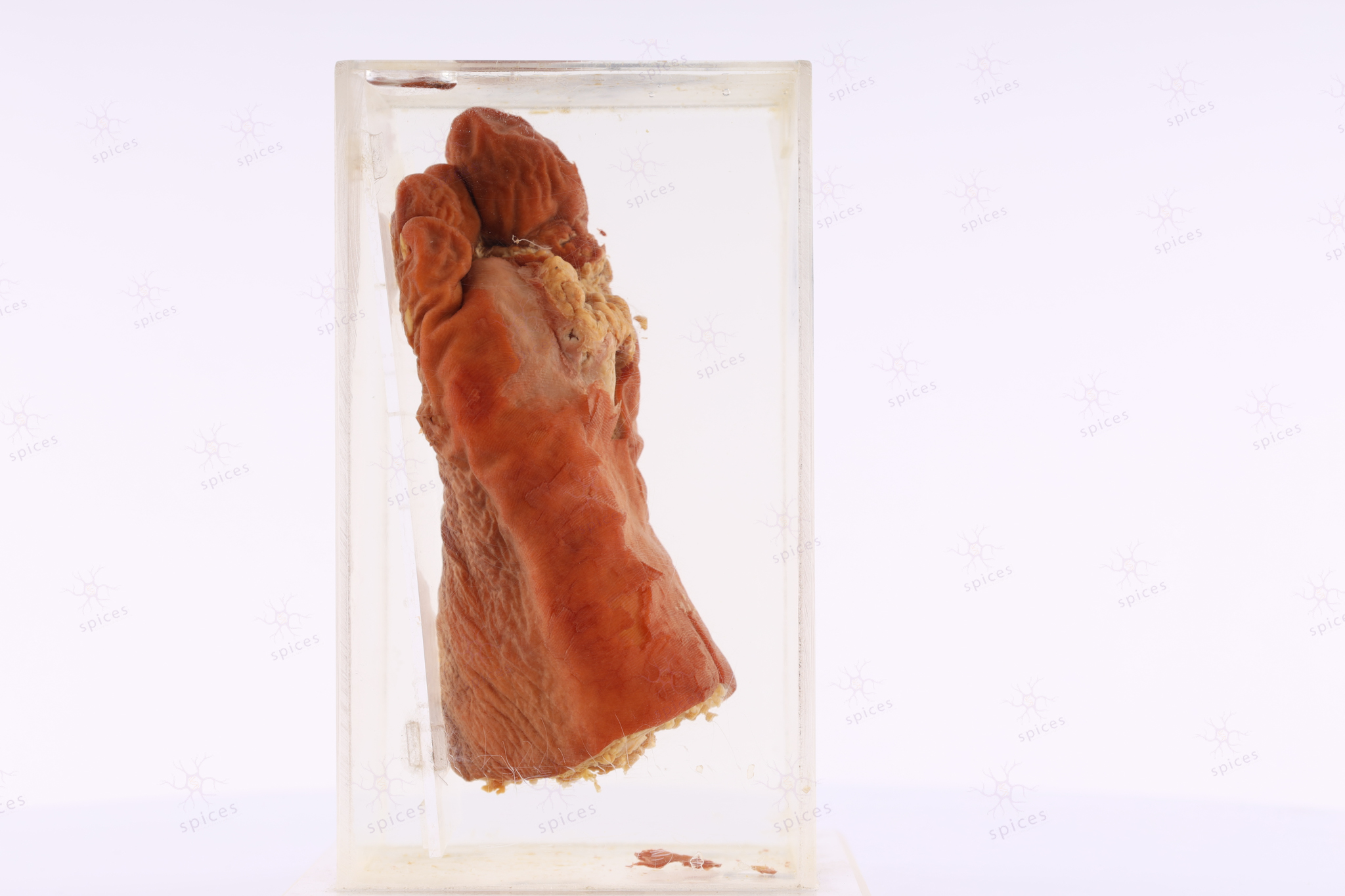
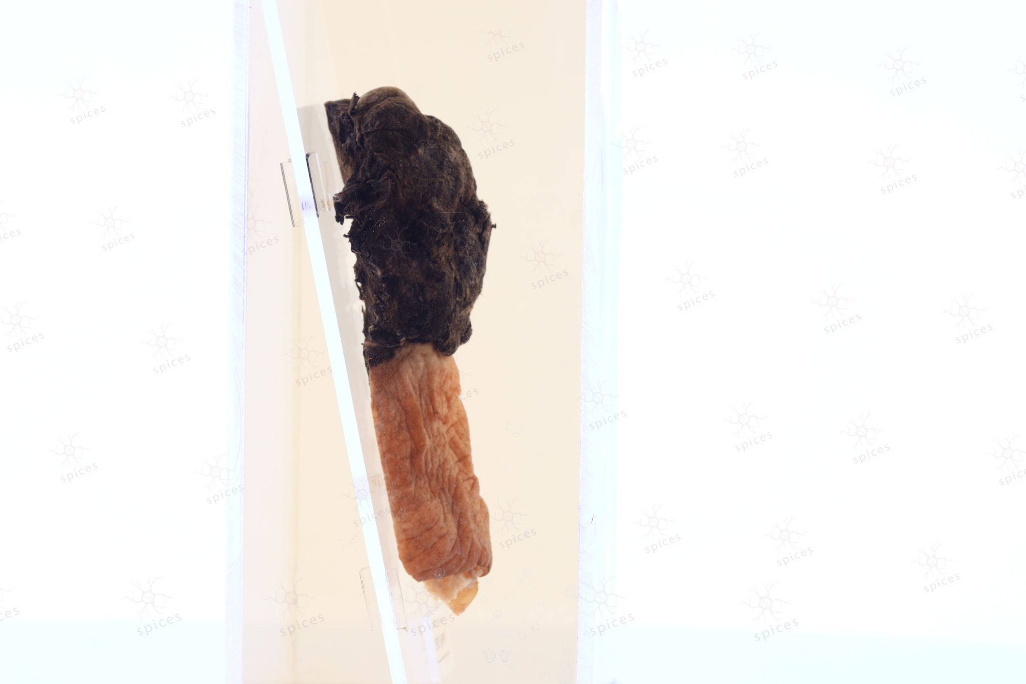
Skin : M647/89
SKIN
Spices No: M647/89

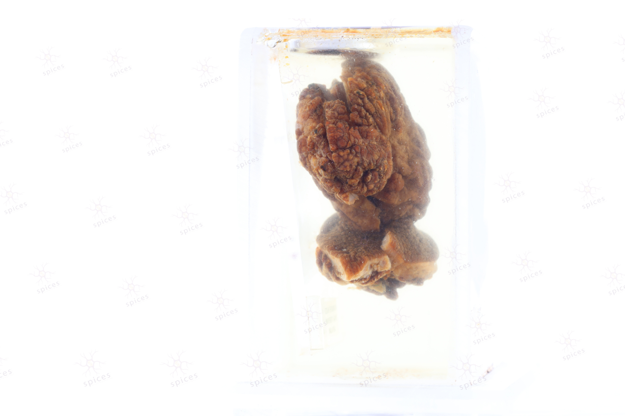
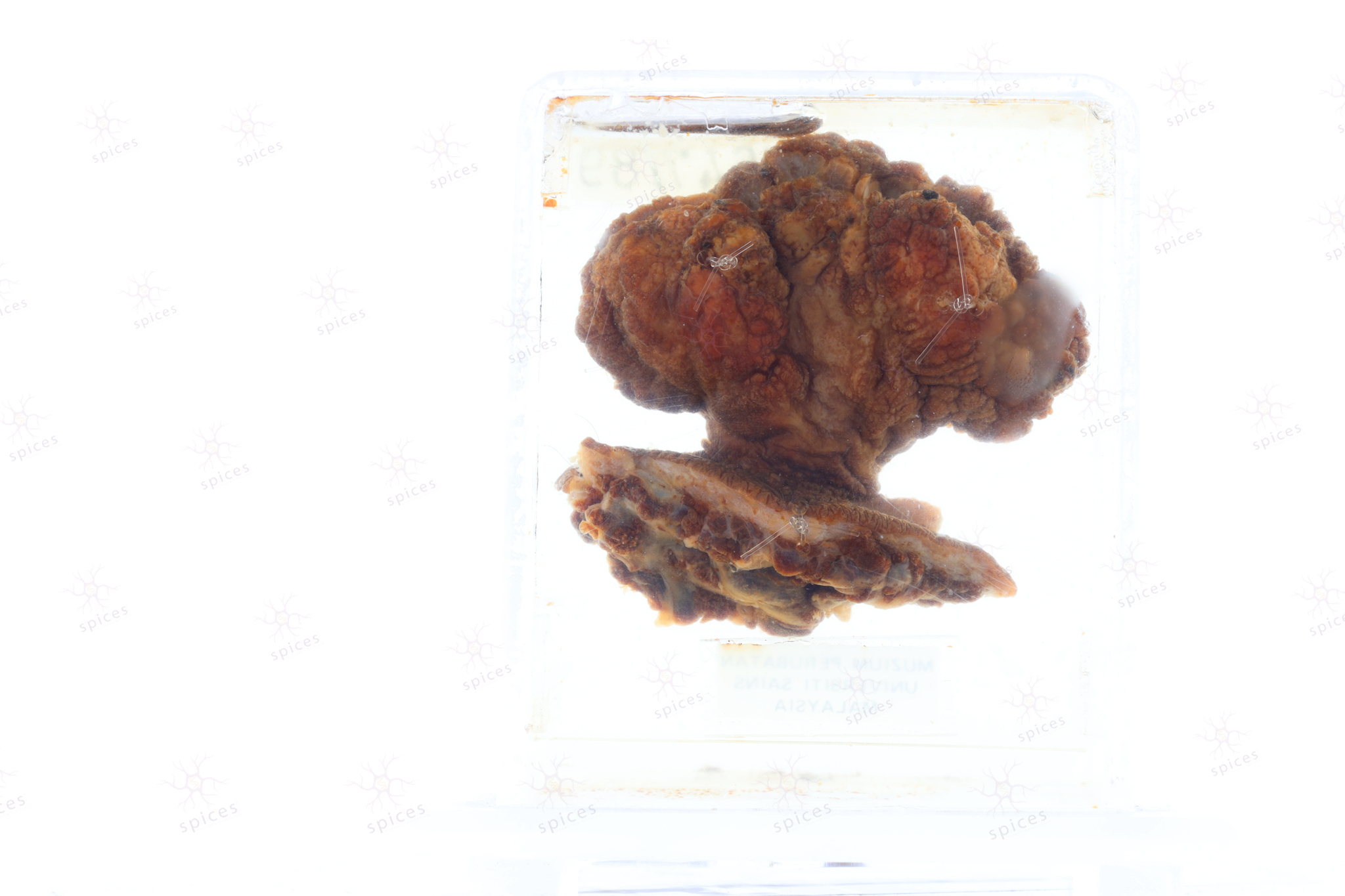
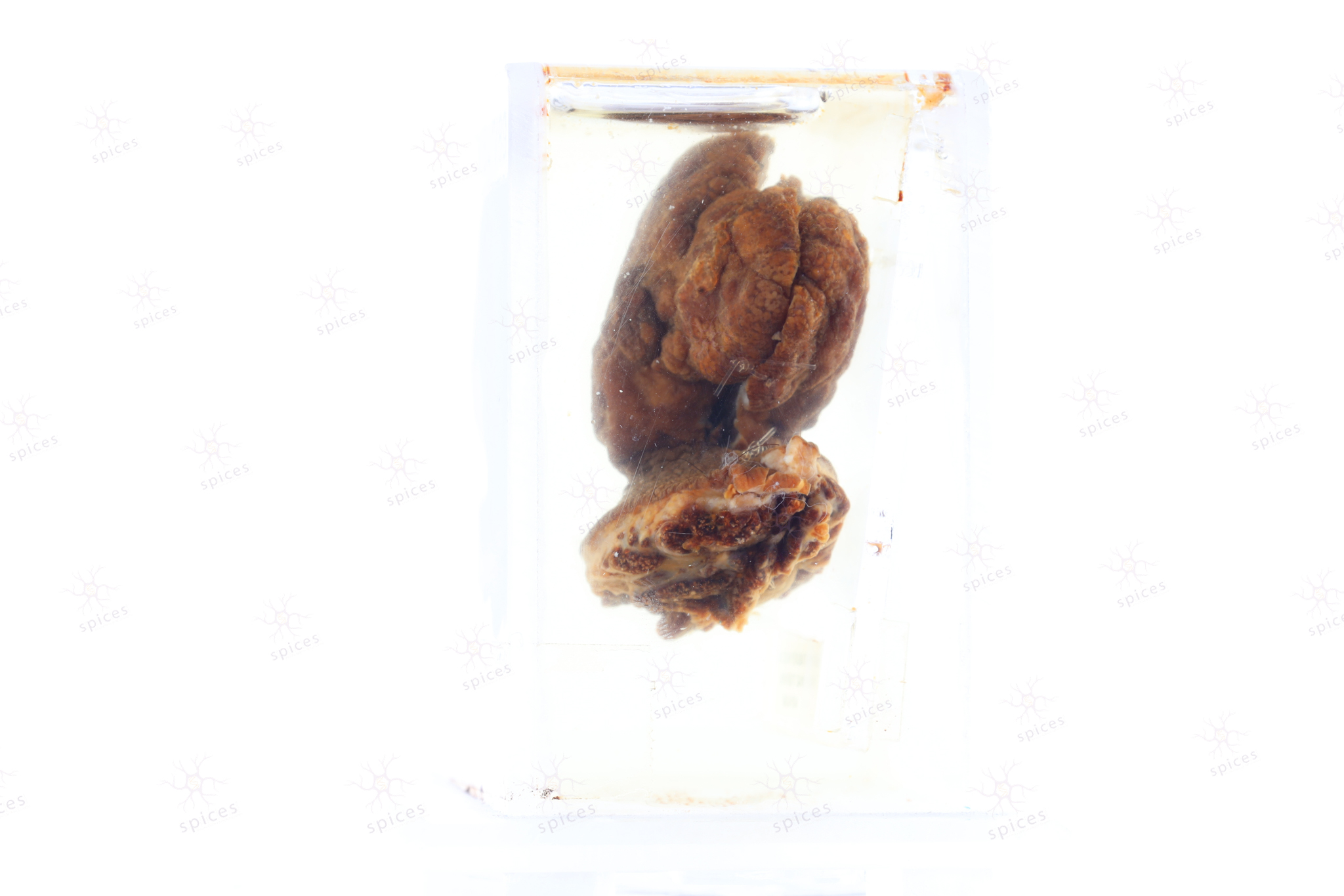
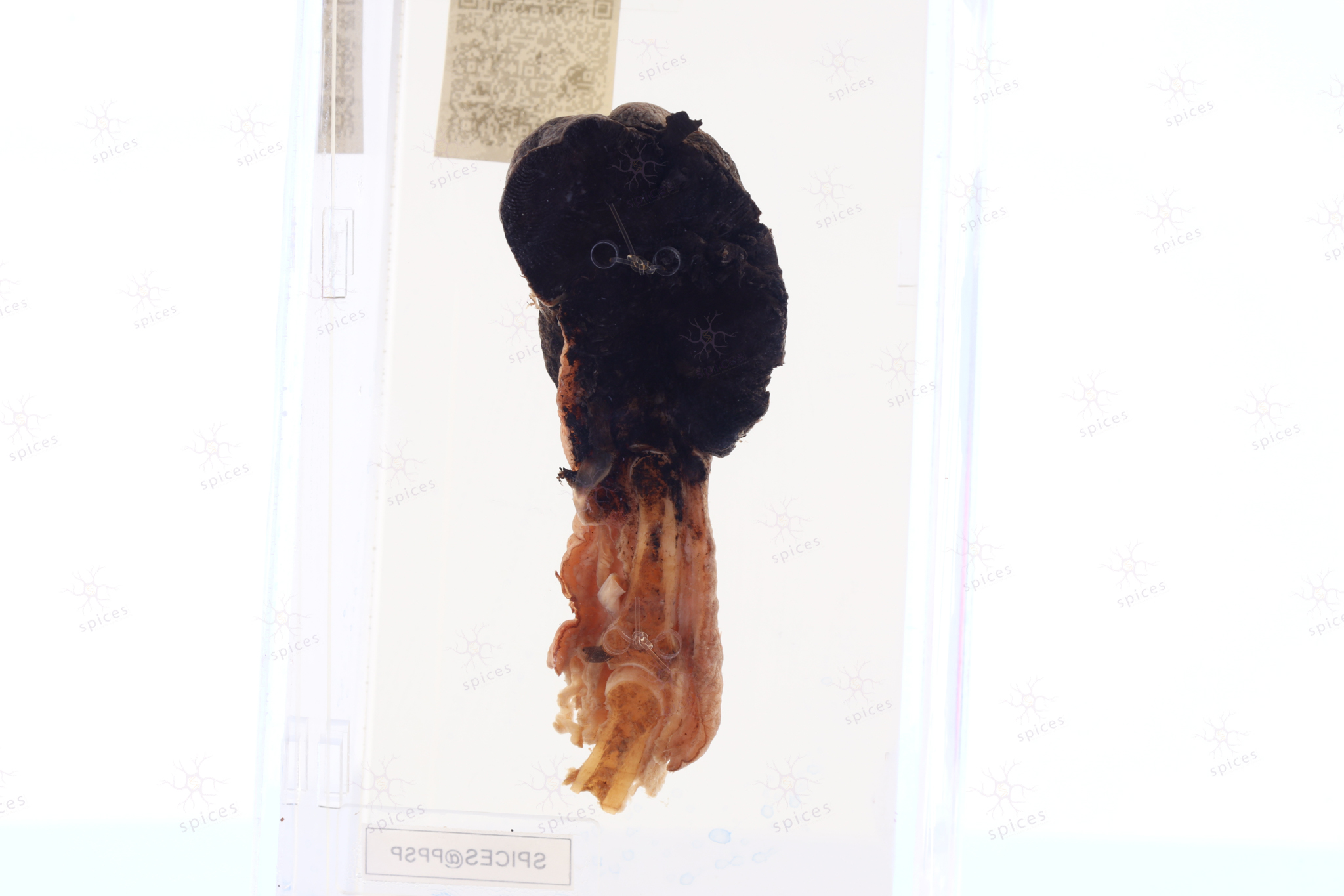
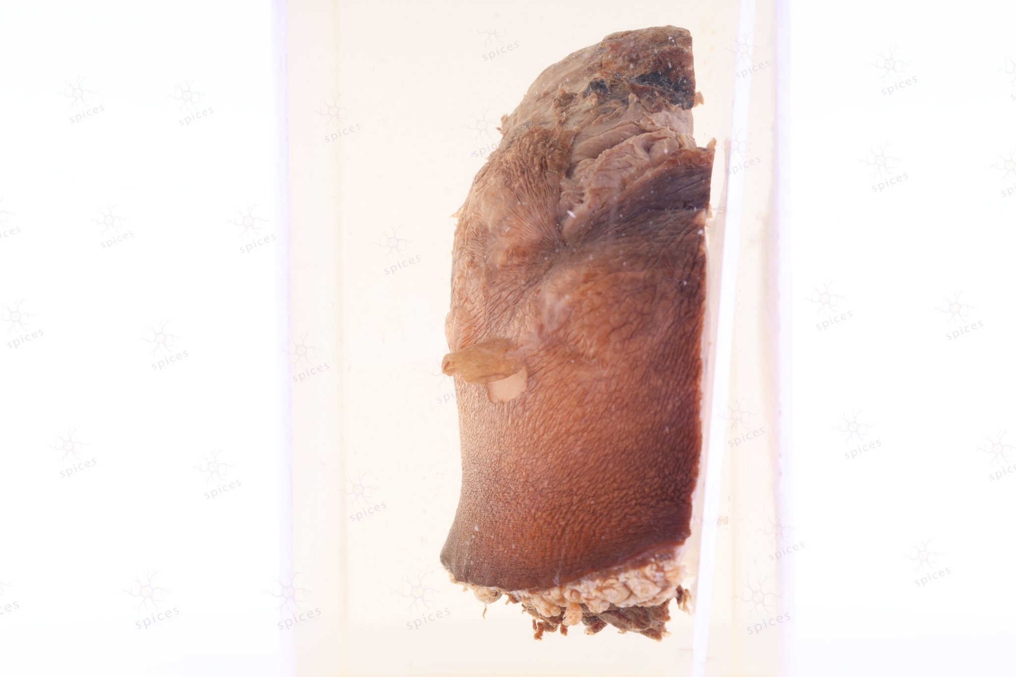
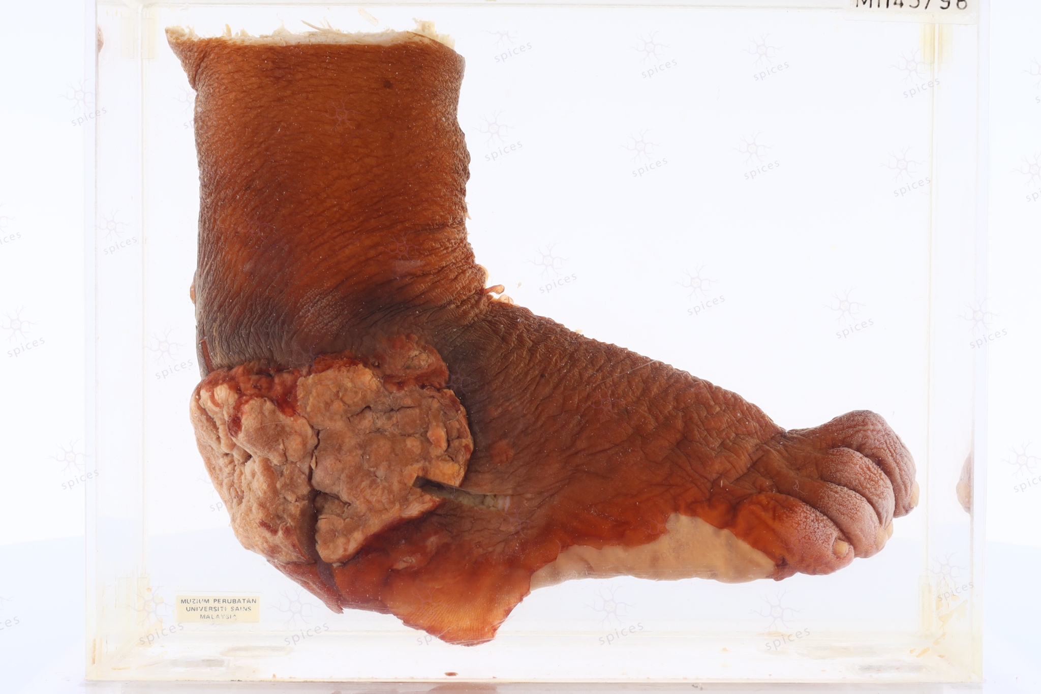
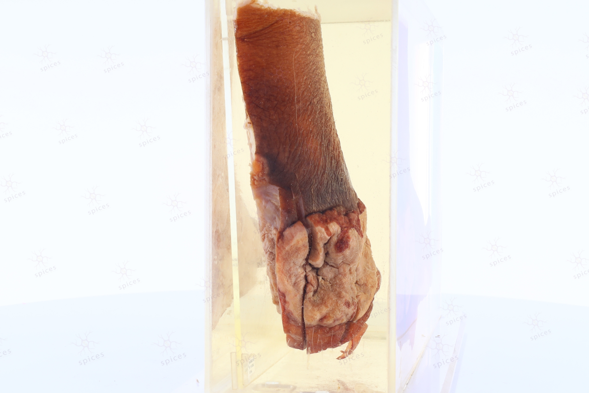
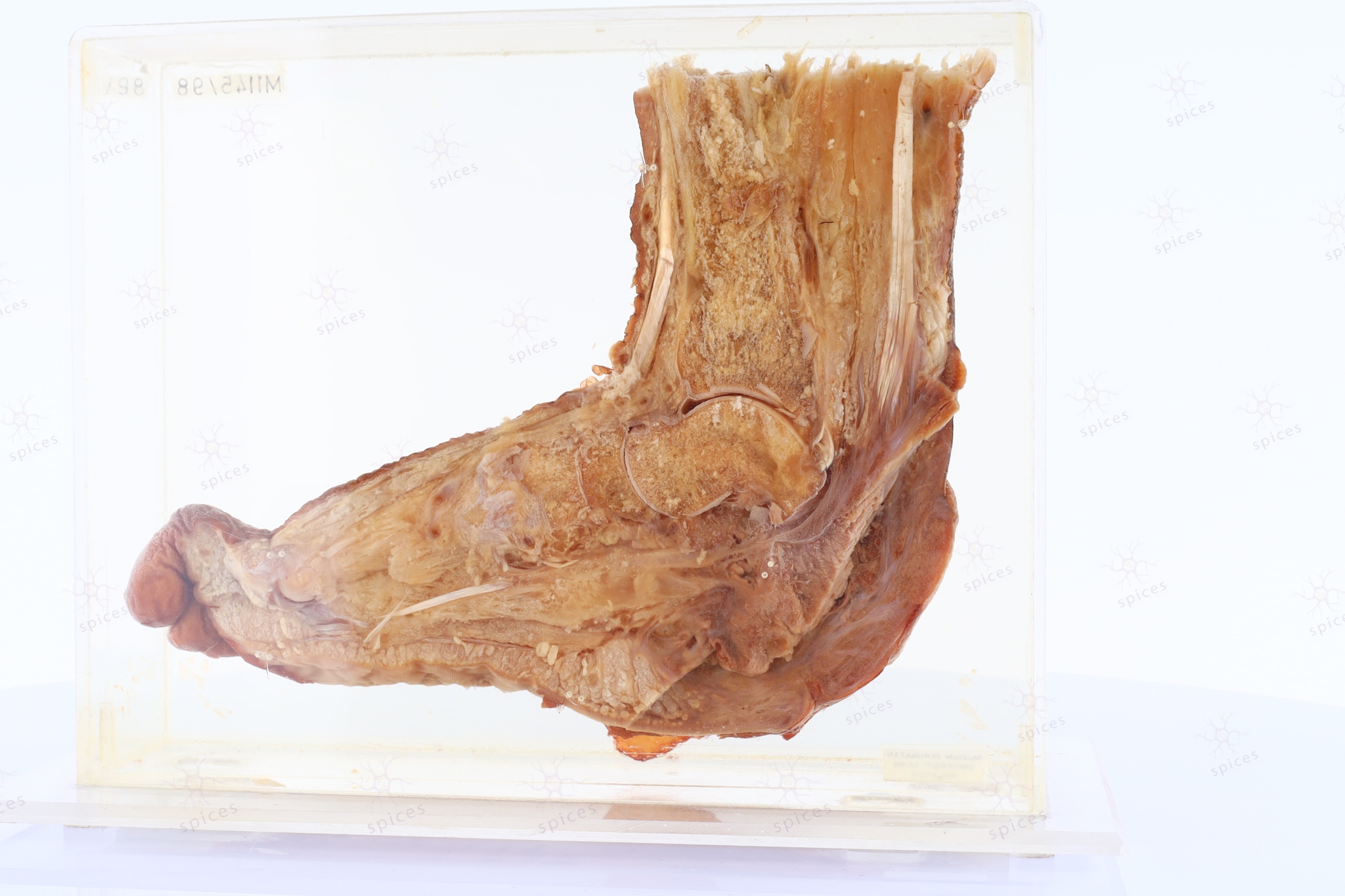
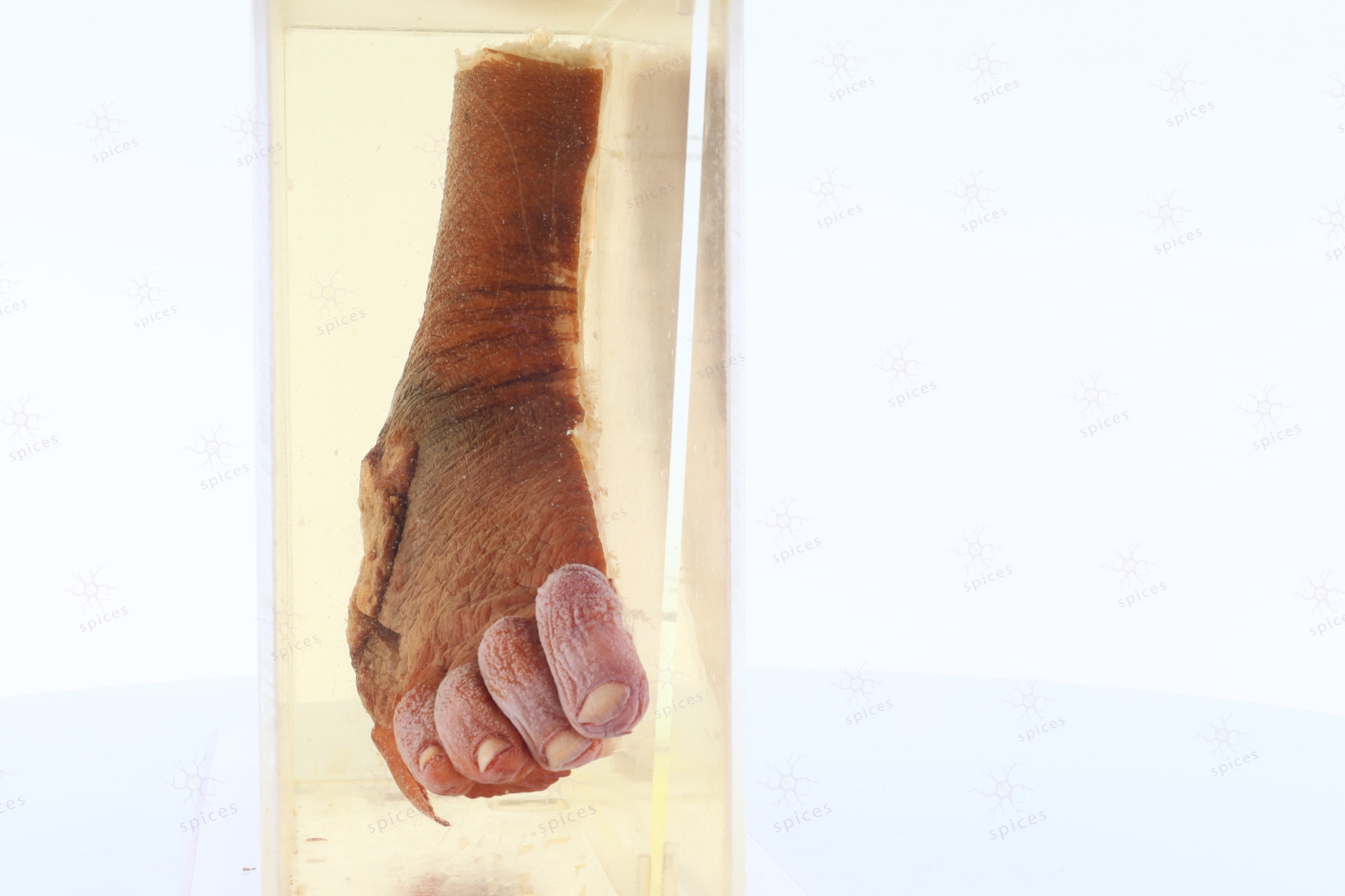
GROSS DESCRIPTION
The exhibit displays a benign exophytic lesion arising from the skin.
HISTOPATHOLOGY DESCRIPTION
Squamous papilloma shows papillary architecture with prominent fibrovascular cores. They exhibit proliferation of stratified squamous epithelium with variable hyperkeratosis or parakeratosis.
DIAGNOSIS
Squamous papilloma