Kidney : M834/91
KIDNEY
Spices No: M834/91

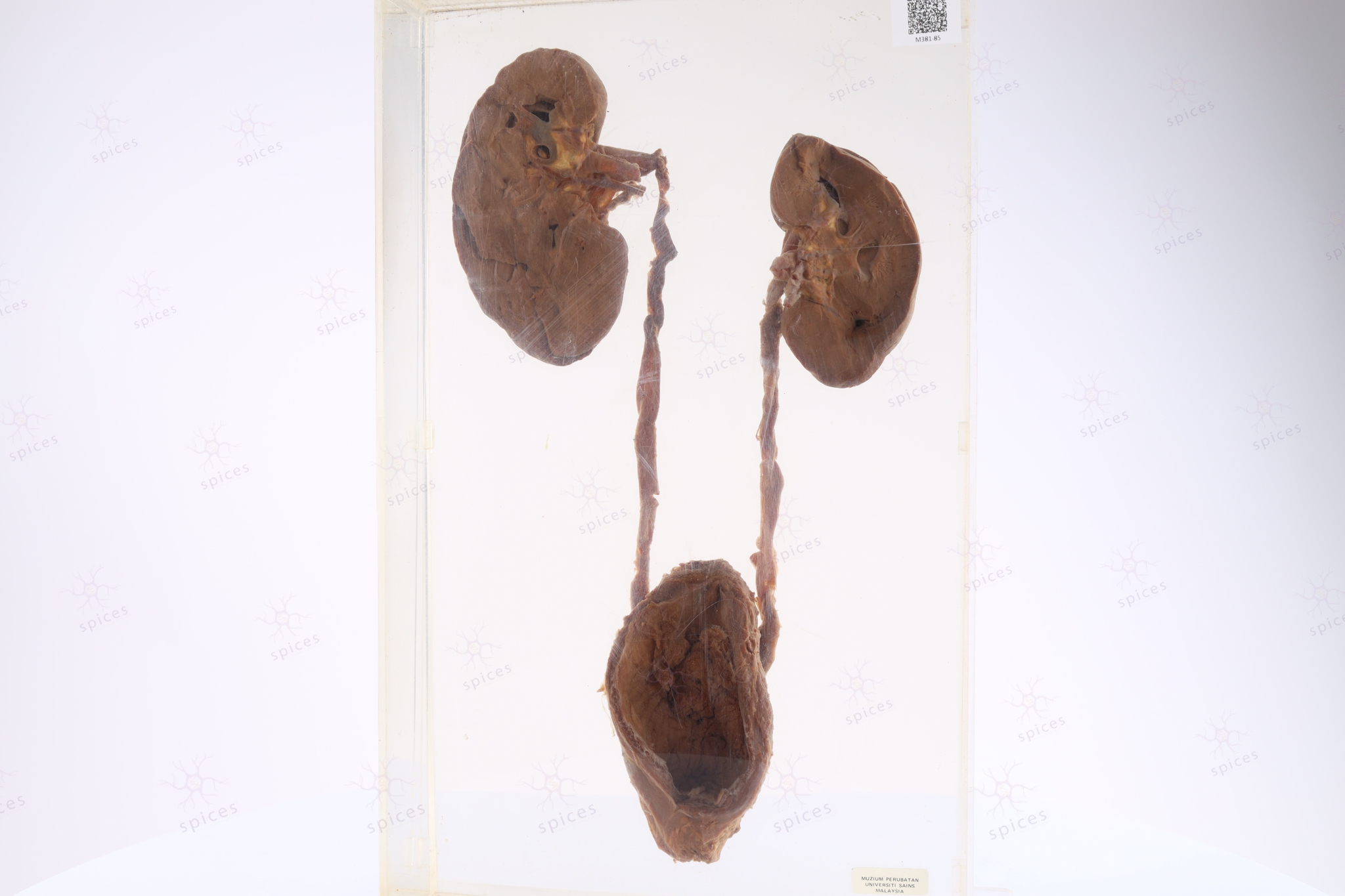
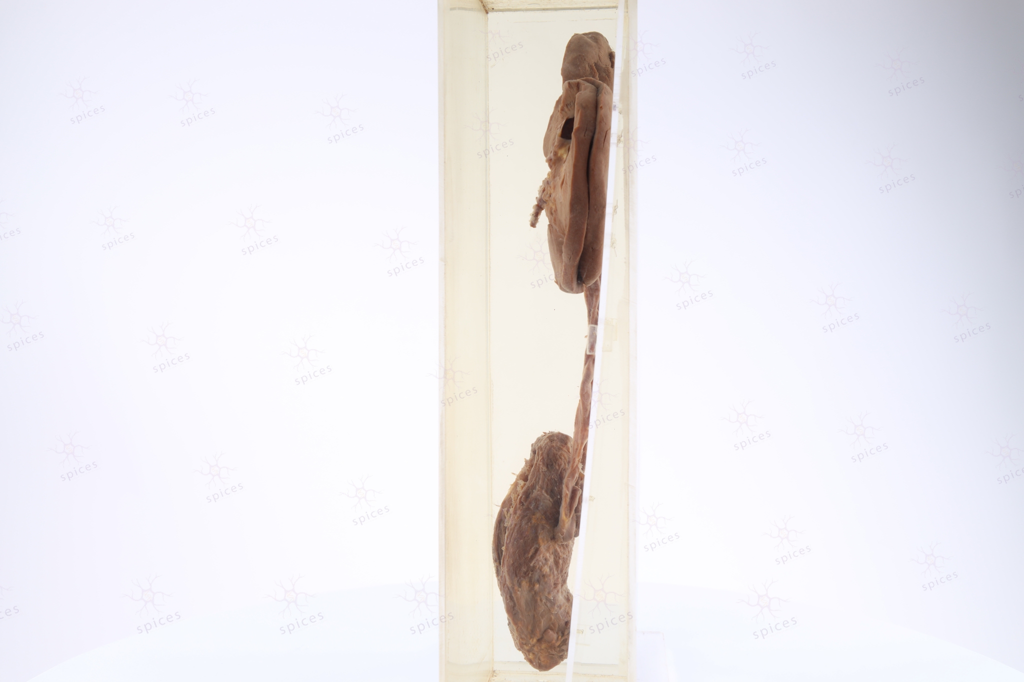
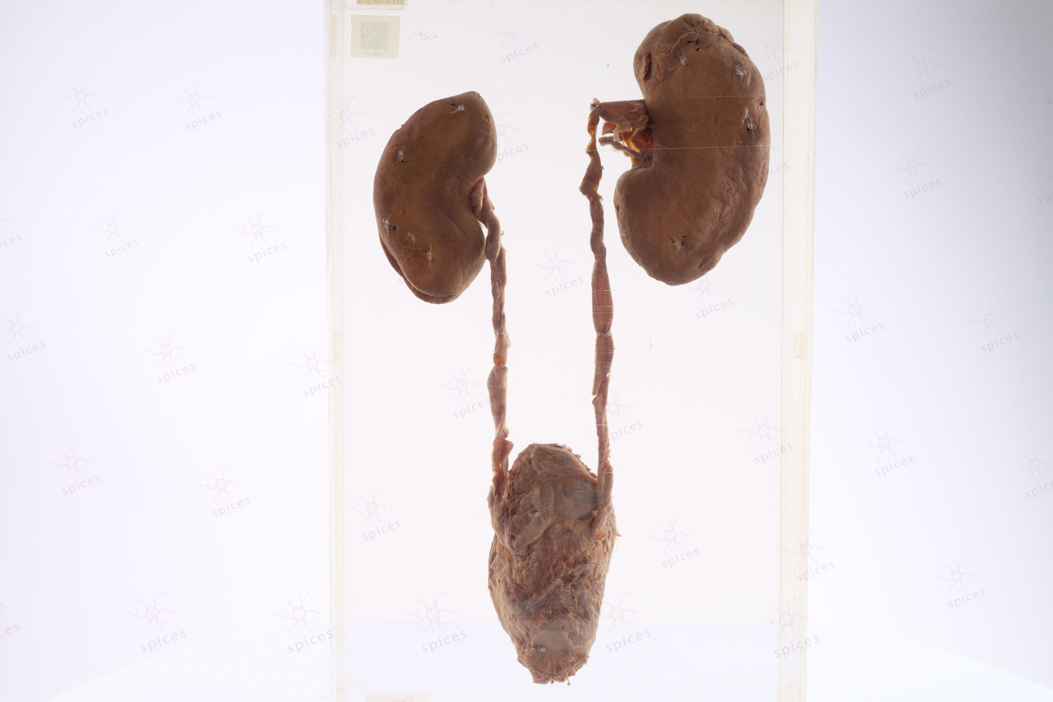
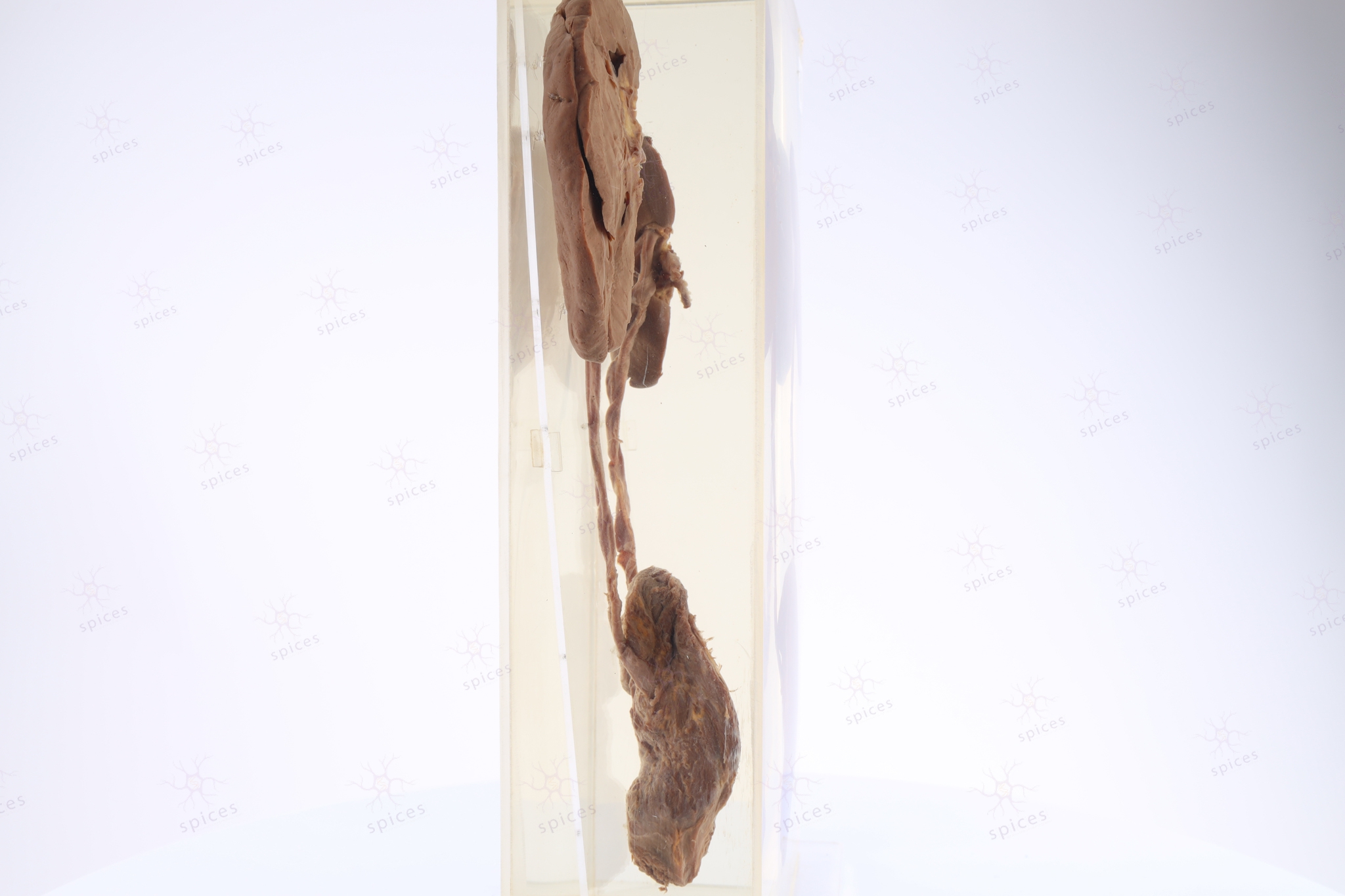
GROSS DESCRIPTION
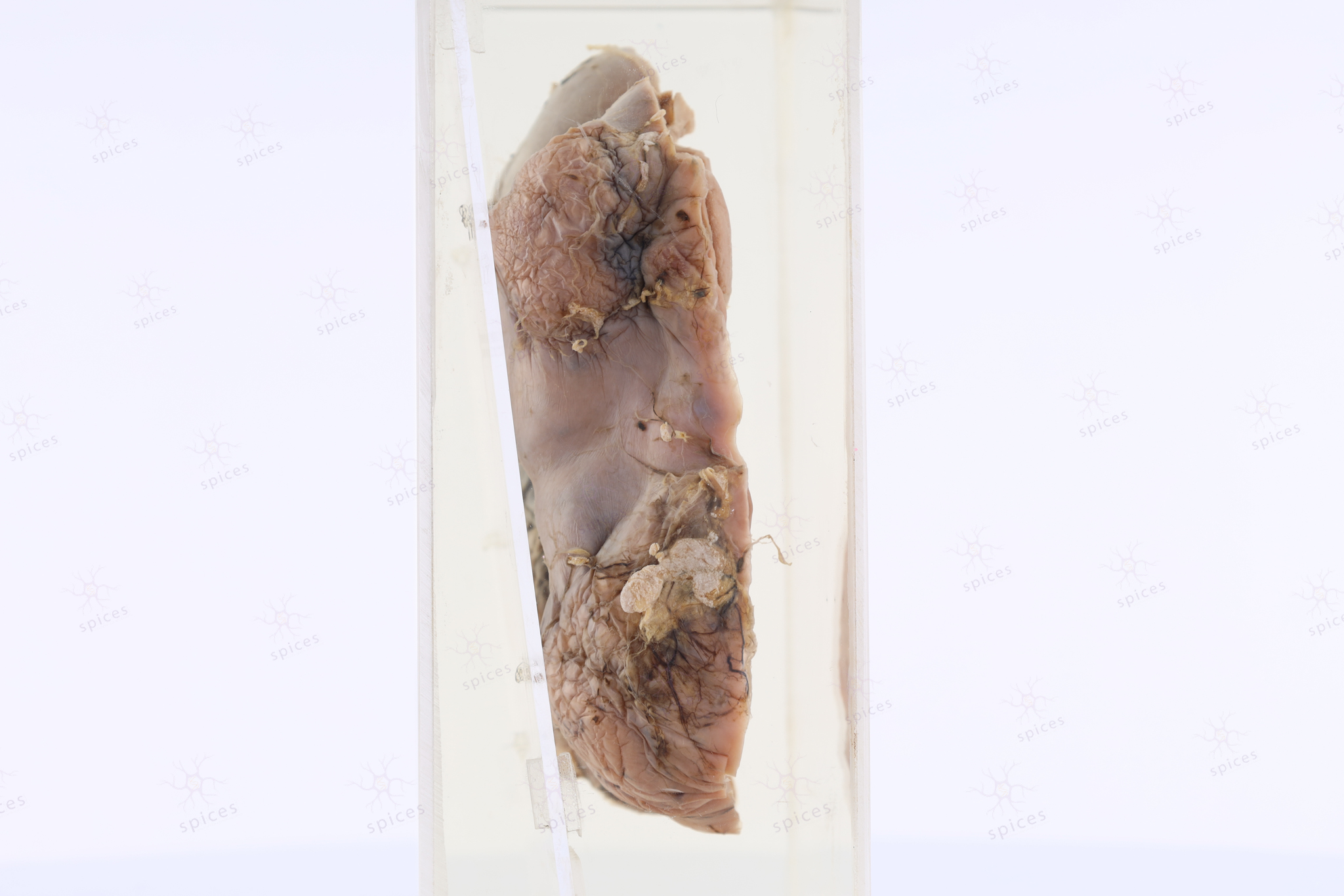
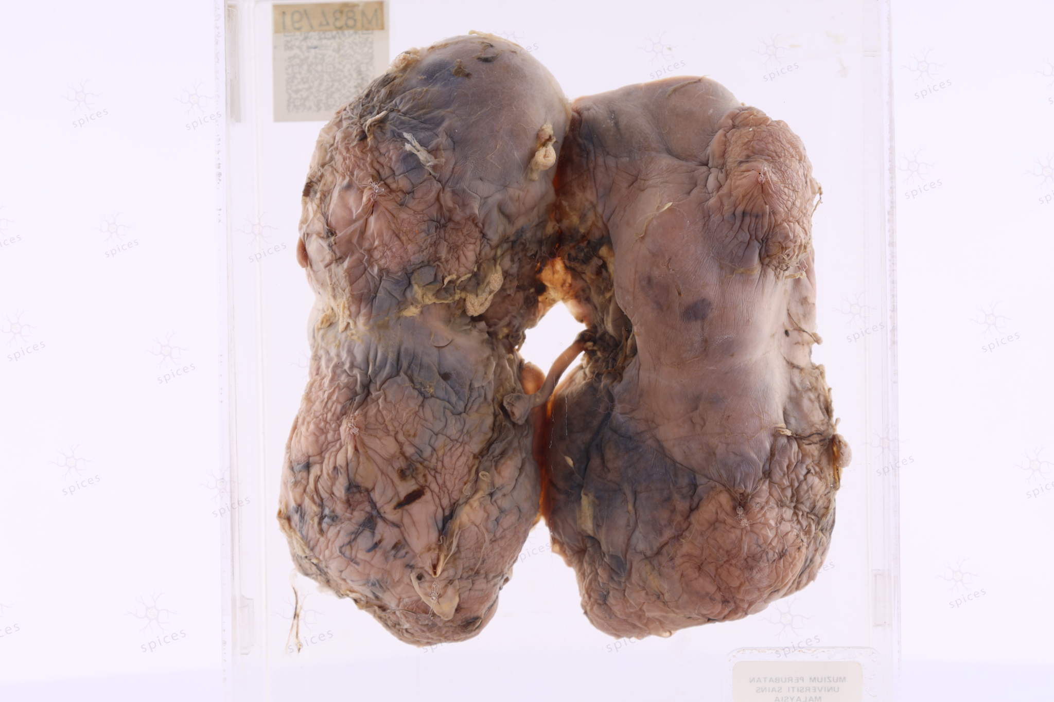
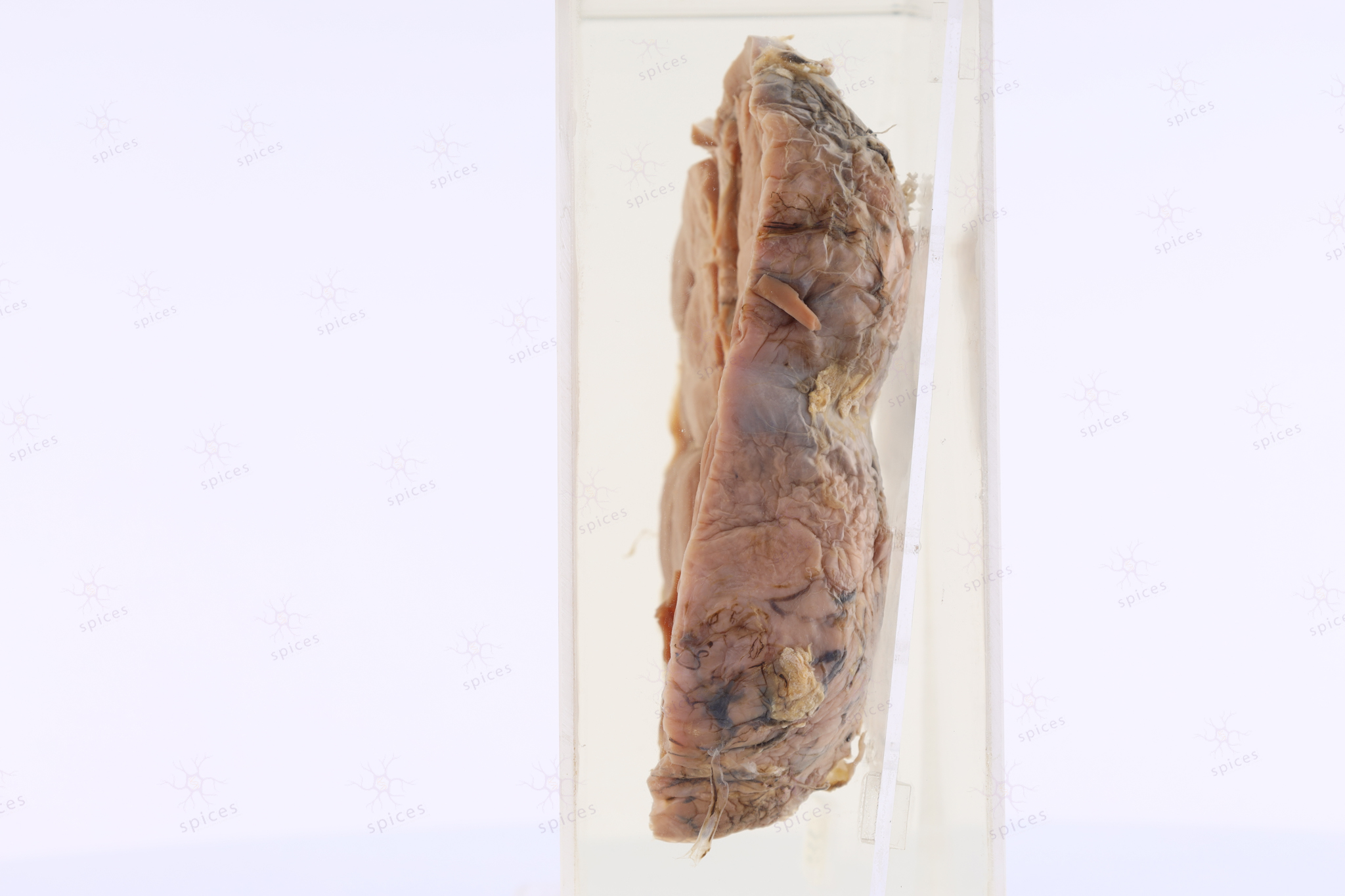
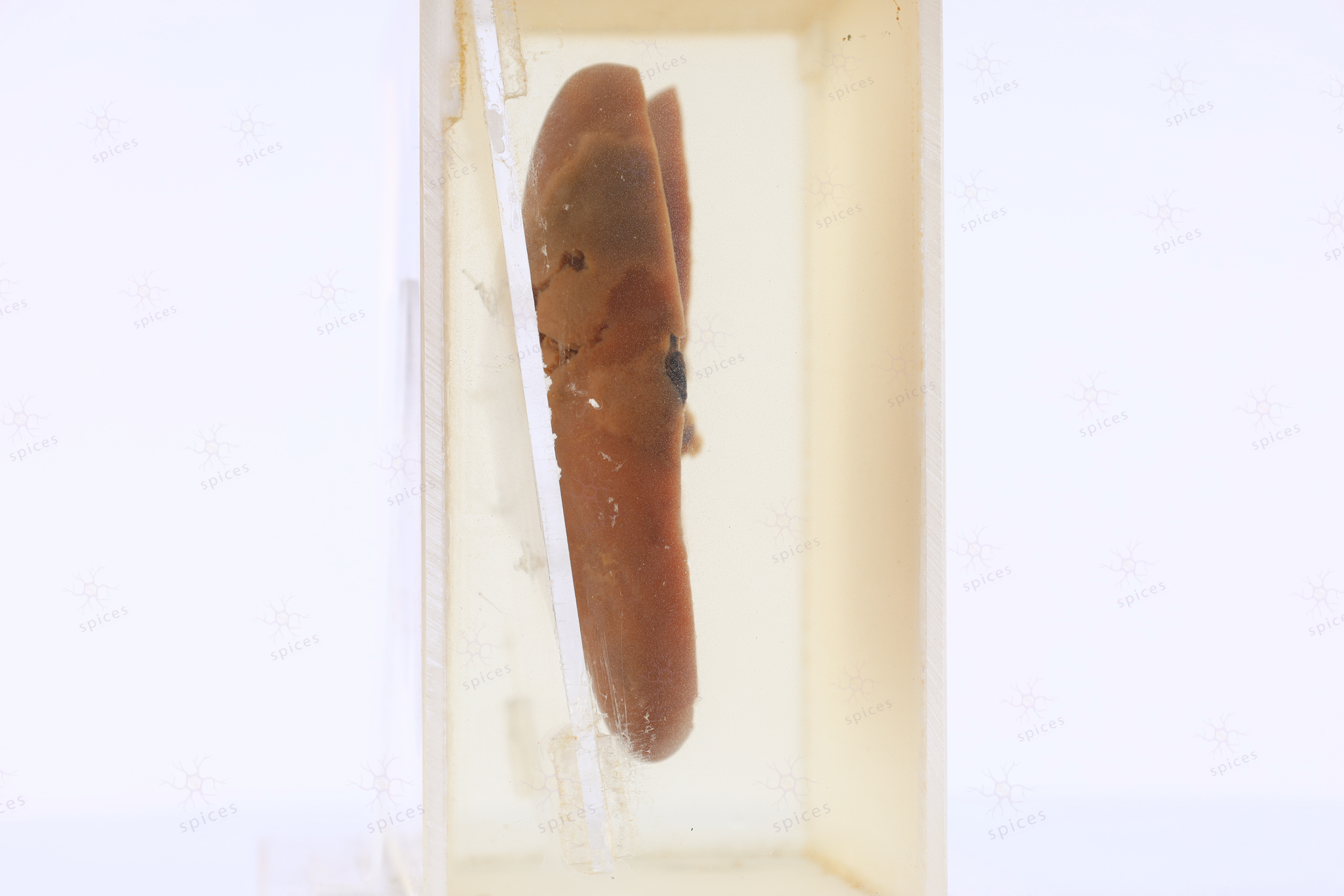
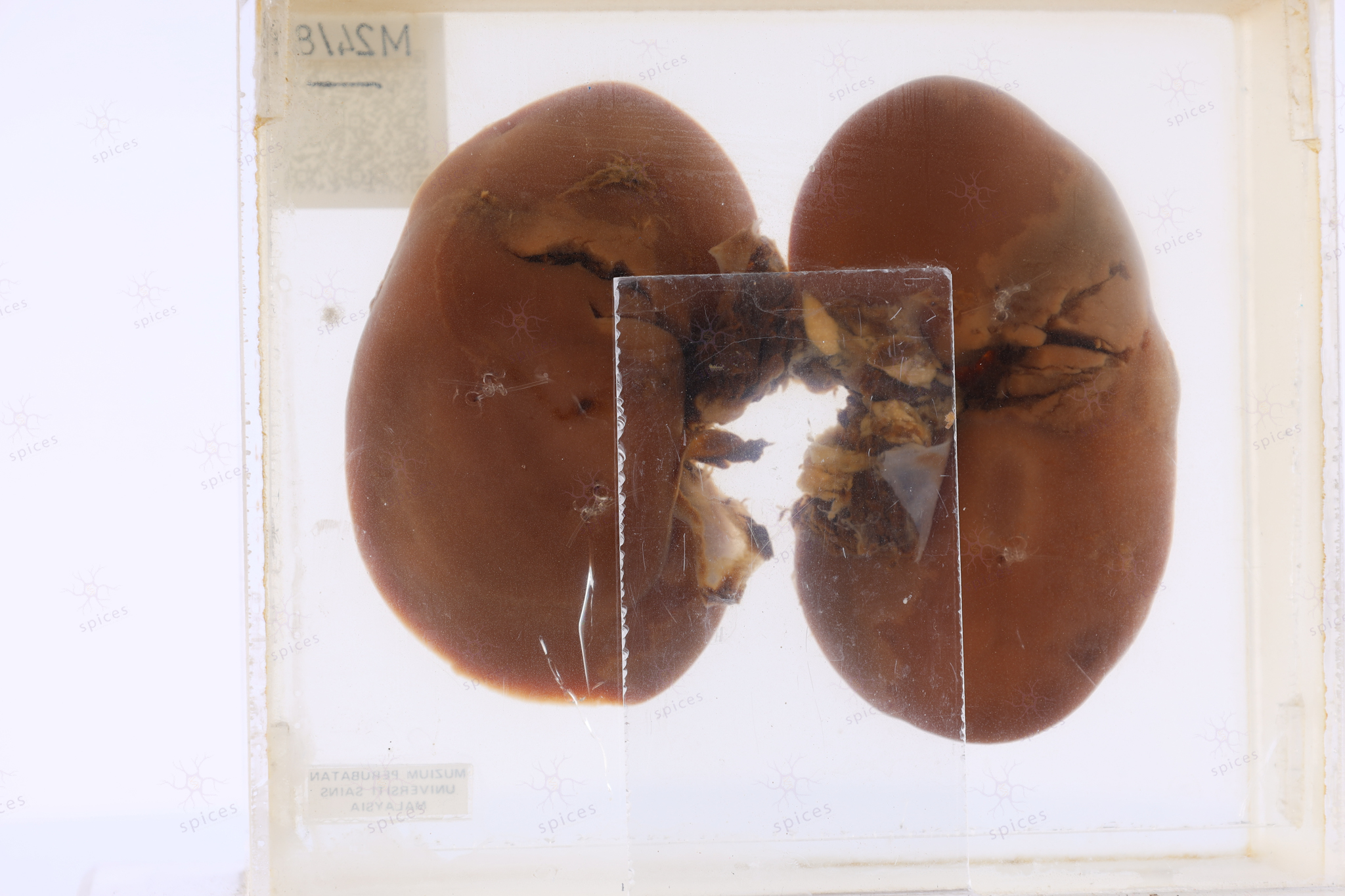
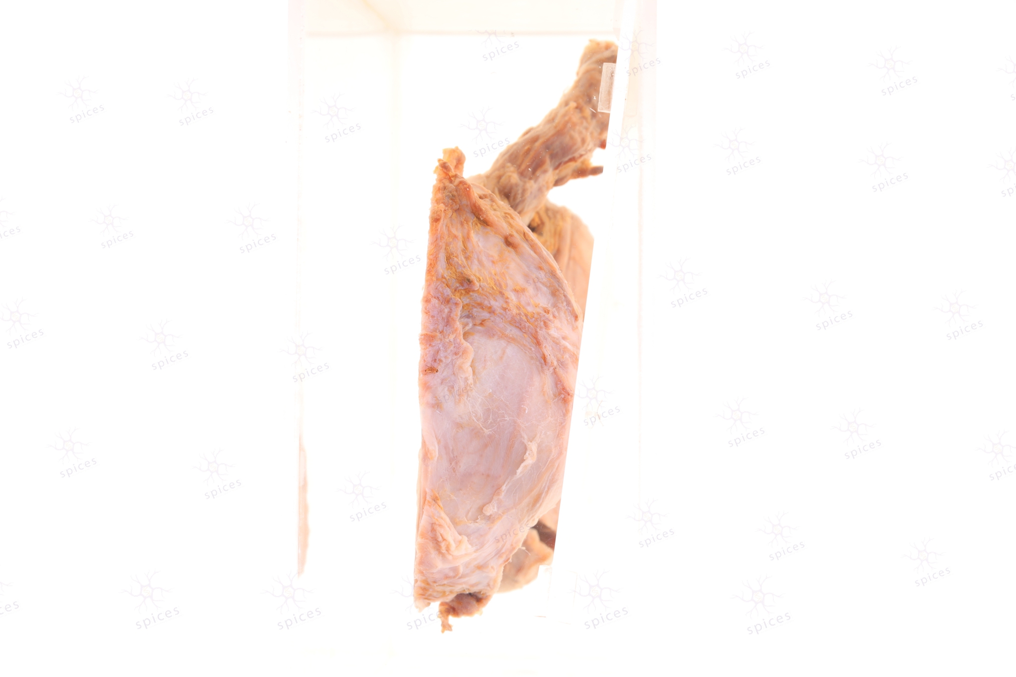
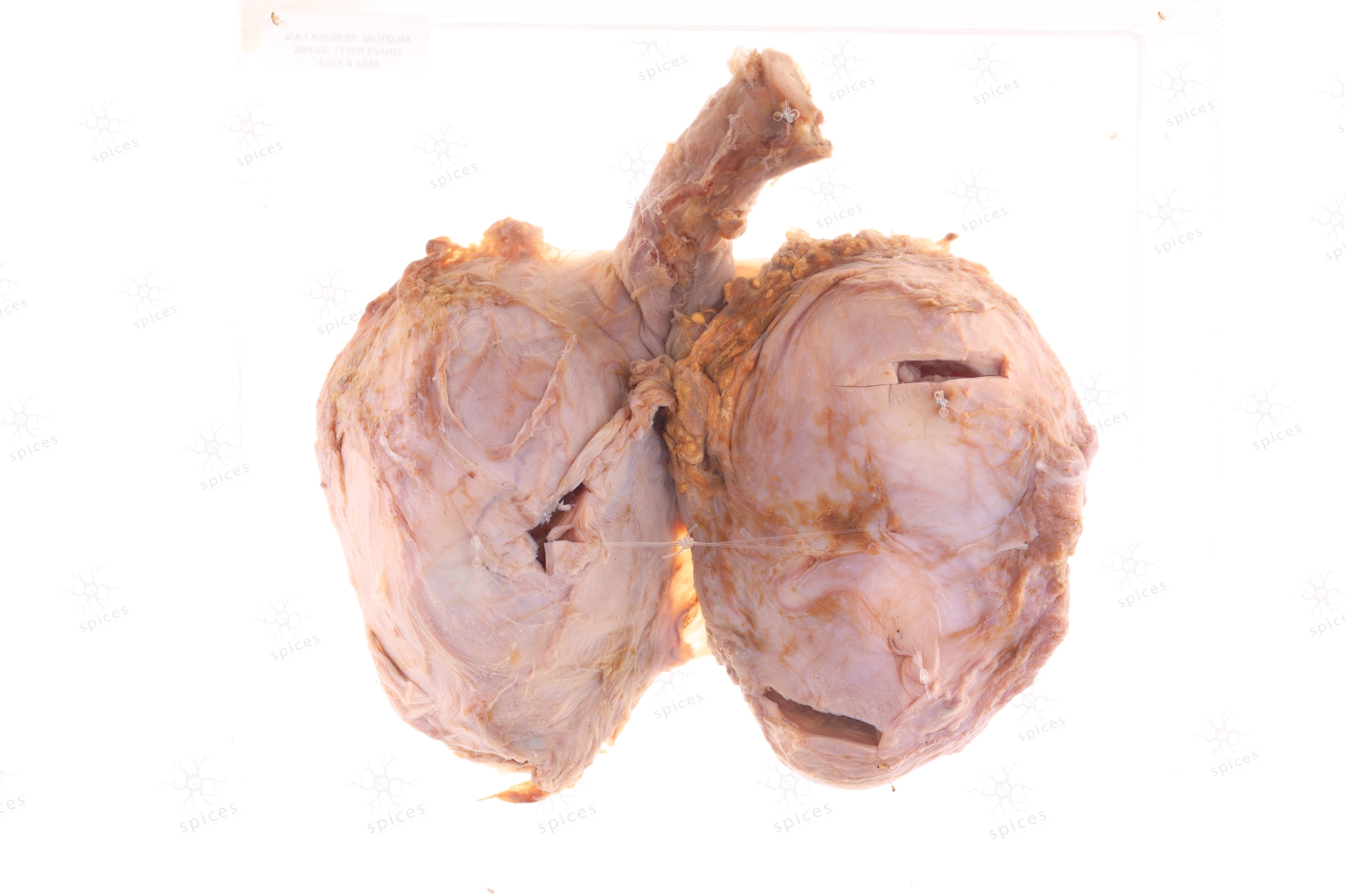
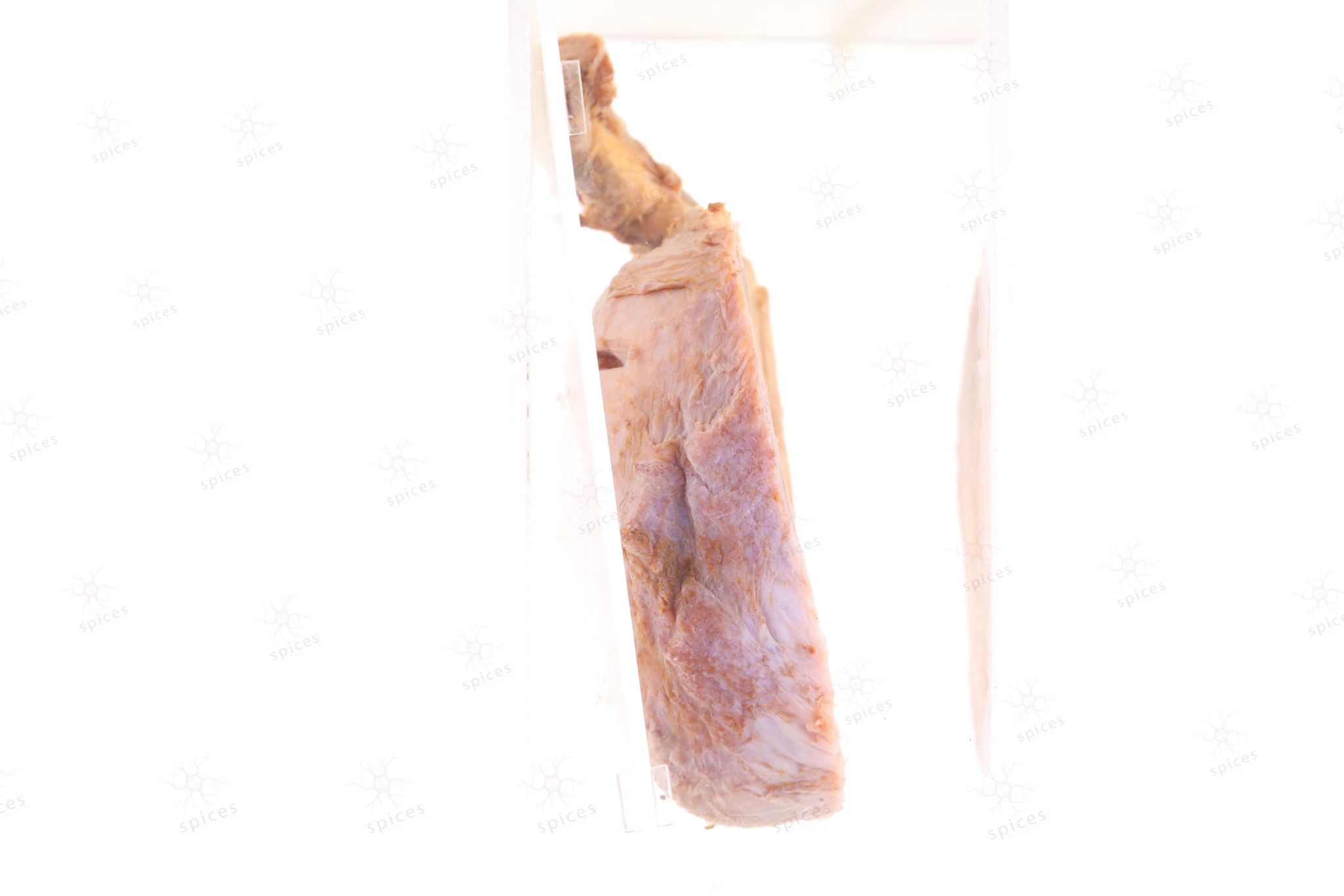
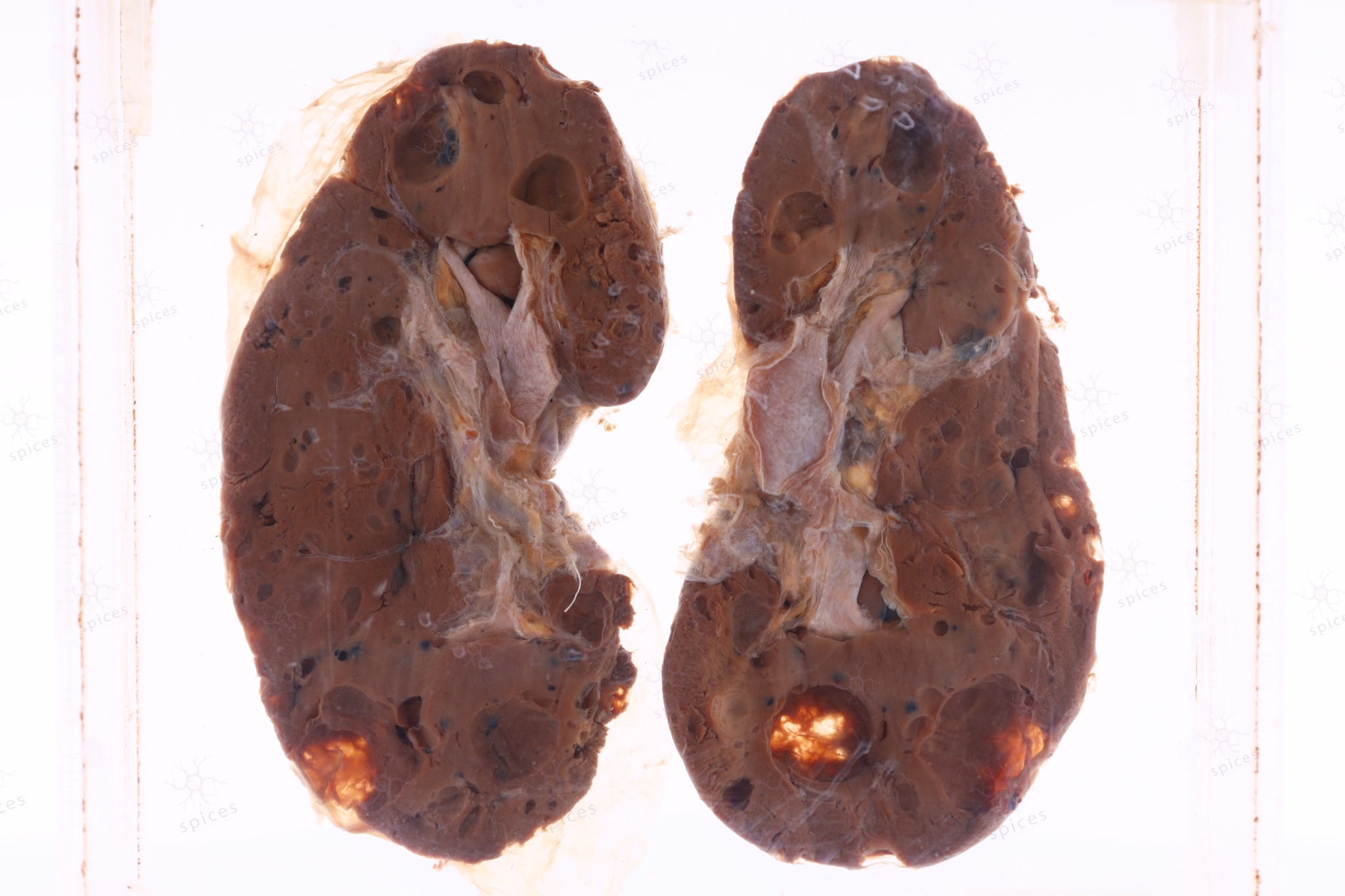
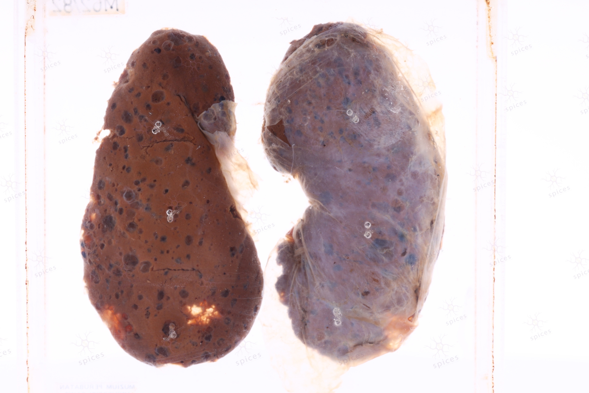
The exhibit displayss solid fleshy mass occupying the whole kidney. It shows homogenous tan, brownish surface.
HISTOPATHOLOGY DESCRIPTION
Nephroblastoma is composed of undifferentiated blastemal cells and cells differentiating to various degreeand variable propotion towards epithelial and stromal lineage. The blastemal cells are small round cells with scant cytoplasm, closely packed and mitotically active. The epithelial component shows early tubular form resemble primitive rosette like structure or glomerular element recapitulate various stages of nephrogenesis. Stromal pattern includes smooth muscle and fibroblastic differentiation.
DIAGNOSIS
Wilms tumour/Nephroblastoma
BAHASA MELAYU
Penerangan kasar: Spesimen menunjukkan ketumbuhan pejal yang mengisi seluruh ginjal. Ia menunjukkan permukaan homogen berwarna coklat kekuningan.


