
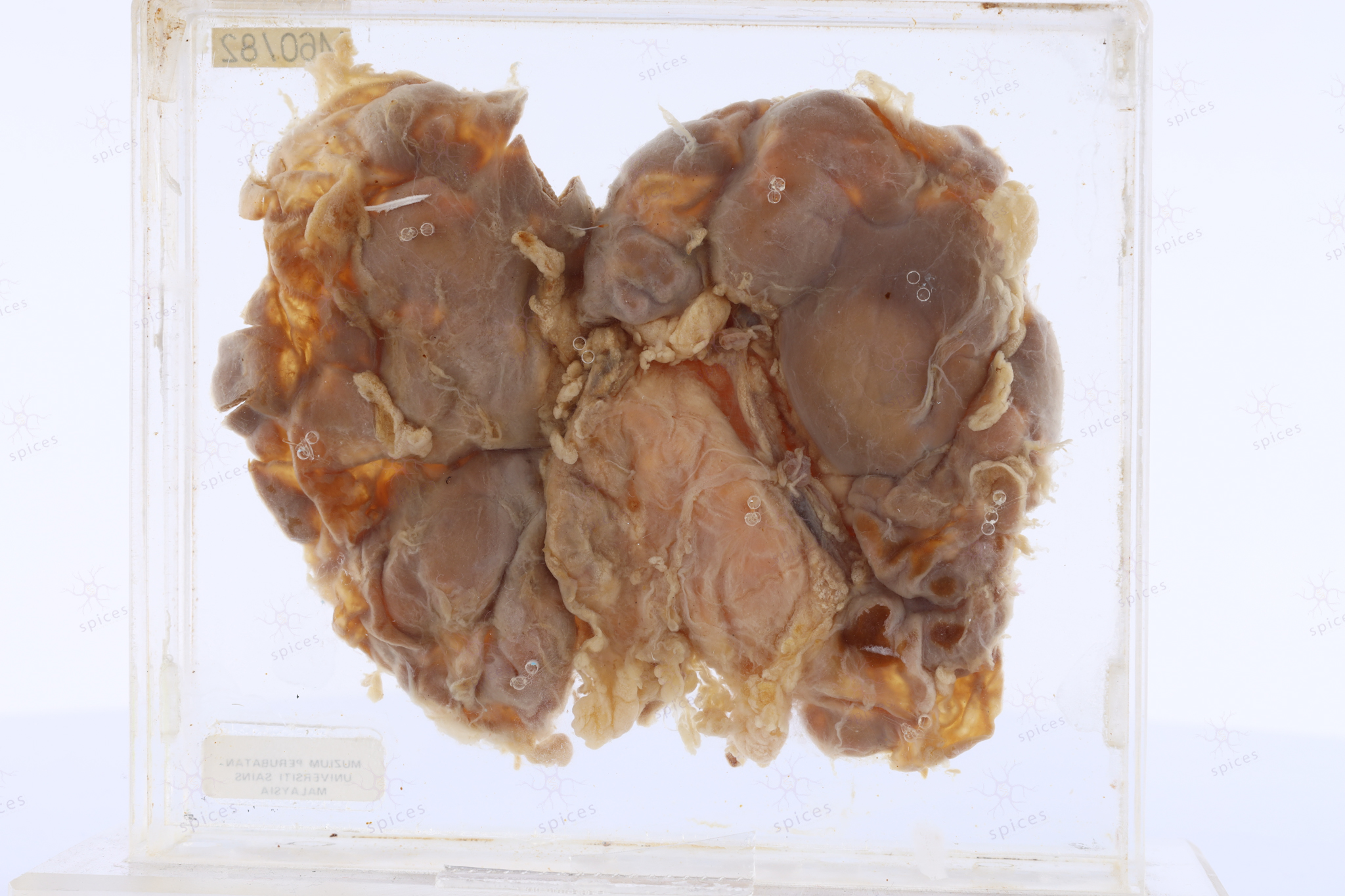
Kidney : M1221/00B
KIDNEY
Spices No: M1221/00B

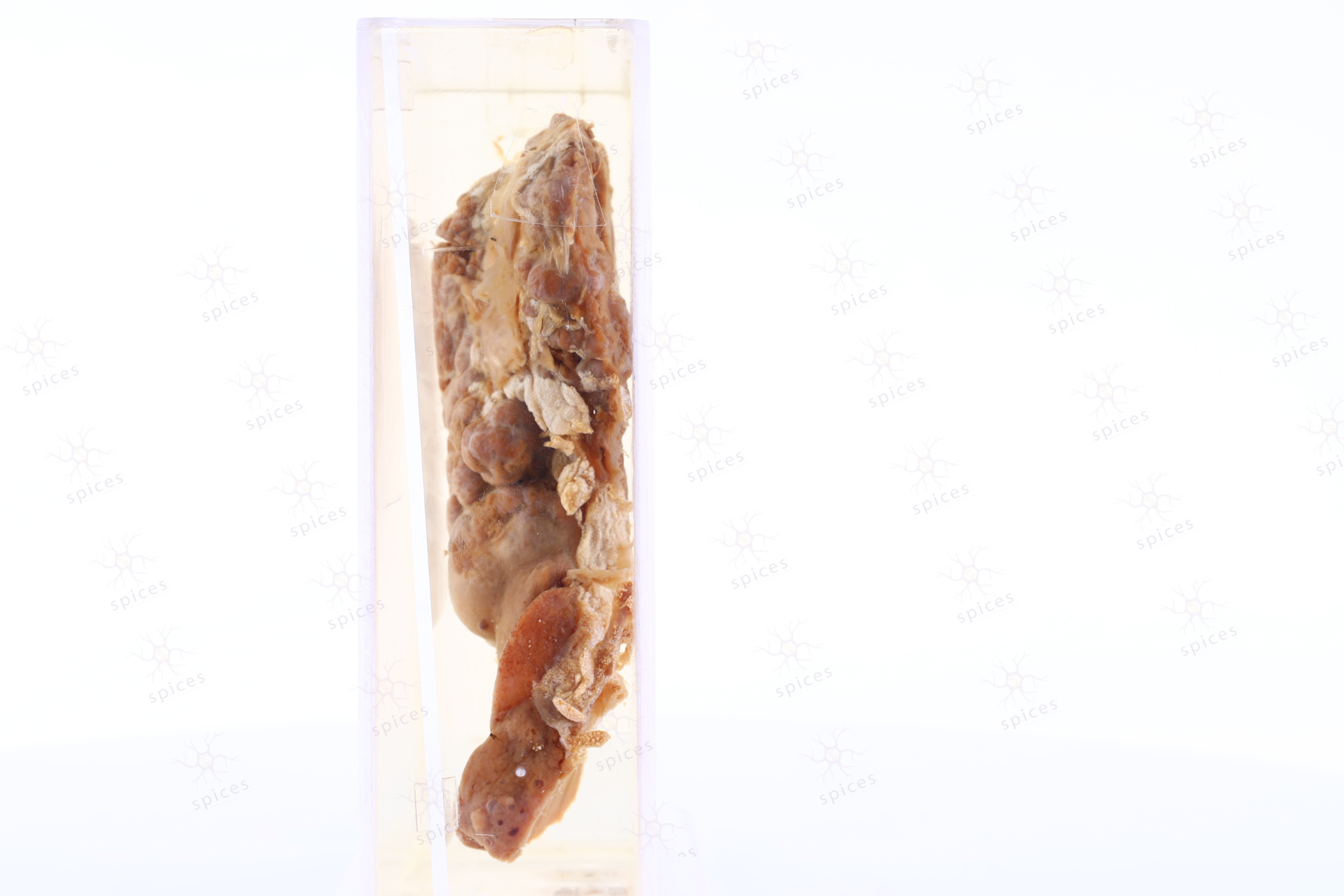
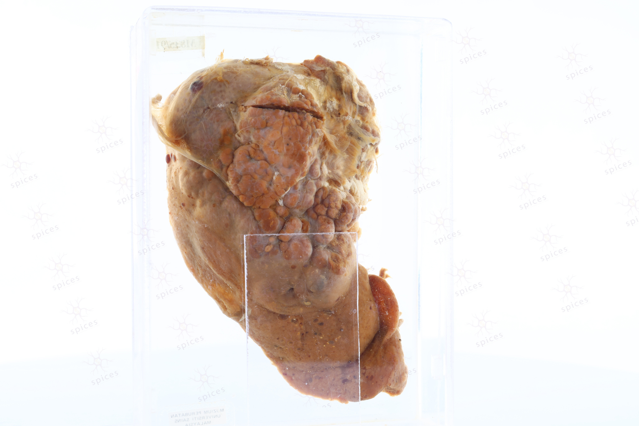
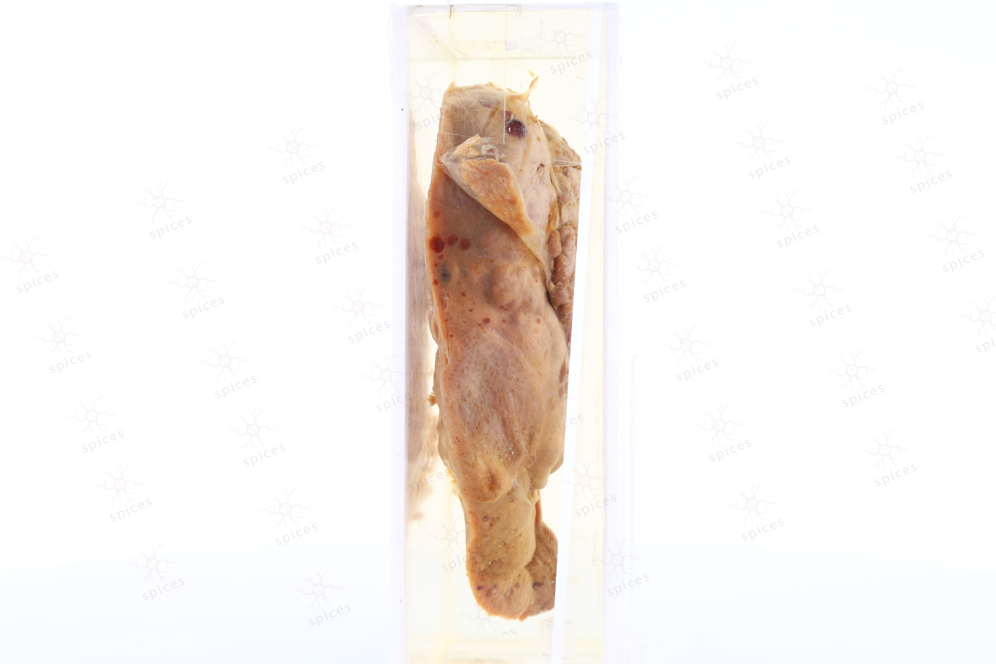
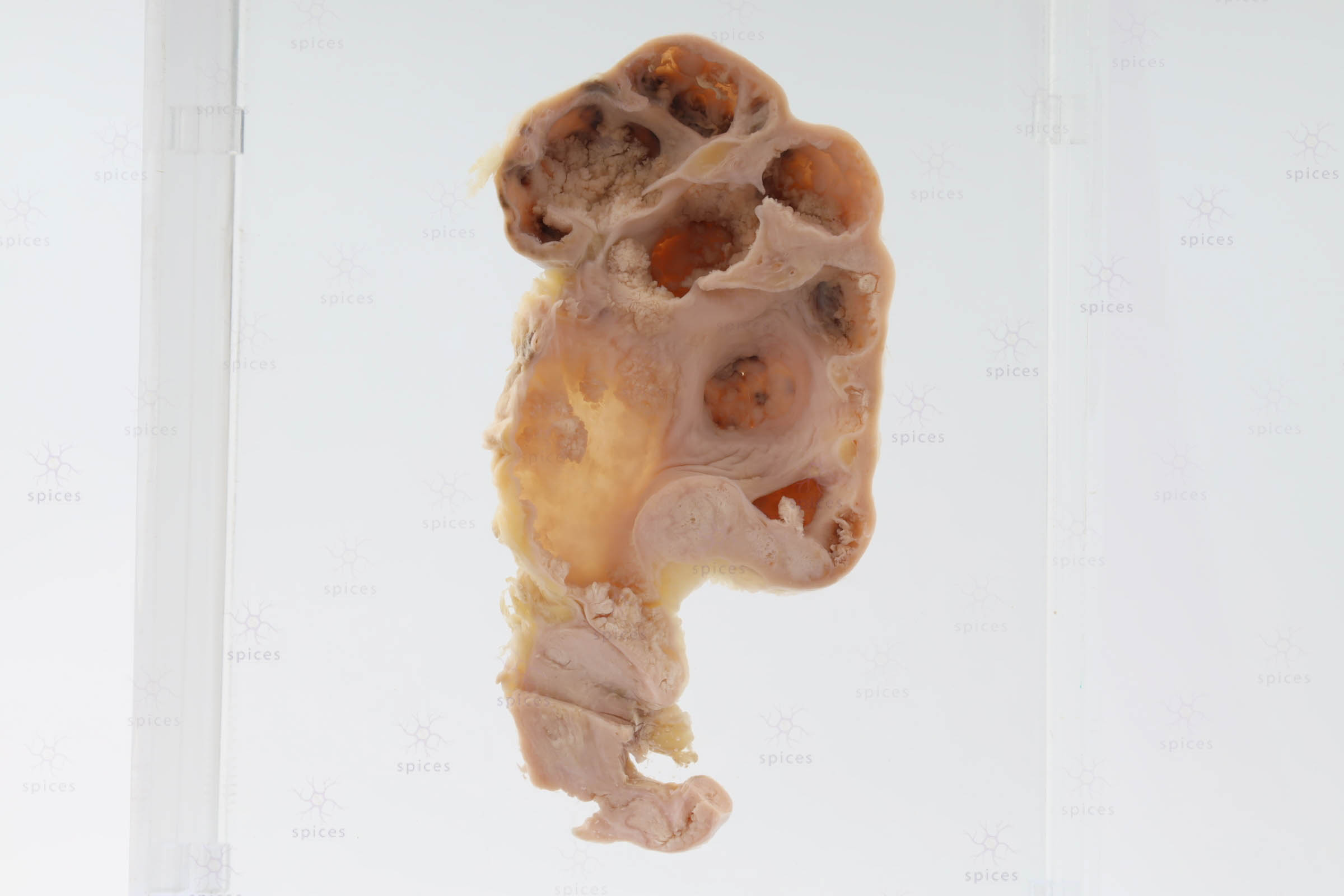
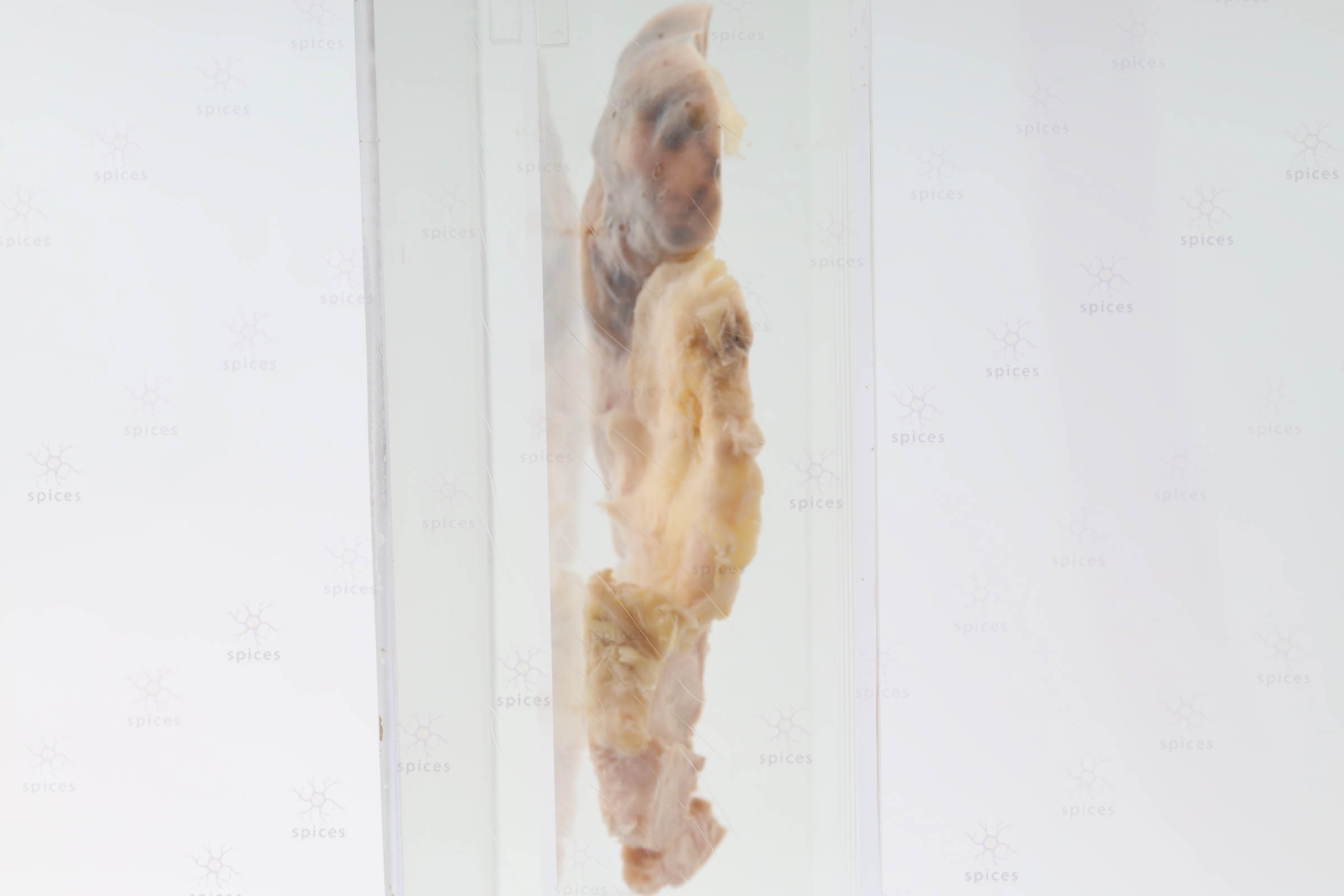
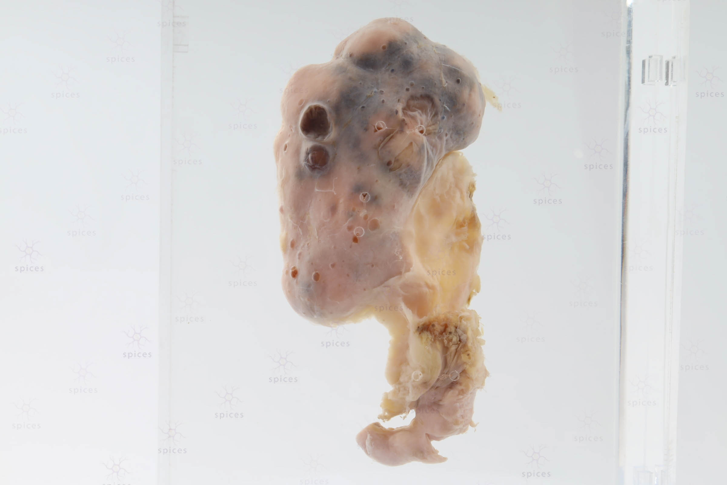
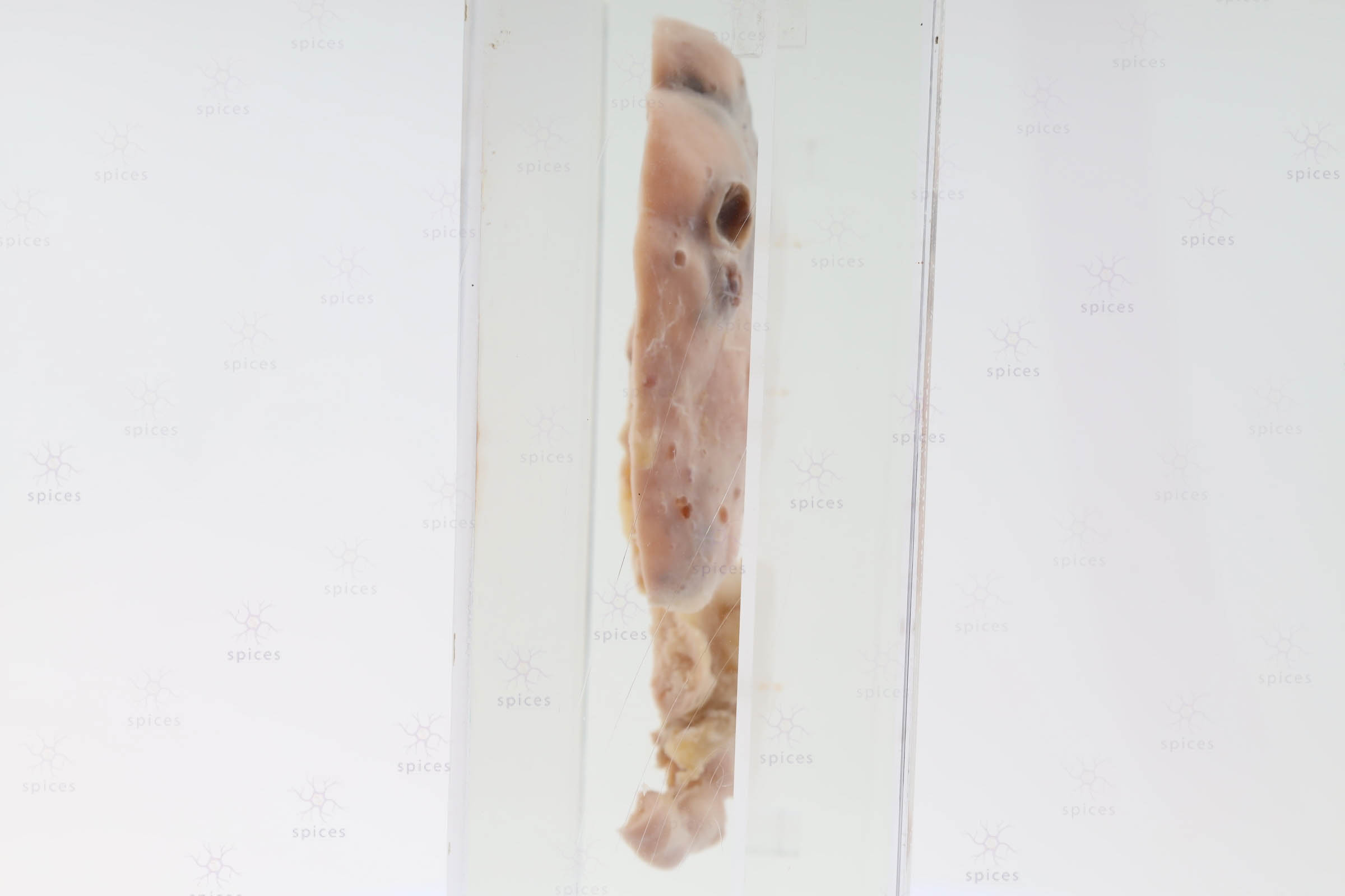
GROSS DESCRIPTION
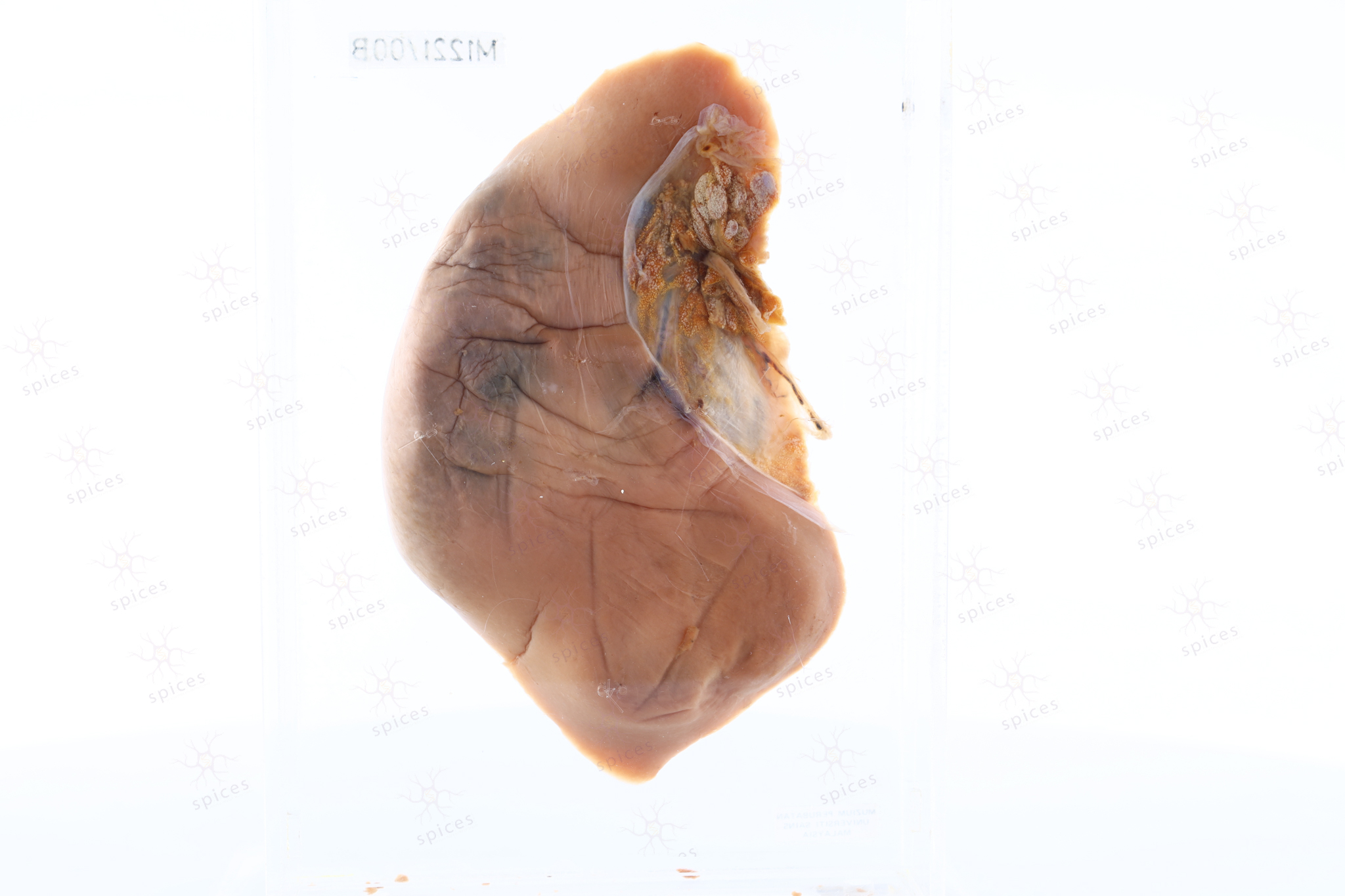
The exhibt display irregular tumour tissue occupying mainly part of the upper pole, middle pole and lower pole of the kidney. It show irregular and infiltrative border with variegated cut surface with areas of haemorrhage and necrosis. Part of the normal kidney parenchymal can be ssen at the upper pole and lower pole.
HISTOPATHOLOGY DESCRIPTION
Renal cell carcinoma from the tumour tissue shows nested pattern of clear cell type surrounded by chicken wire vasculature. The malignant cells are polygonal with round nuclei and prominent nucleoli. The cytoplasm is clear and eosinophilic
DIAGNOSIS
Renal cell carcinoma - clear cell type
QUIZ
State the name of the gene that commonly associated with this histological type of renal cell carcinoma.
BAHASA MELAYU
Penerangan kasar: Spesimen memaparkan tisu ketumbuhan tidak sekata yang mengisi sebahagian besar bahagian kutub atas, kutub tengah, dan kutub bawah ginjal. Ia menunjukkan sempadan yang tidak sekata dan bersifat menyerap serta mempunyai permukaan potongan yang pelbagai dengan kawasan pendarahan dan nekrosis. Sebahagian dari parenkim ginjal normal dapat dilihat di kutub atas dan kutub bawah.
Peneranganhistopatologi: Karsinoma sel ginjal dari tisu tumor menunjukkan sel dengan corak bersarang yang dikelilingi oleh chicken wire vasculature. Sel-sel malignan ini berbentuk segi empat dengan nukleus bulat dan nukleolus yang jelas. Sitoplasma kelihatan jernih dan berwarna eosinofilik.
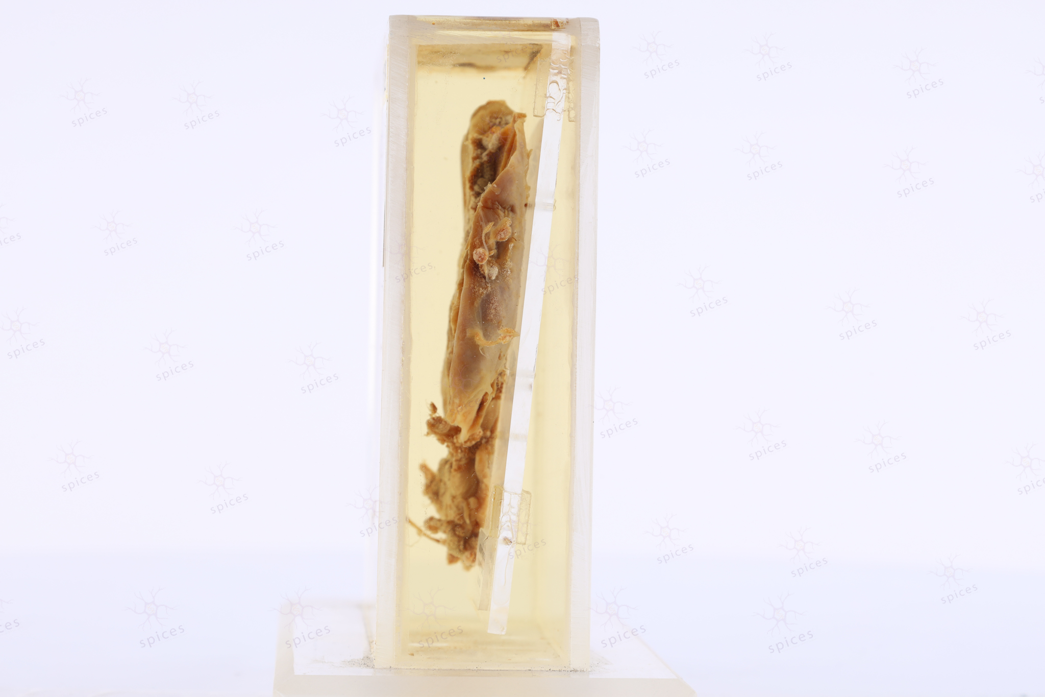
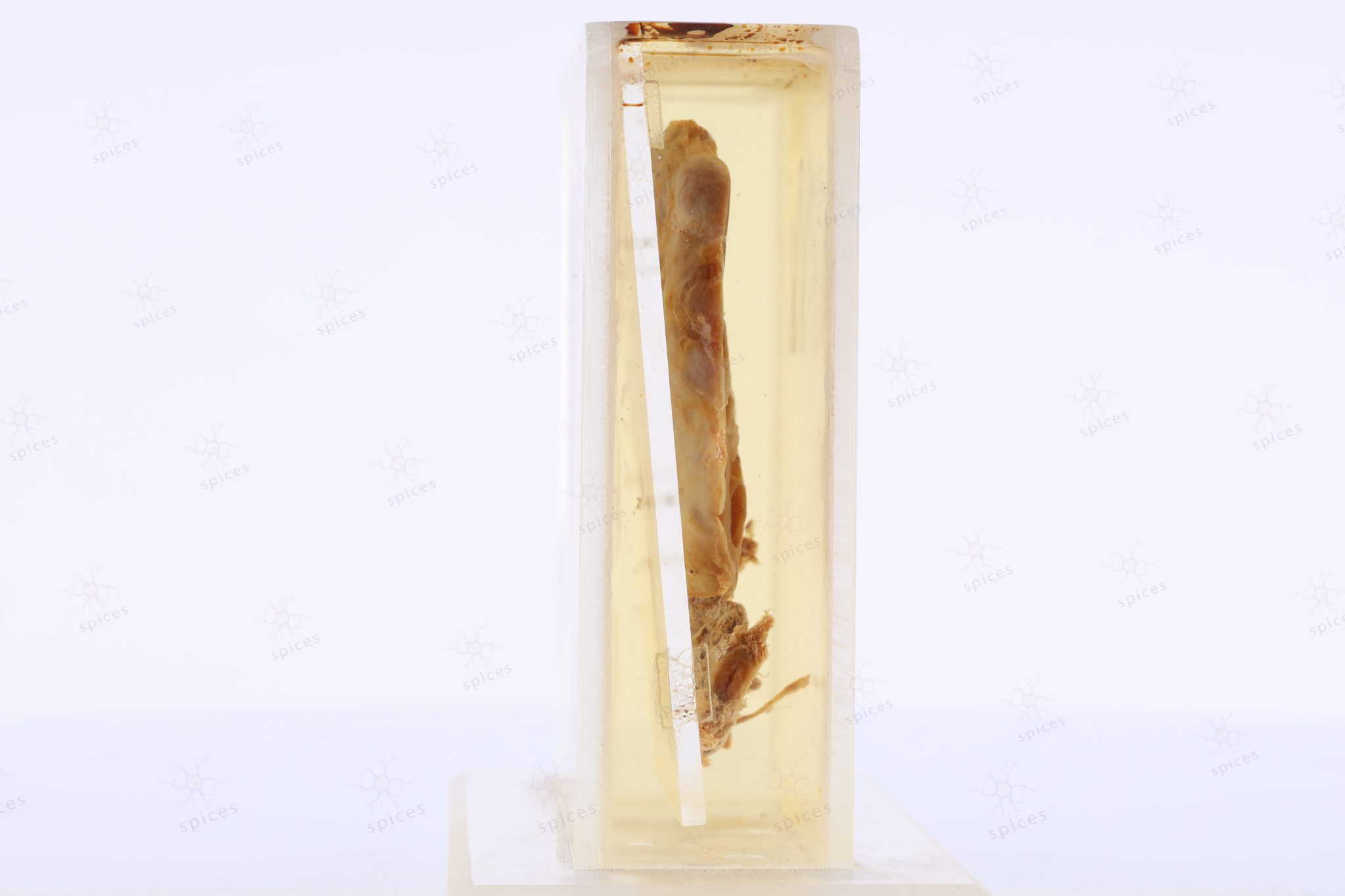
Kidney : M60/82
KIDNEY
Spices No: M60/82


HISTOPATHOLOGY DESCRIPTION
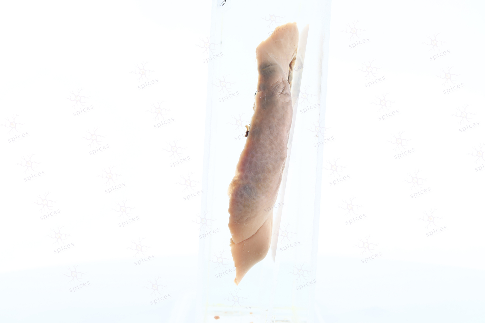
Section shows cystic dilation of renal pelvis and calyces. Interstitial inflammatory infiltrate with chronic changes displaying cortical atrophy, diffuse interstitial fibrosis and blunting of calyces is seen.
DIAGNOSIS
Hydronephrosis
BAHASA MELAYU
Penerangan histopatologi: Bahagian tisu ini menunjukkan pelvis dan calyces ginjal yang mengalami pengembangan sistik. Terdapat infiltrat inflamasi interstisial dengan perubahan kronik yang menunjukkan cortical atrophy, fibrosis interstisial meluas, dan pengecilan calyces terlihat.
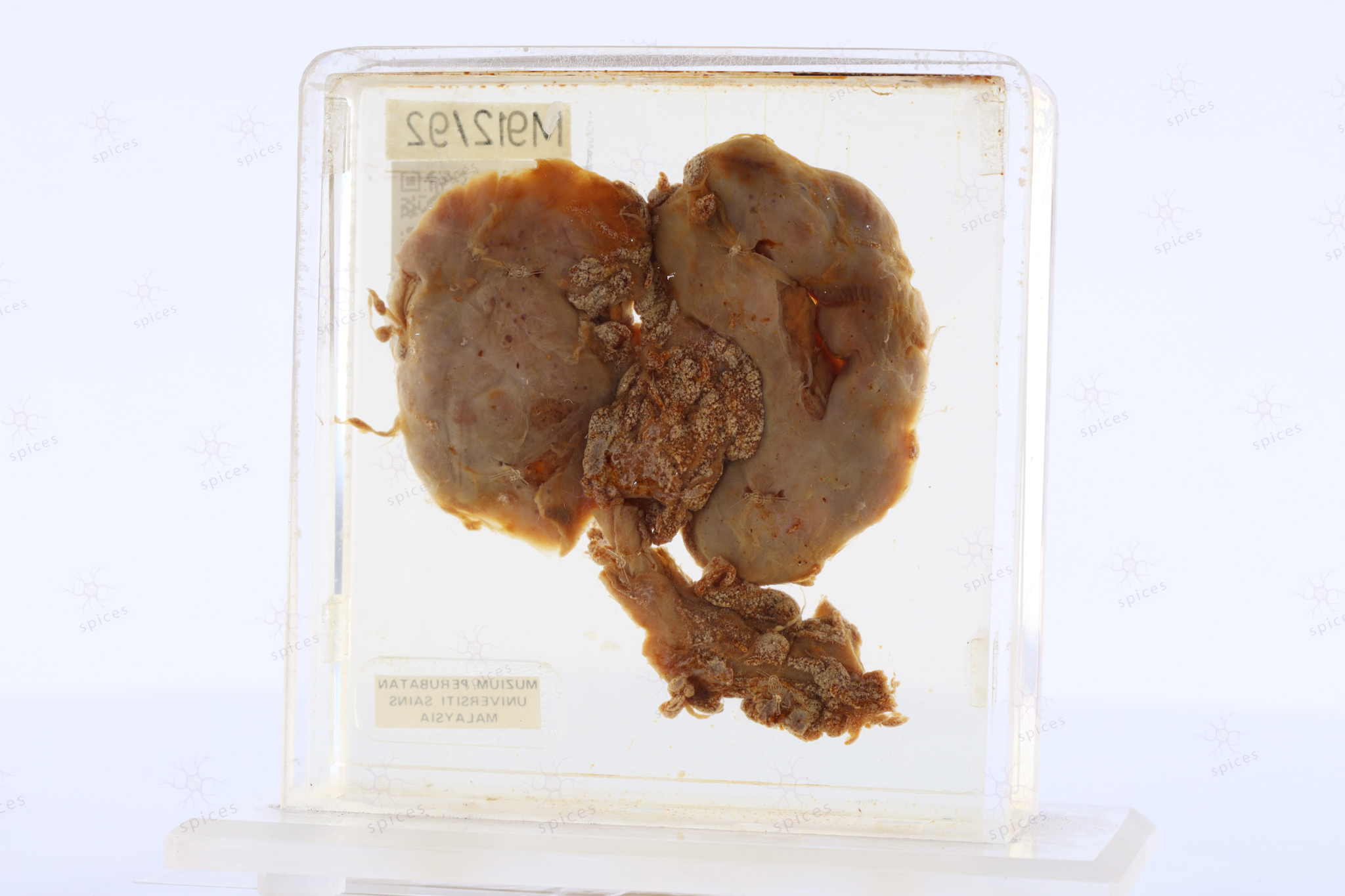
Kidney : M912/92
KIDNEY
Spices No: M912/92

GROSS DESCRIPTION
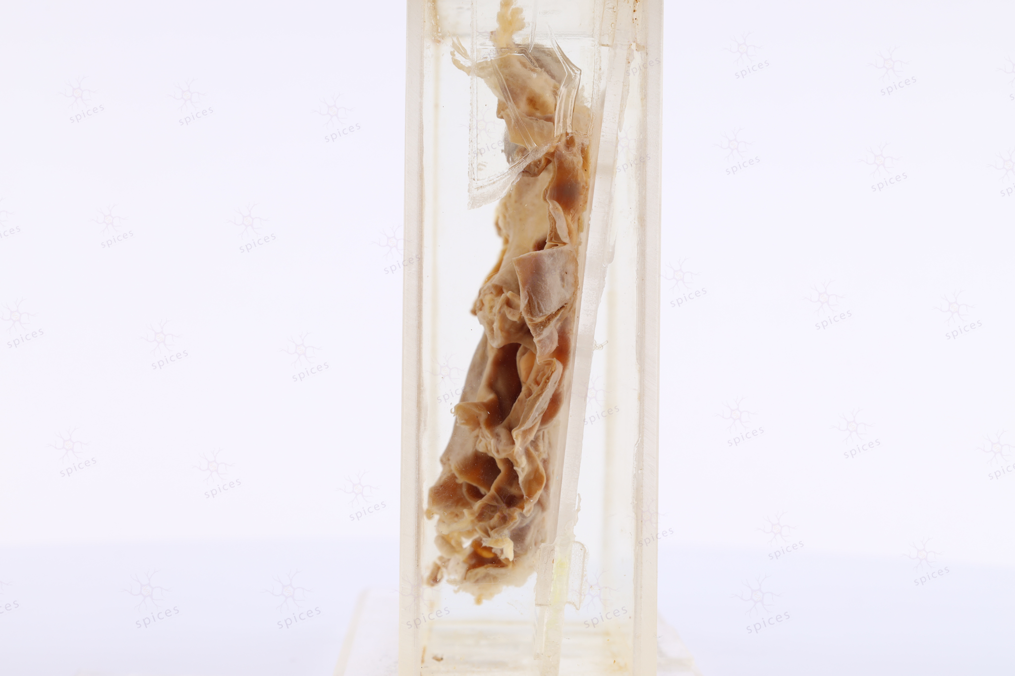
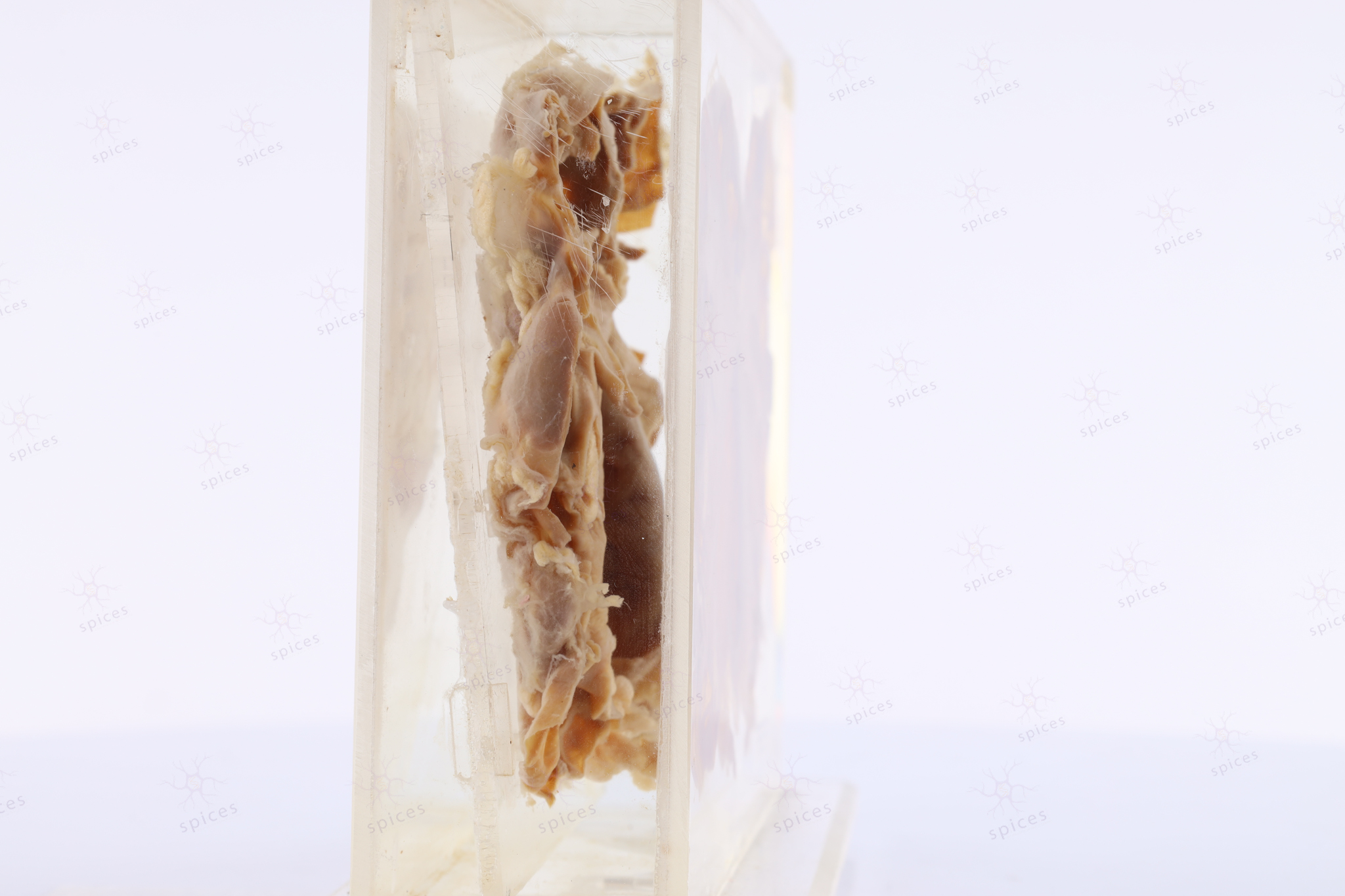
The exhibit displays kidney with presence of multiple cystic spaces.
HISTOPATHOLOGY DESCRIPTION
Renal parenchyma in chronic pyelonephritis shows tubular atrophy and interstitial and periglomerular fibrosis. Lymphoplasmacytic infiltration can be observed.
DIAGNOSIS
Chronic pyelonephritis
BAHASA MELAYU
Penerangan kasar: Spesimen menunjukkan ginjal dengan ruang sistik yang banyak.