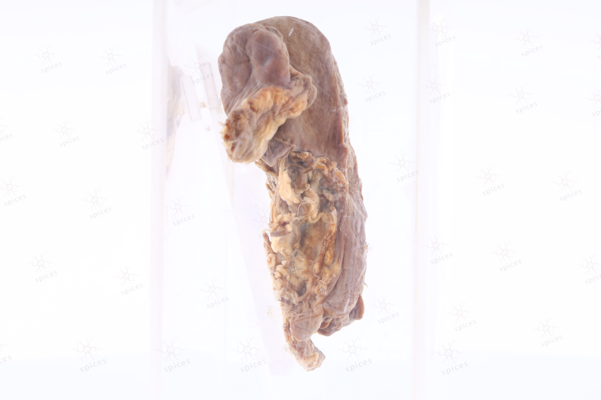
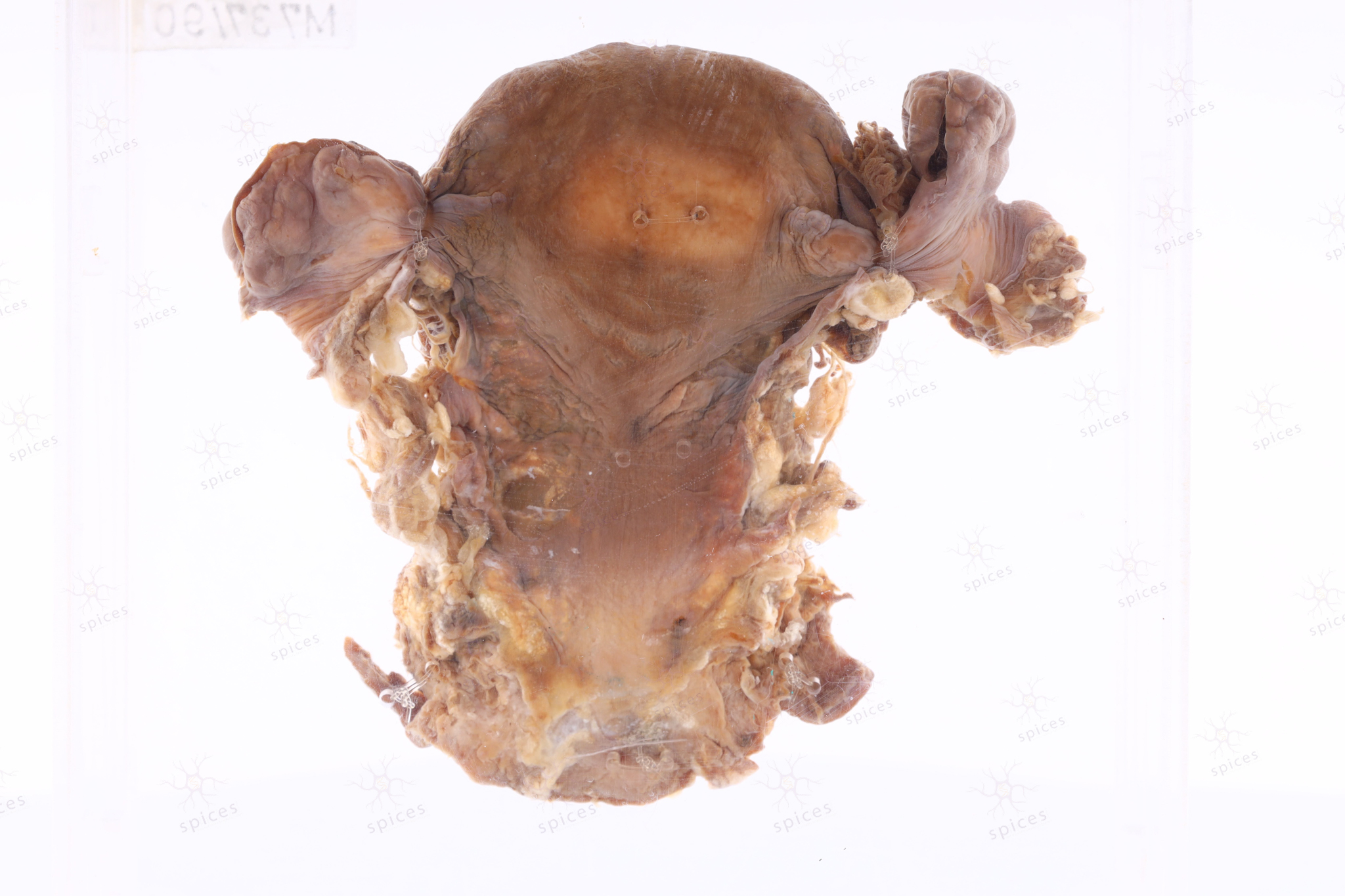
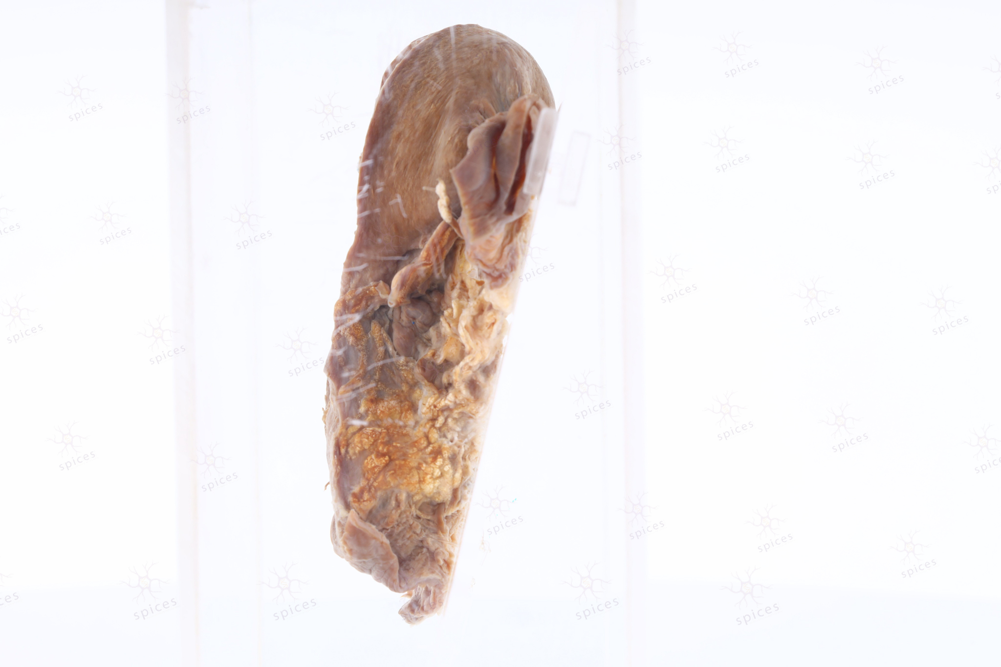
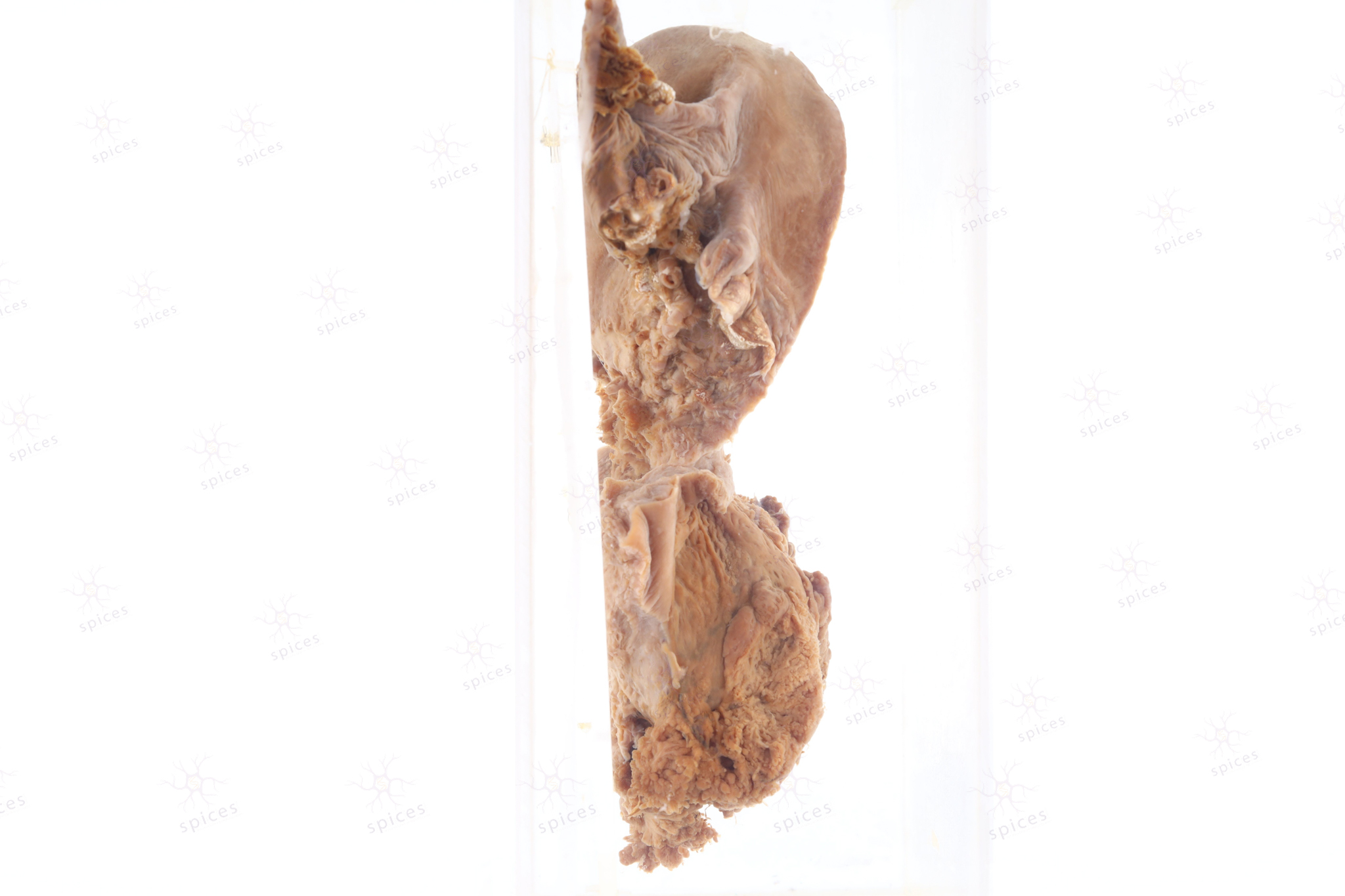
Uterus : M753/90
UTERUS
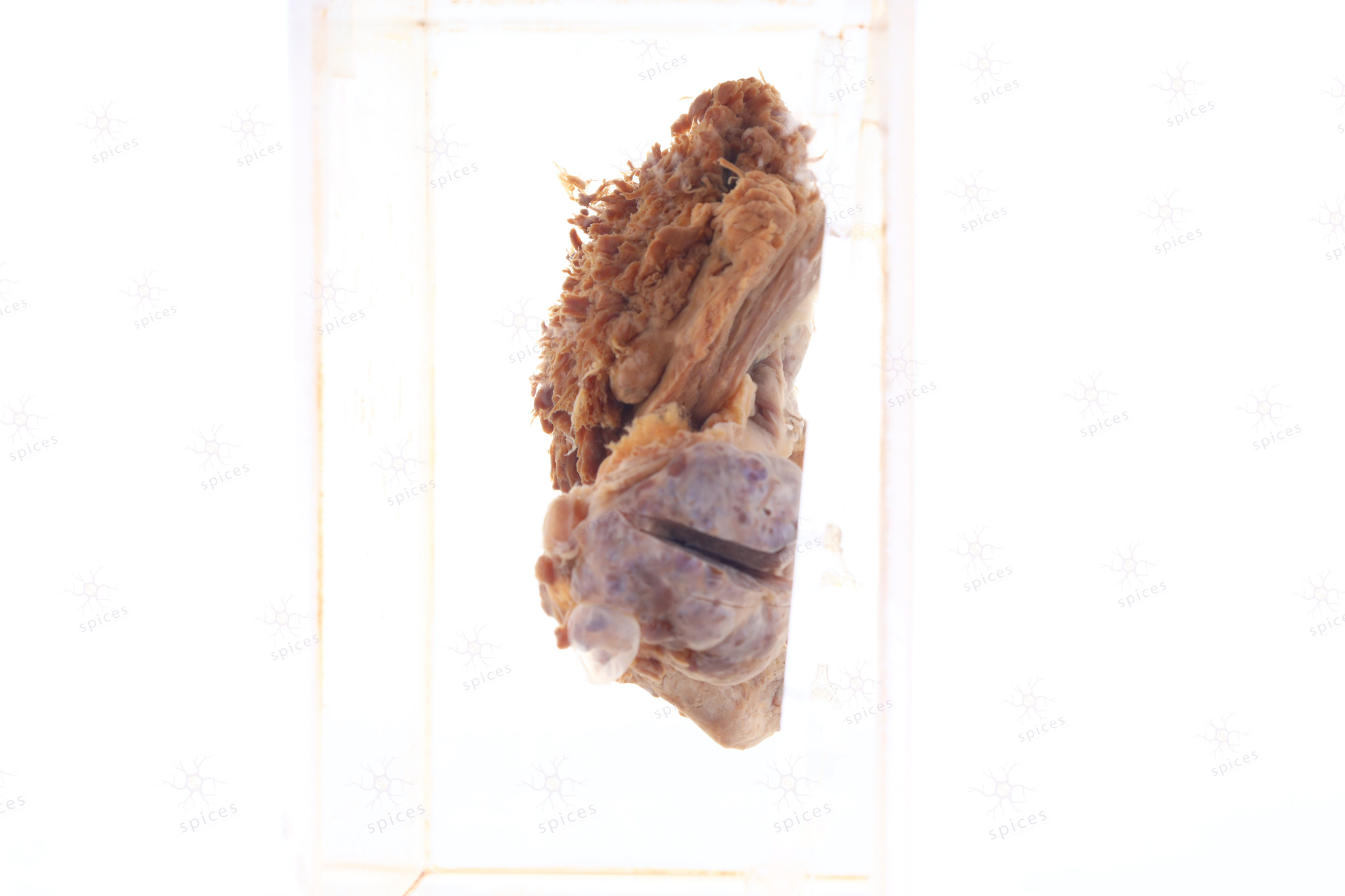
SPICES No. M753/90
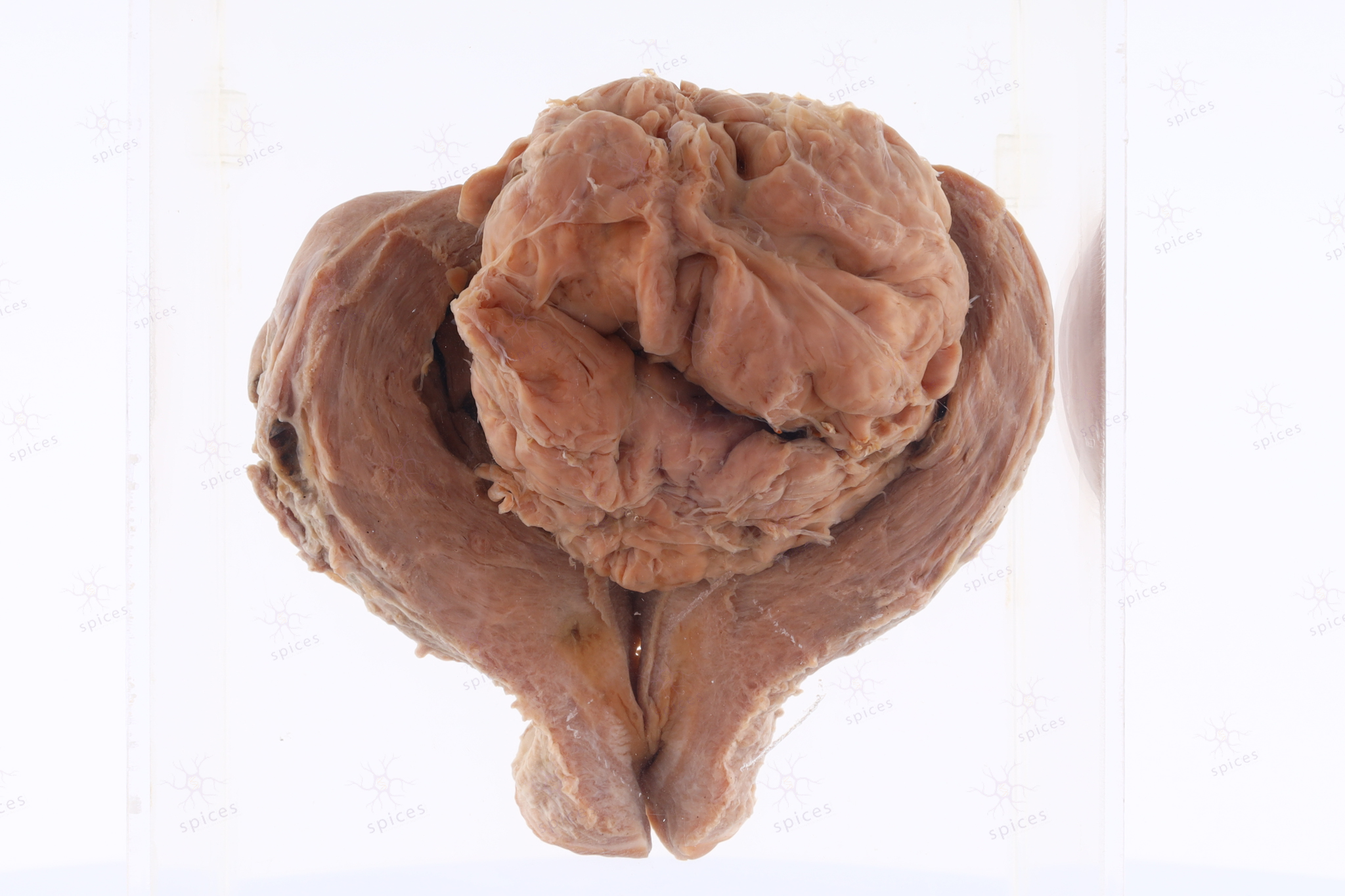
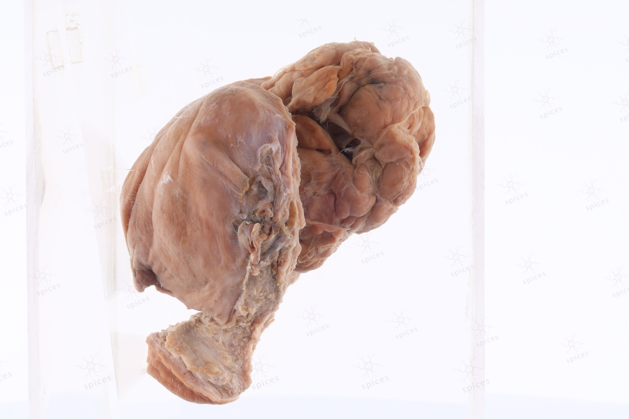
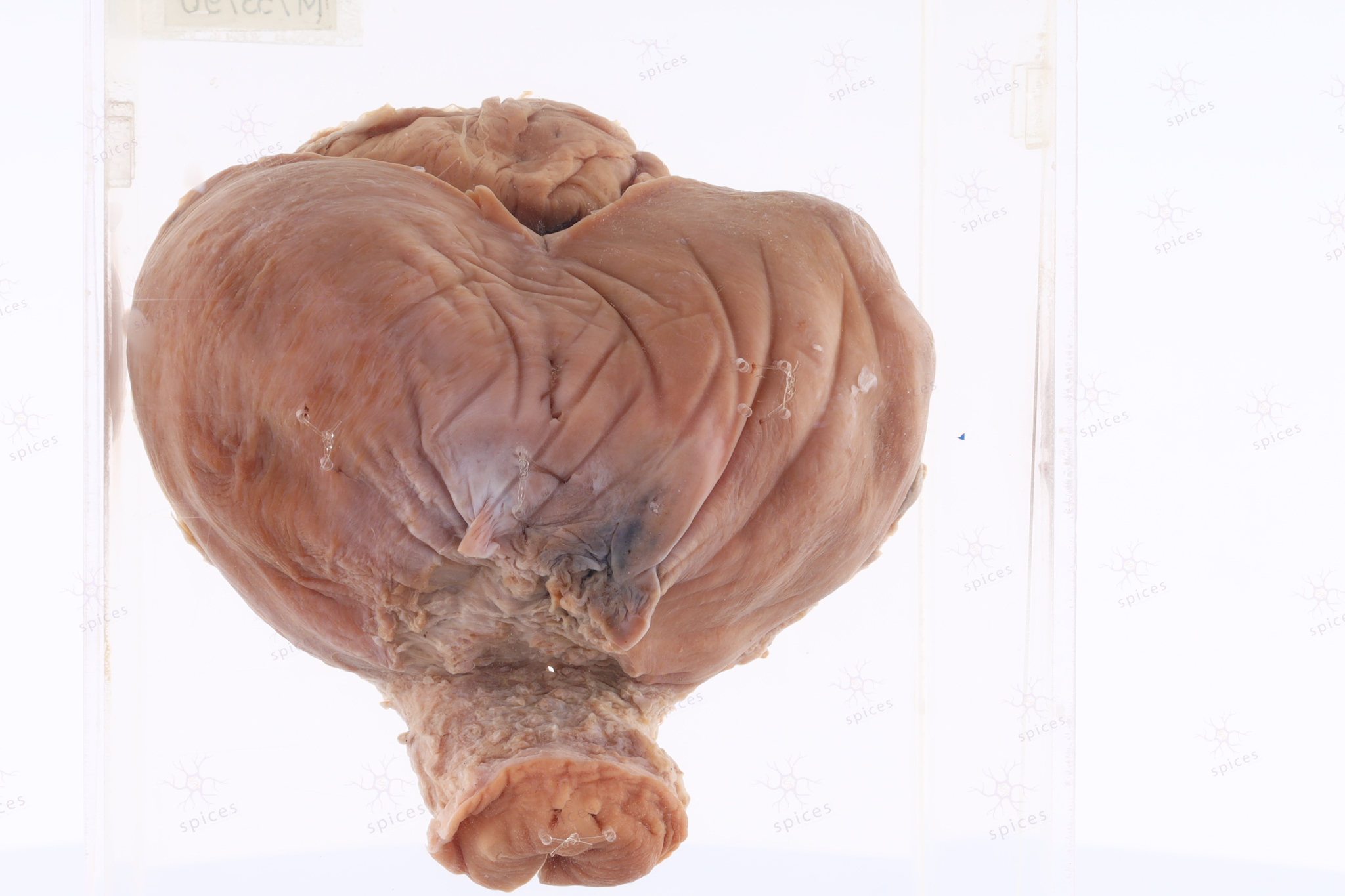
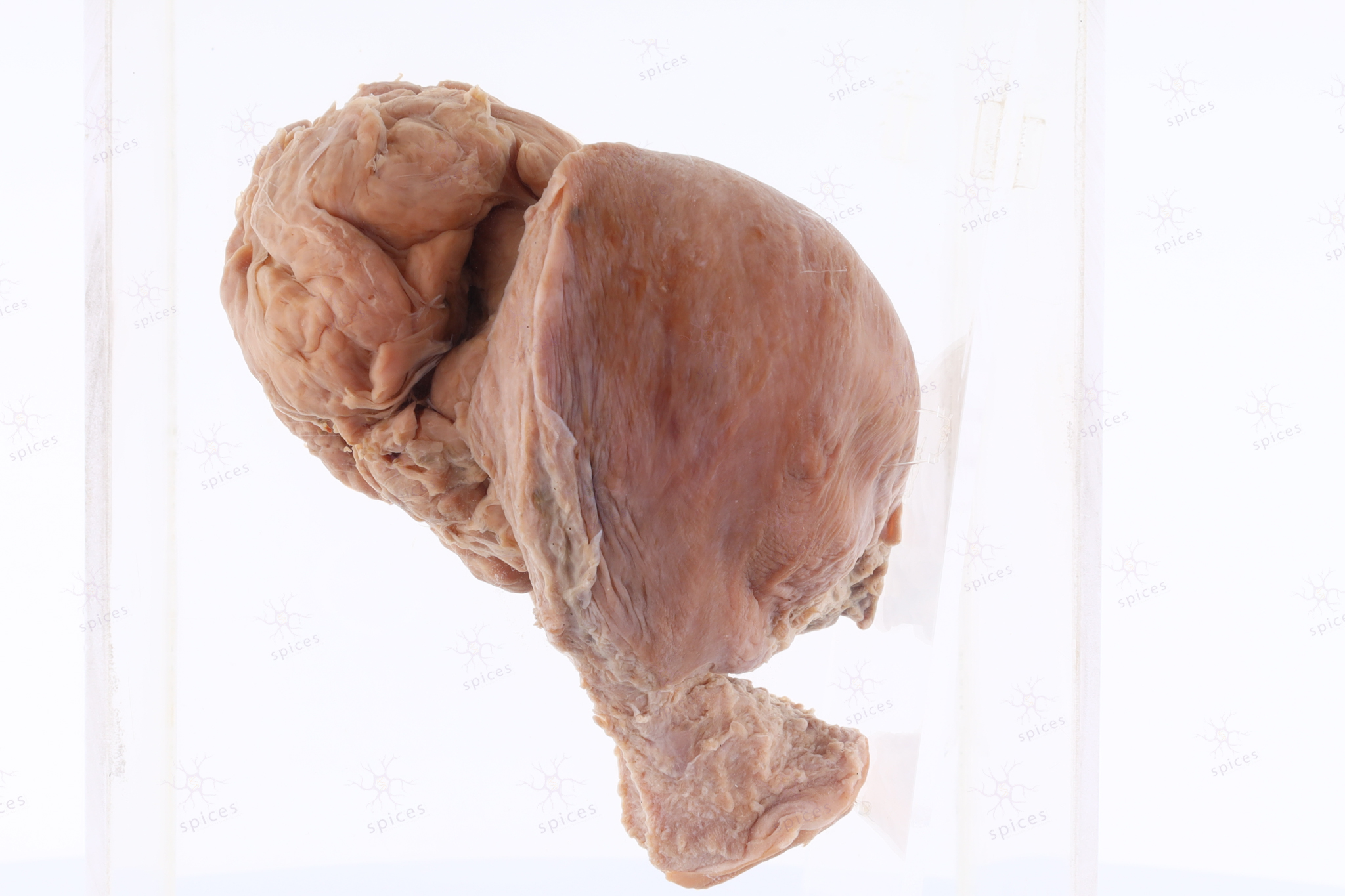
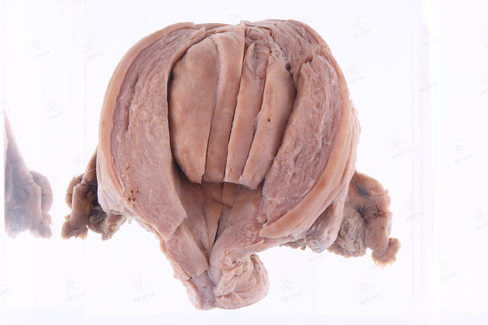
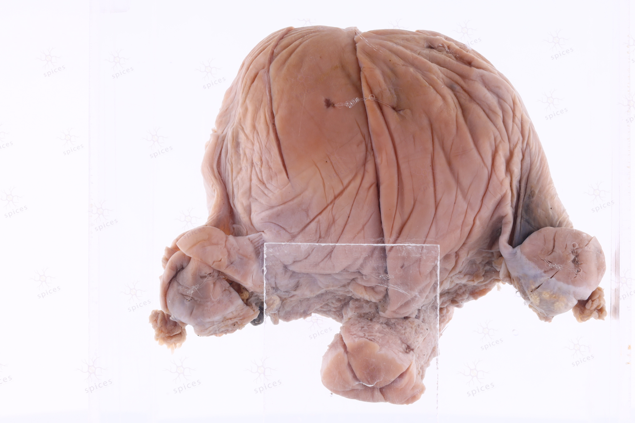
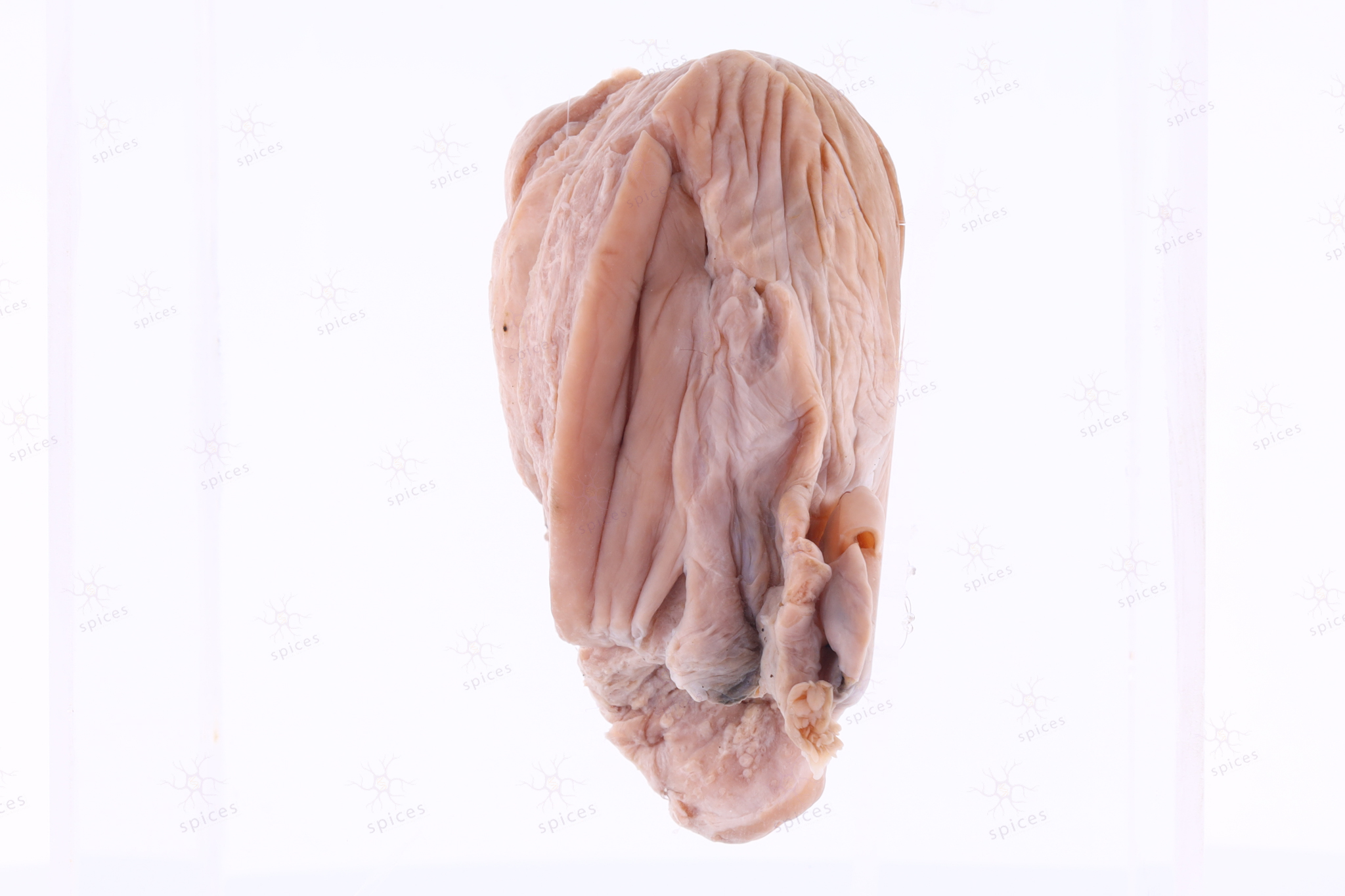
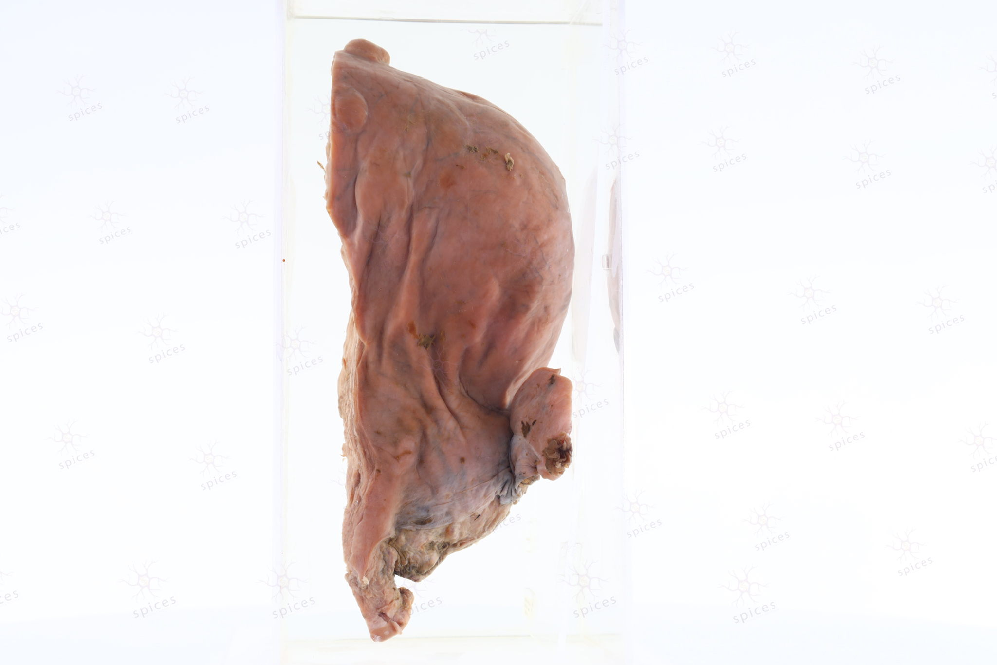
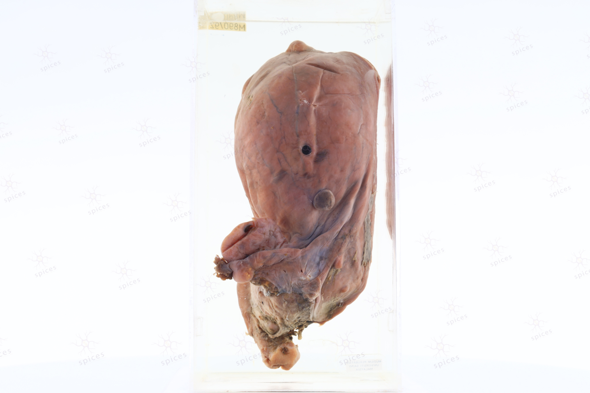
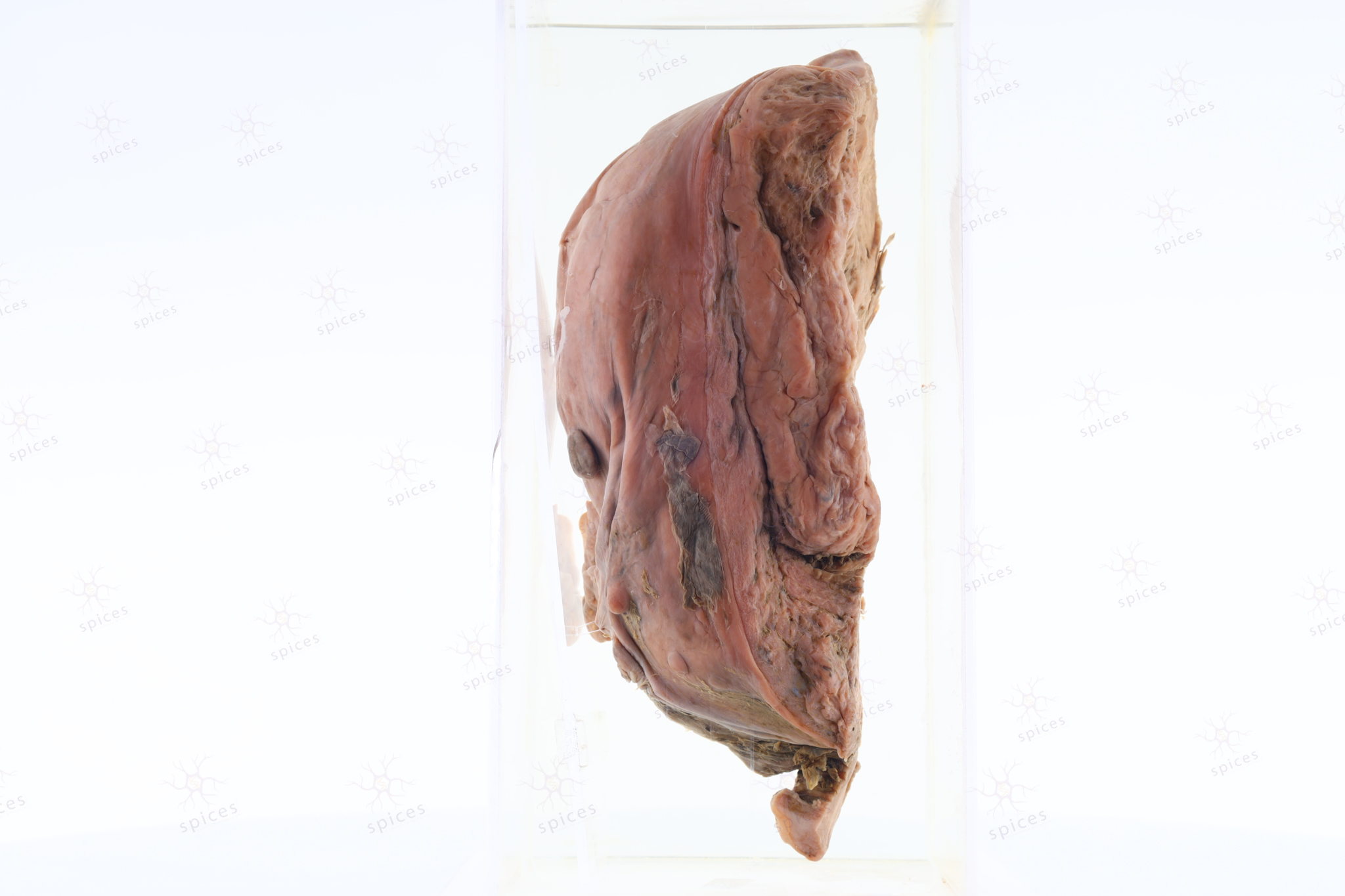
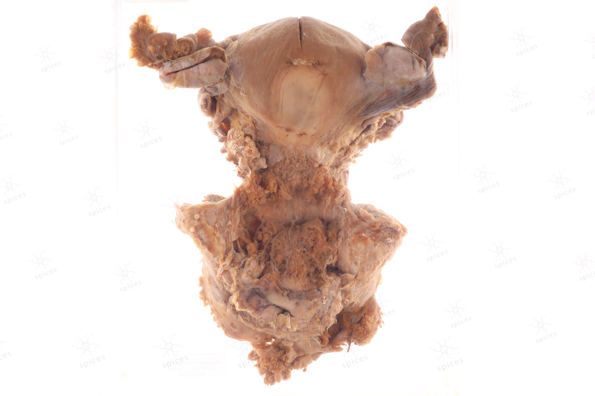
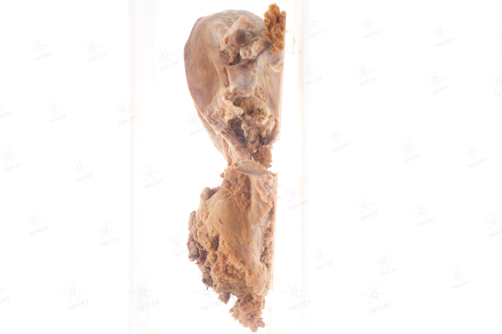
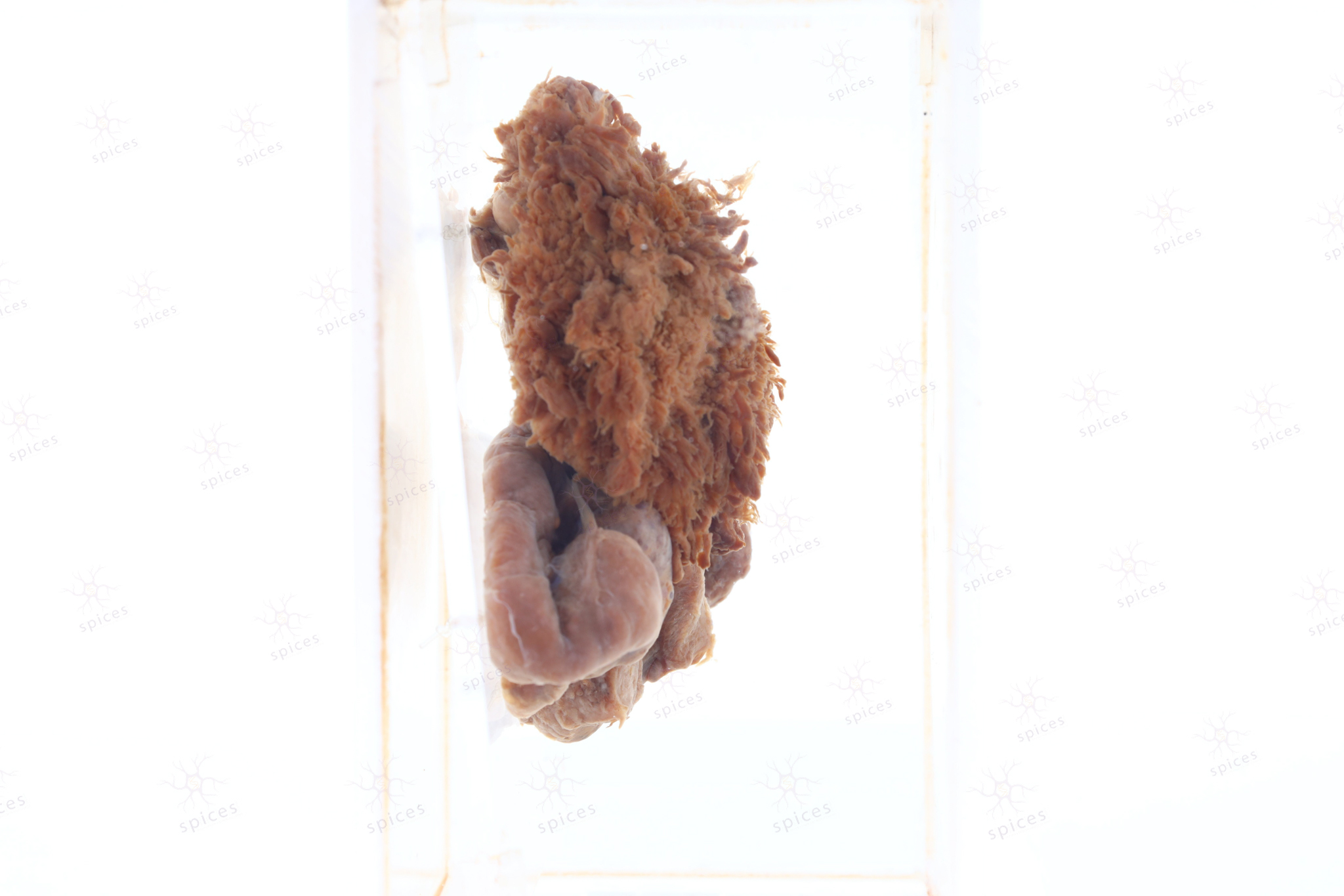
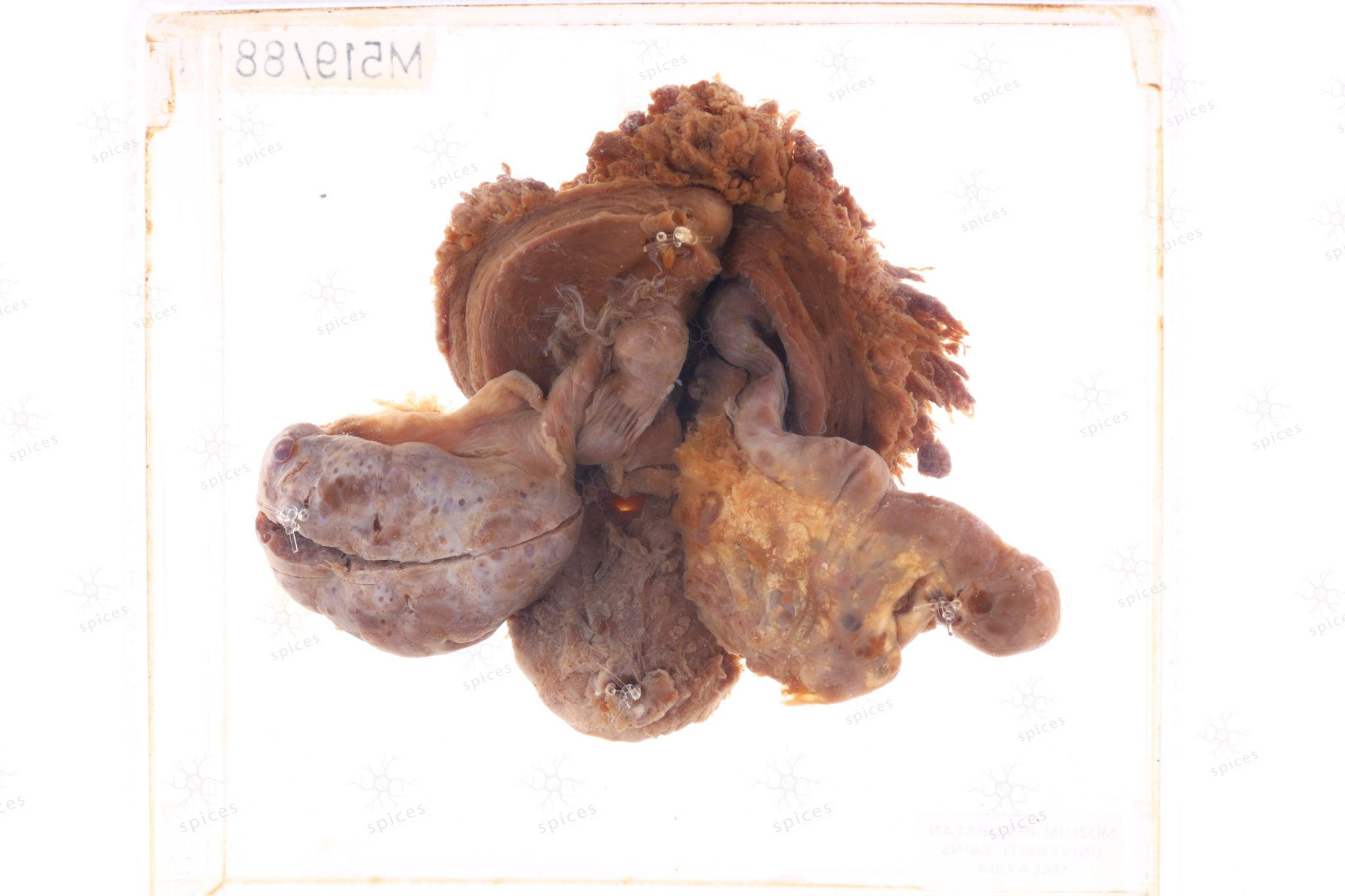
GROSS DESCRIPTION
The exhibit diplay large submucosal fibroid bulging into the endometrial cavity. It show well circumscribed margin, with lobulated appearance. The surface is homogenous. Trabeculation can be observed.
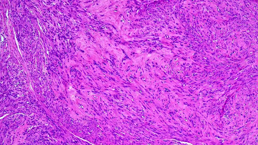
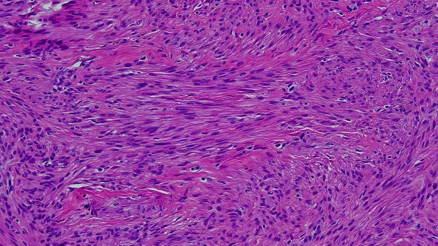
HISTOPATHOLOGY DESCRIPTION
Leimyoma is well circumscribed and unencapsulated. It is composed of spindle cells arranged in intersecting fascicles. The cells have cigar-shaped nuclei, indistinct cytoplasmic borders and eosinophilic cytoplasm.
DIAGNOSIS
Leiomyoma


