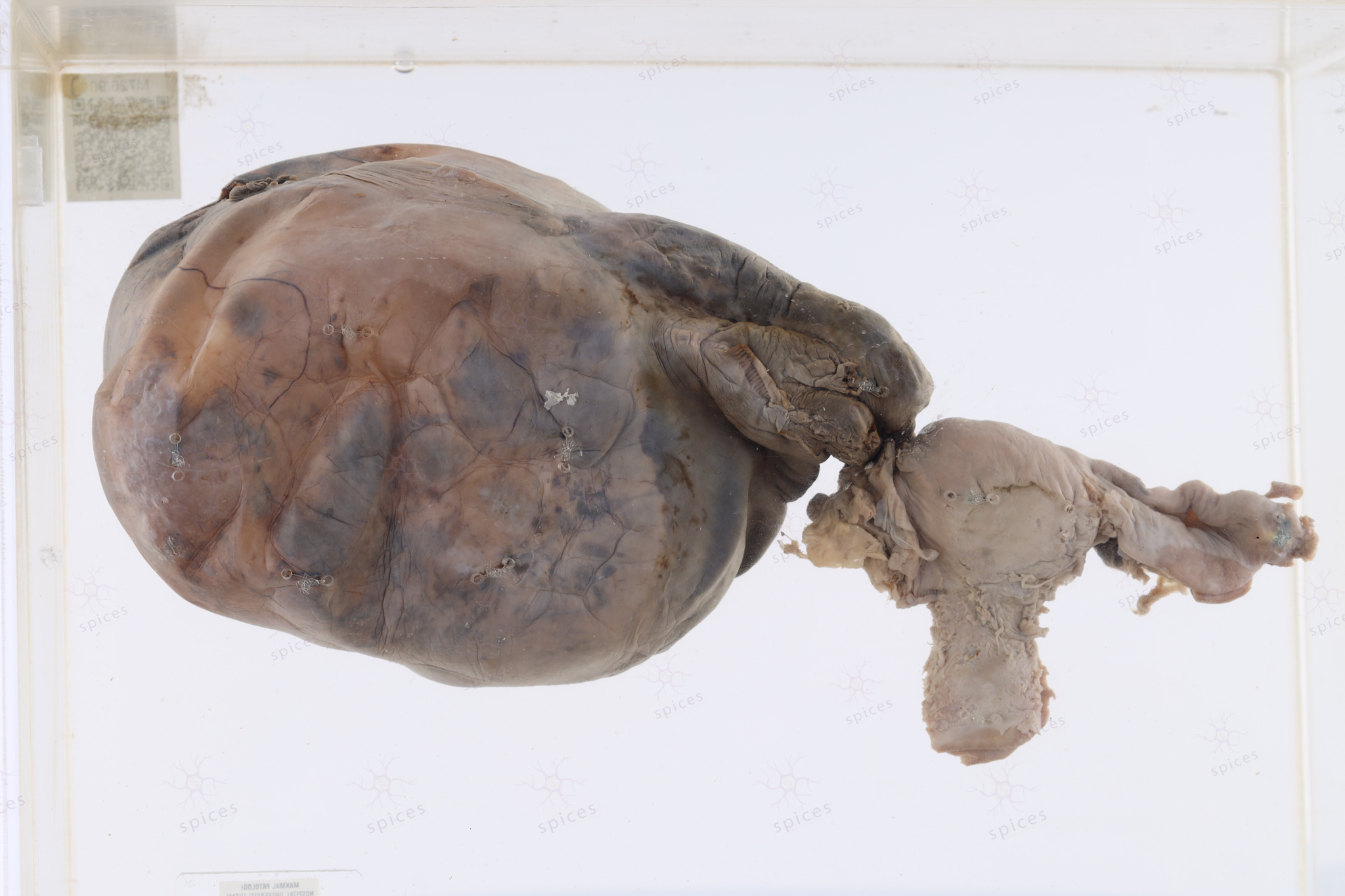
Ovary : M425/86
OVARY
Spices No: M425/86
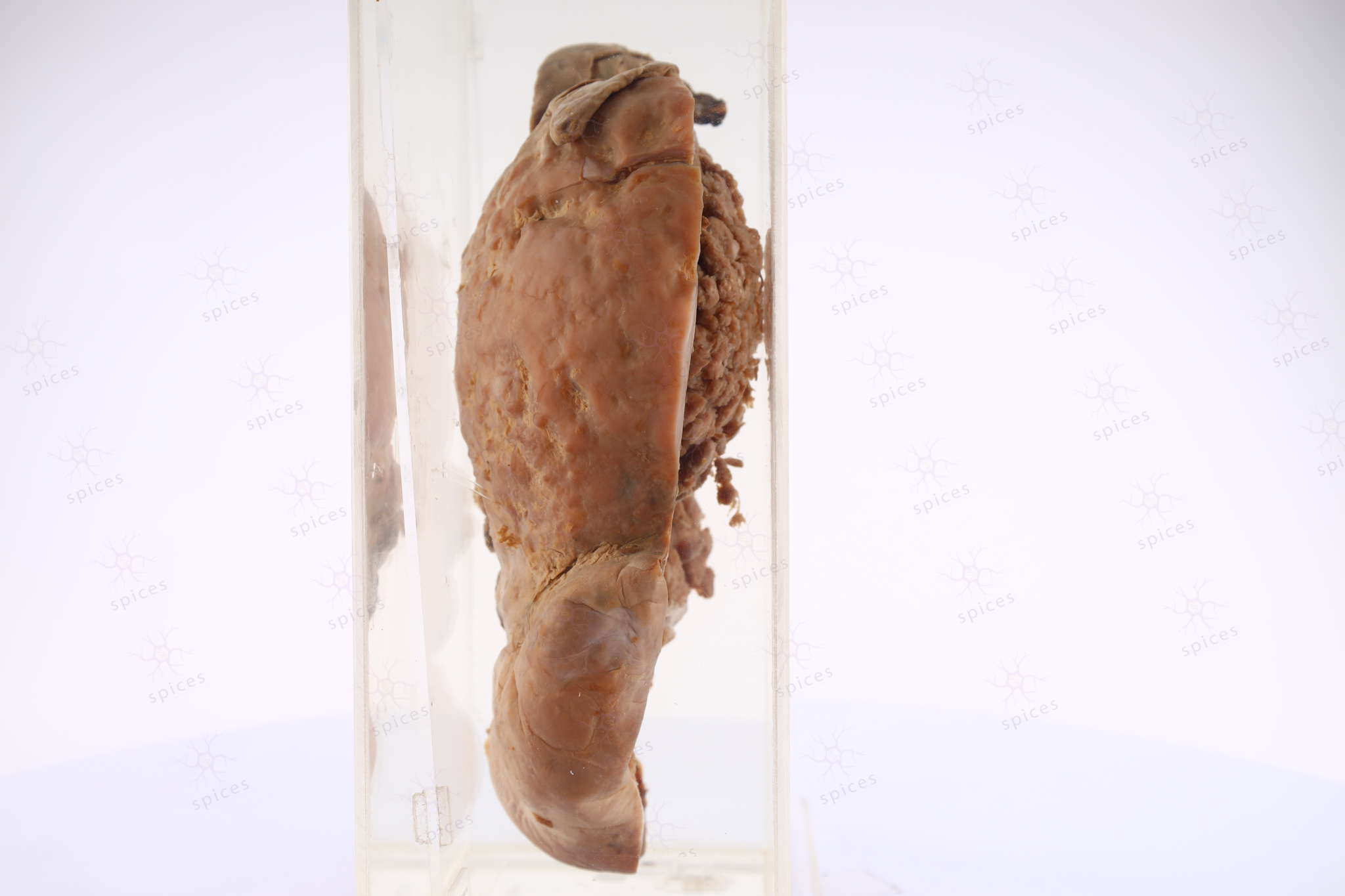
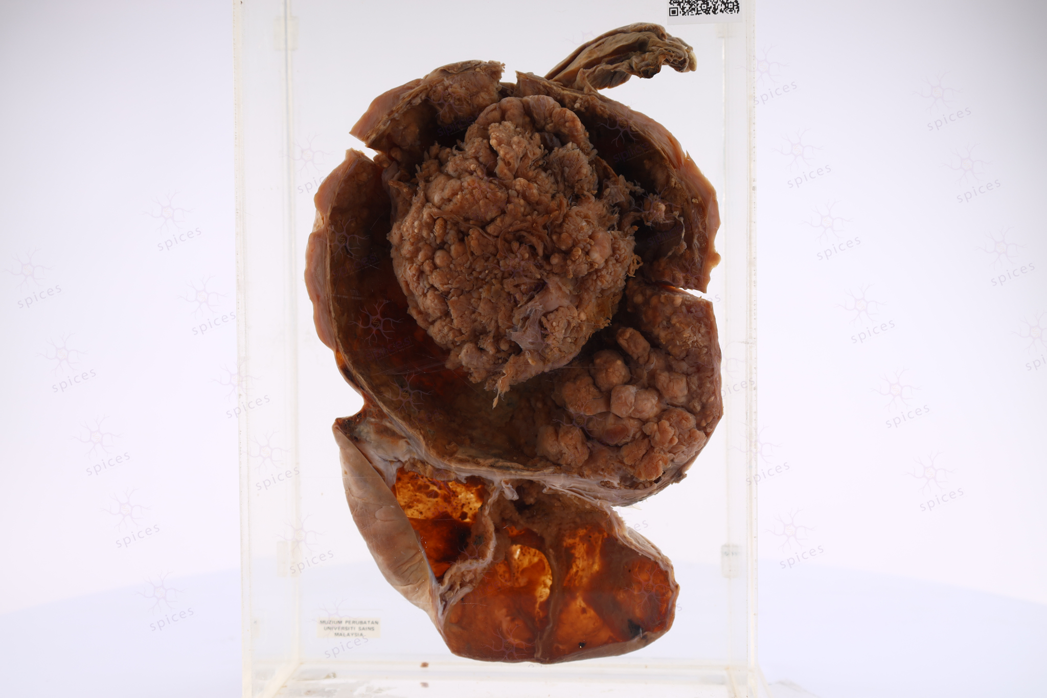
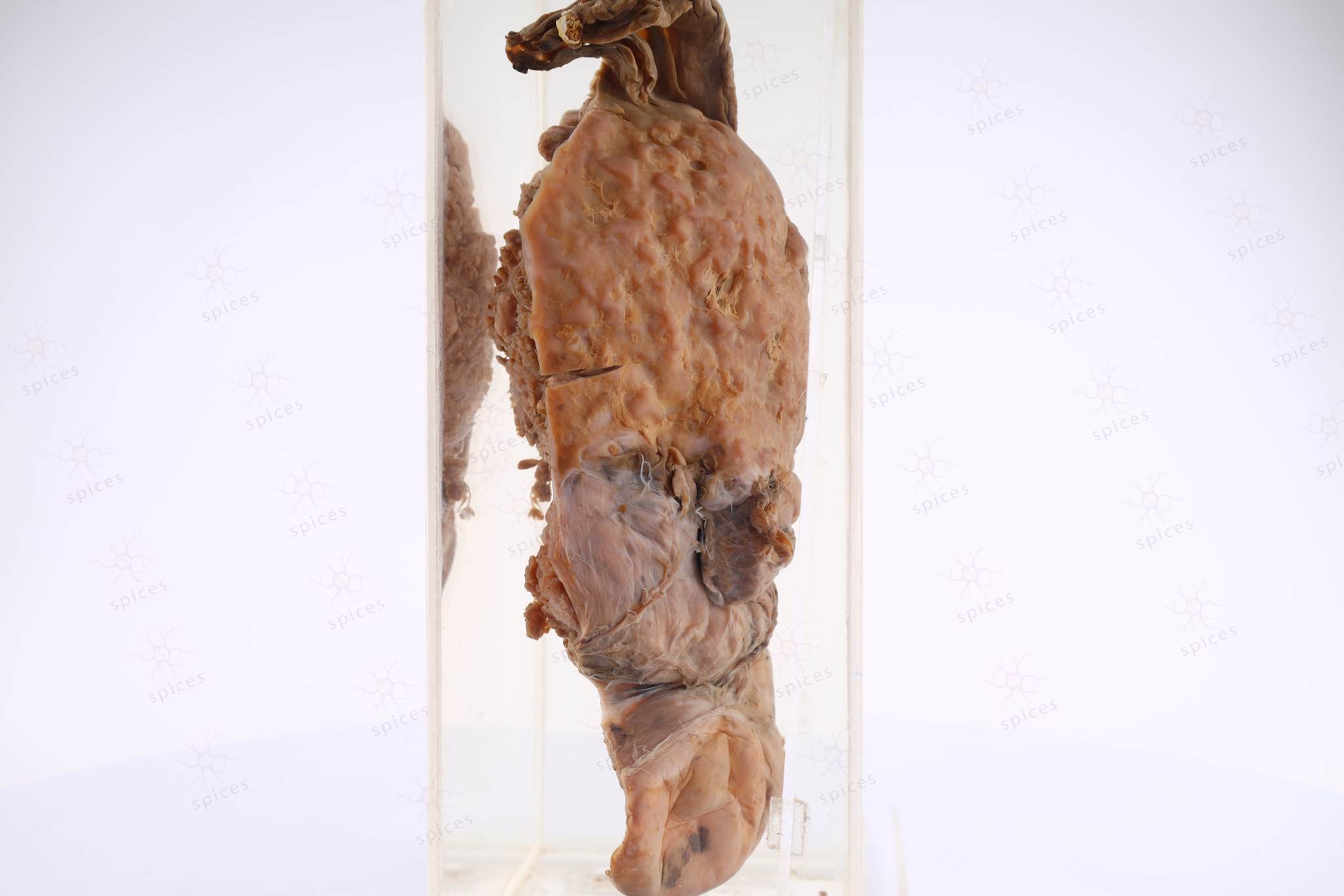
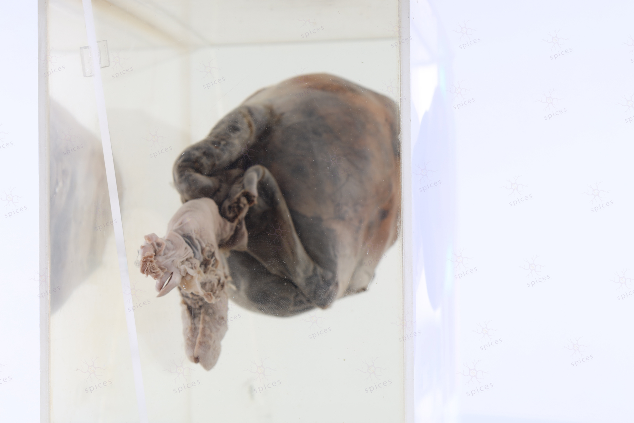
GROSS DESCRIPTION
The exhibit displays a cystic mass with multiloculation and septation. The wall is thin, however there is thick areas with foci of simple papillary structure project into the lumen. Cystadenoma exhibit smooth outer and inner surface.
HISTOPATHOLOGY DESCRIPTION
Serous cystadenoma show fibrocollagenous wall lined by single layer non stratified cuboidal to columnar bland tubal epithelium. Epithelial proliferation should be less than 10%.
DIAGNOSIS
Serous cystadenoma
BAHASA MELAYU
Penerangan kasar: Spesimen menunjukkan ketumbuhan dalam rupa bentuk seperti kantung/sistika yang mempunyai dinding yang memisahkan dan membentuk beberapa ruang. Dinding ketumbuhan sistika ini nipis. Terdapat kawasan dinding yang tebal dan juga mempunyai unjuran dalam bentuk papilari.