Thigh - M1152/98
THIGH
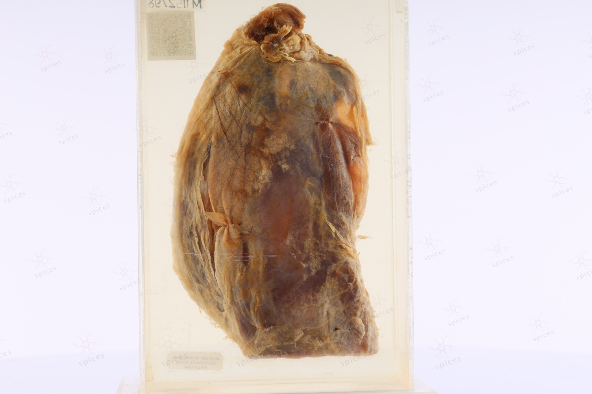
Spices No: M1152/98

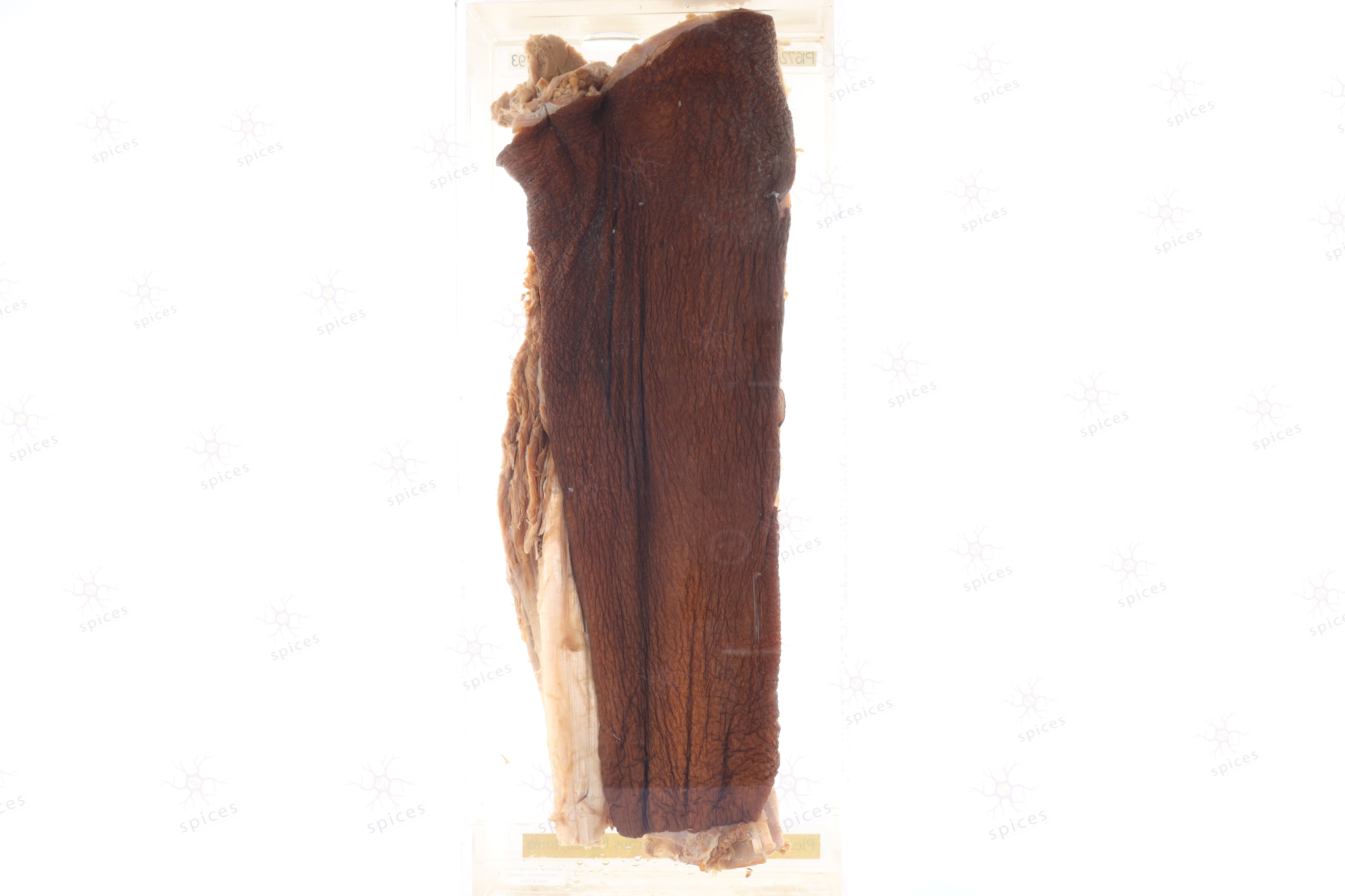
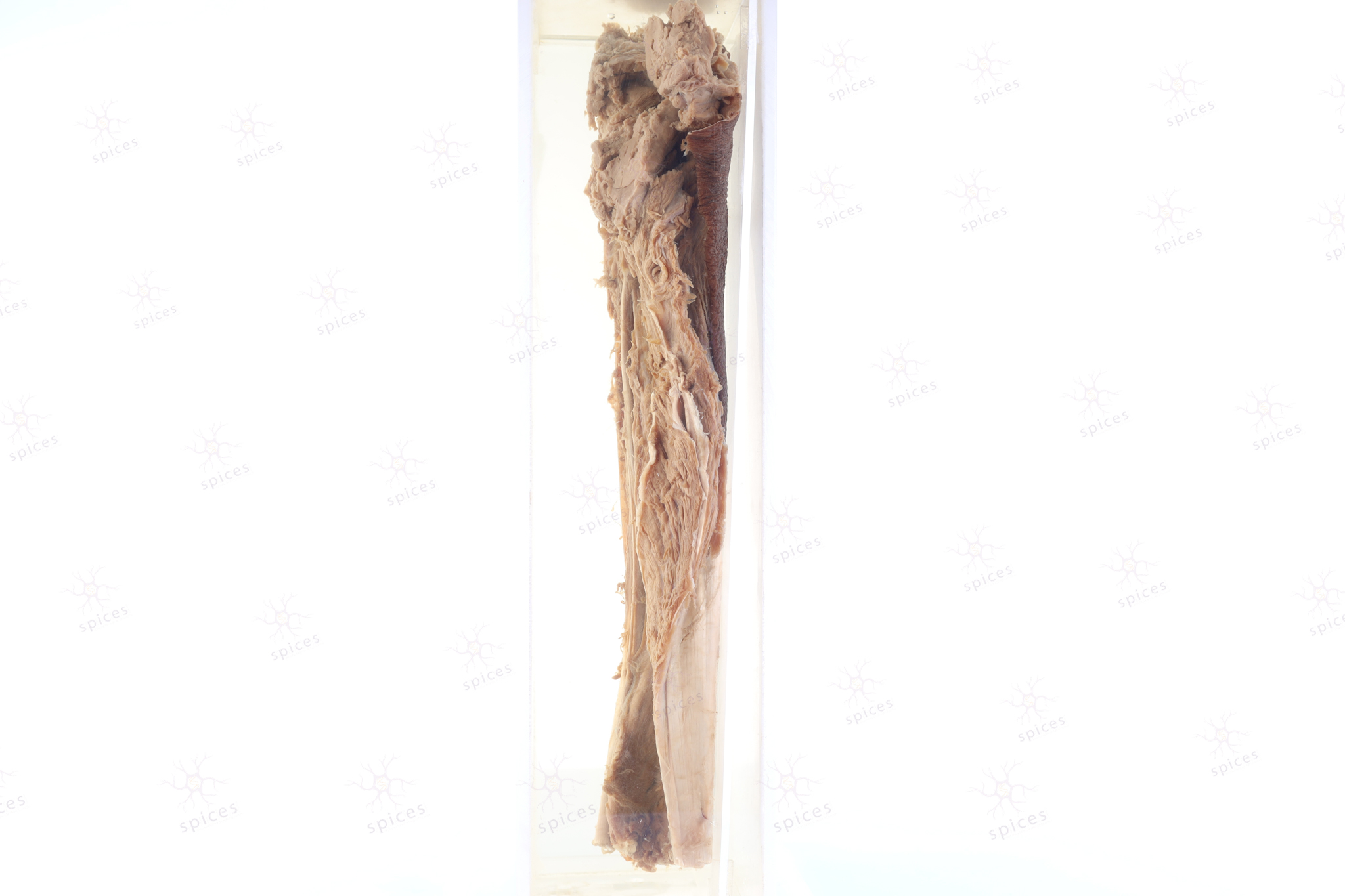
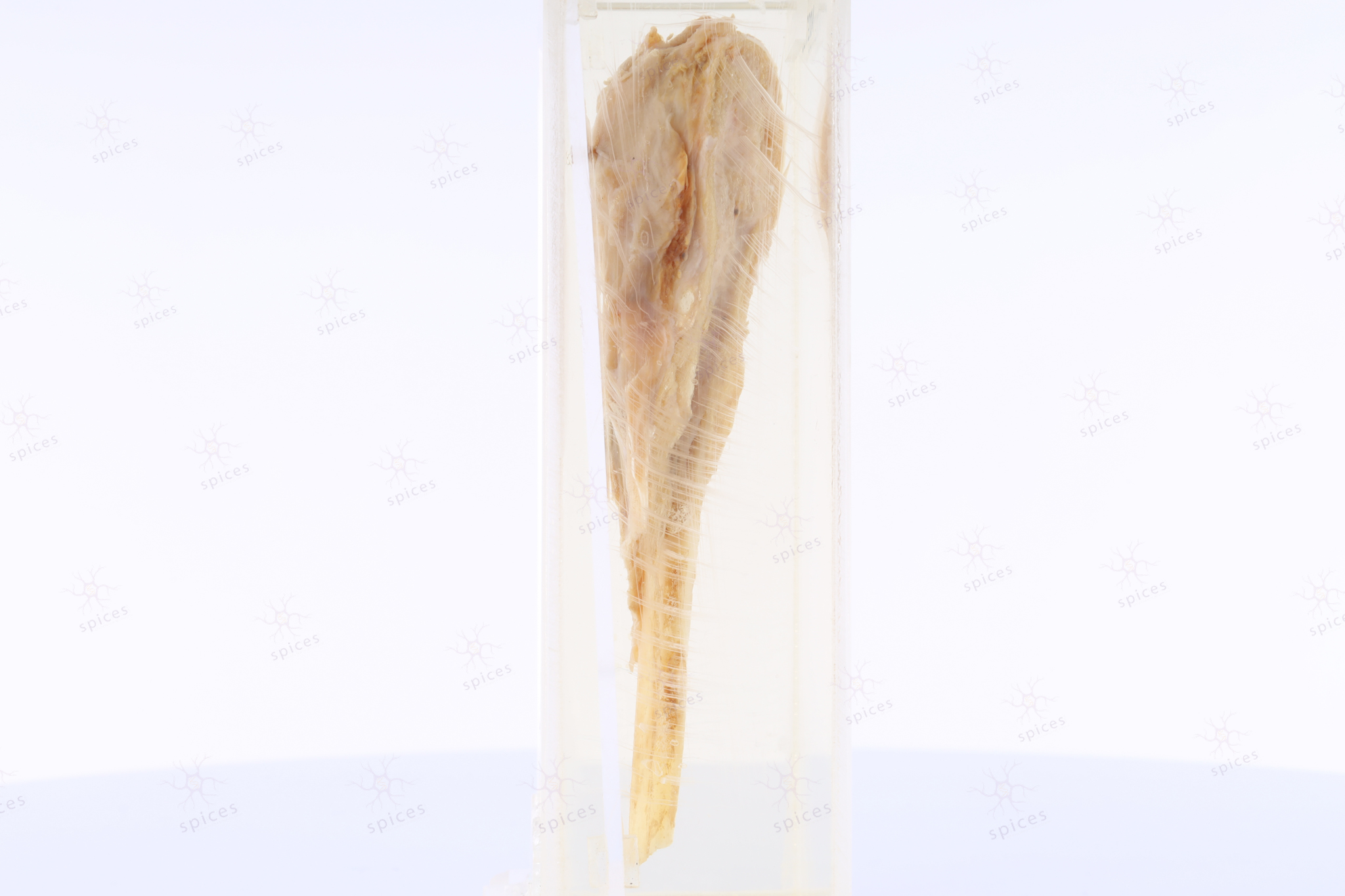
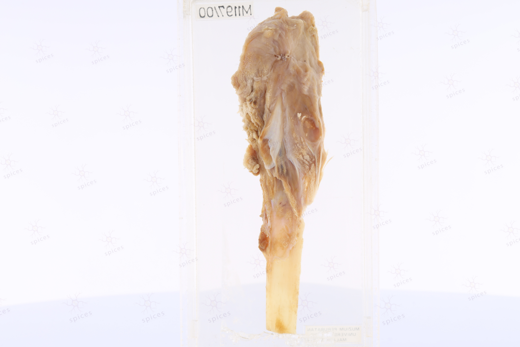
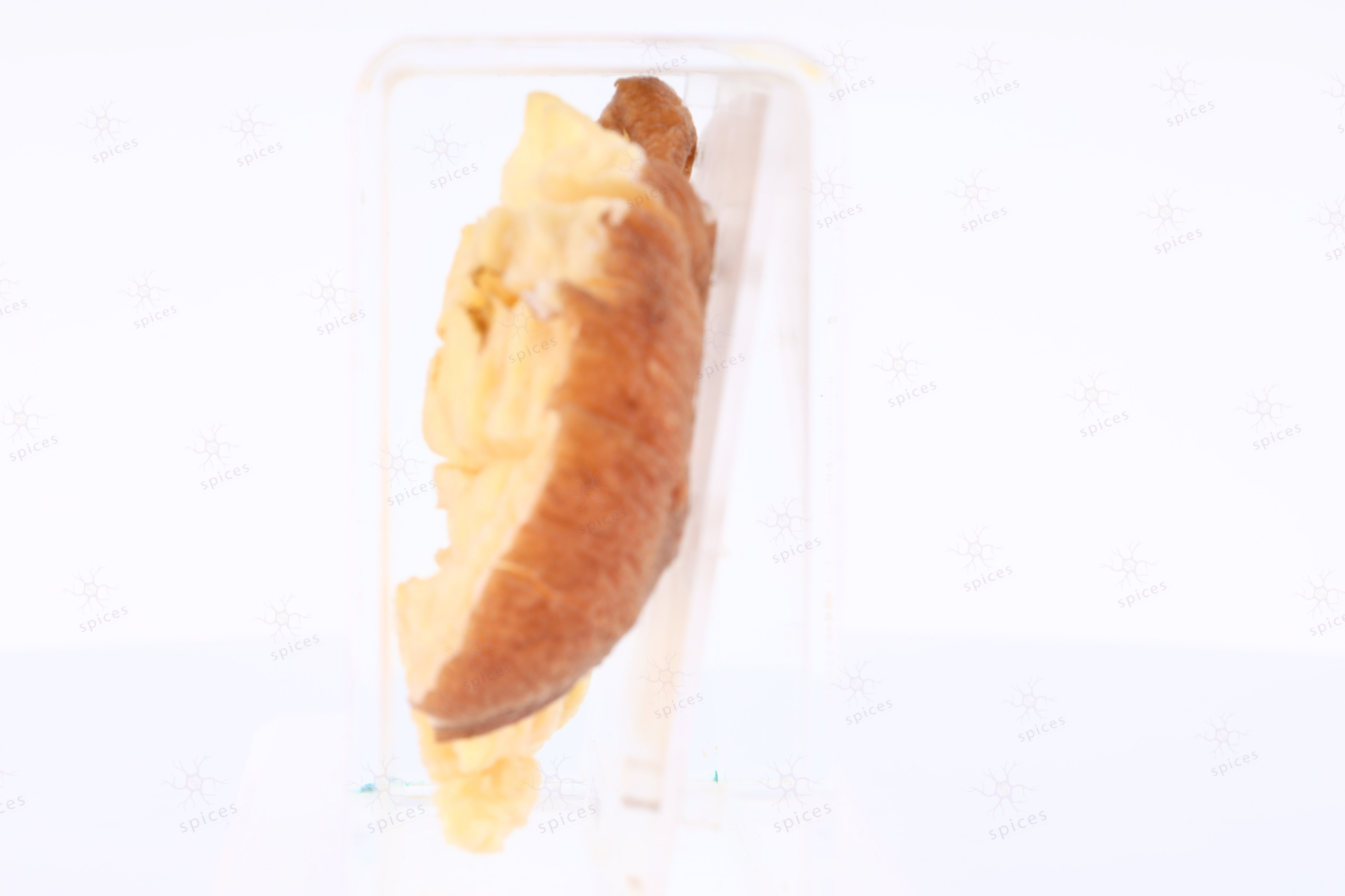
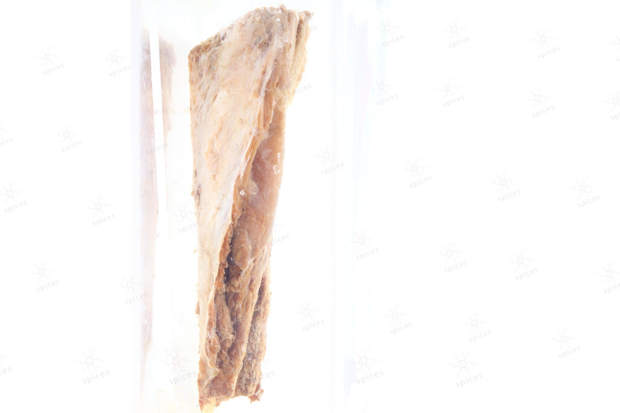
GROSS DESCRIPTION
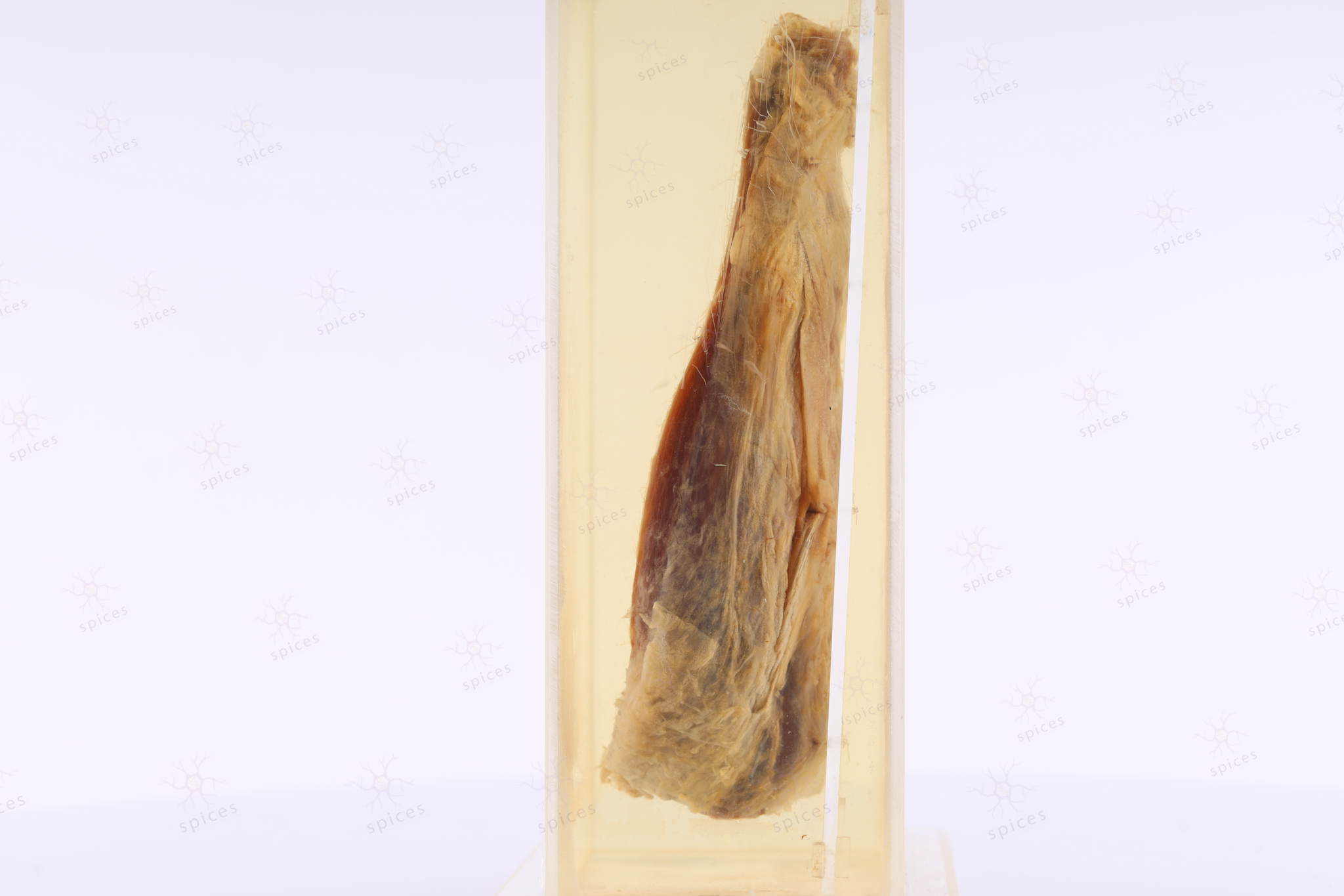
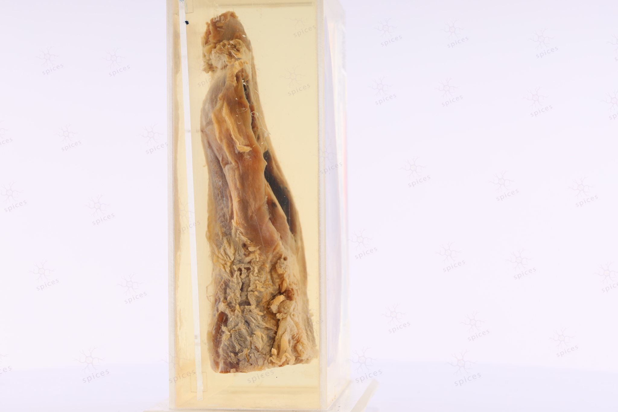
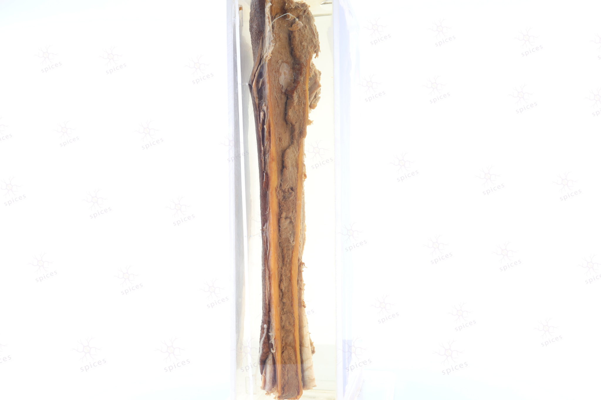
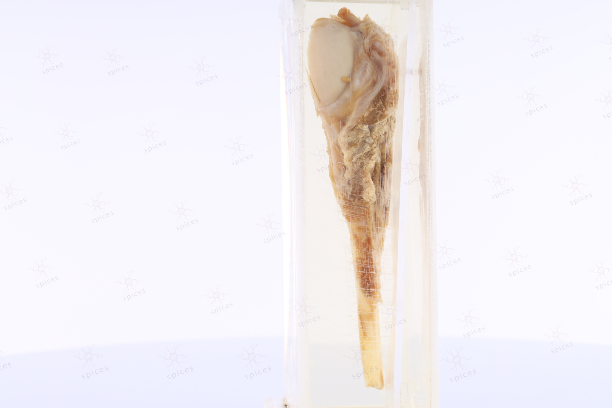
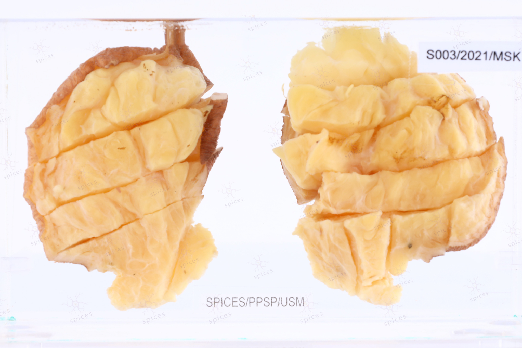
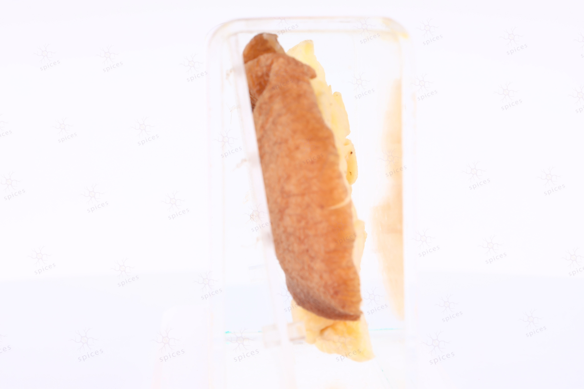
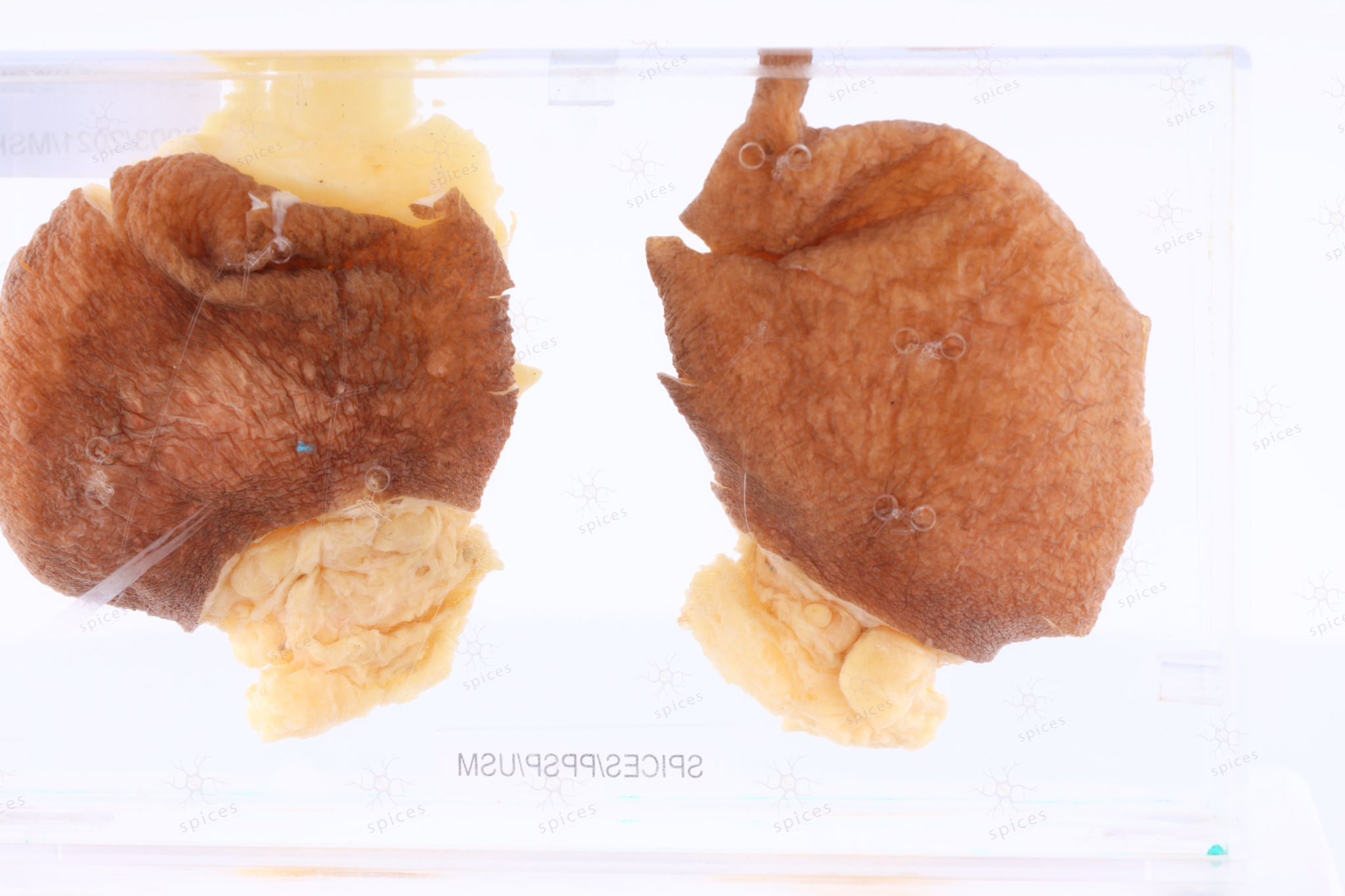
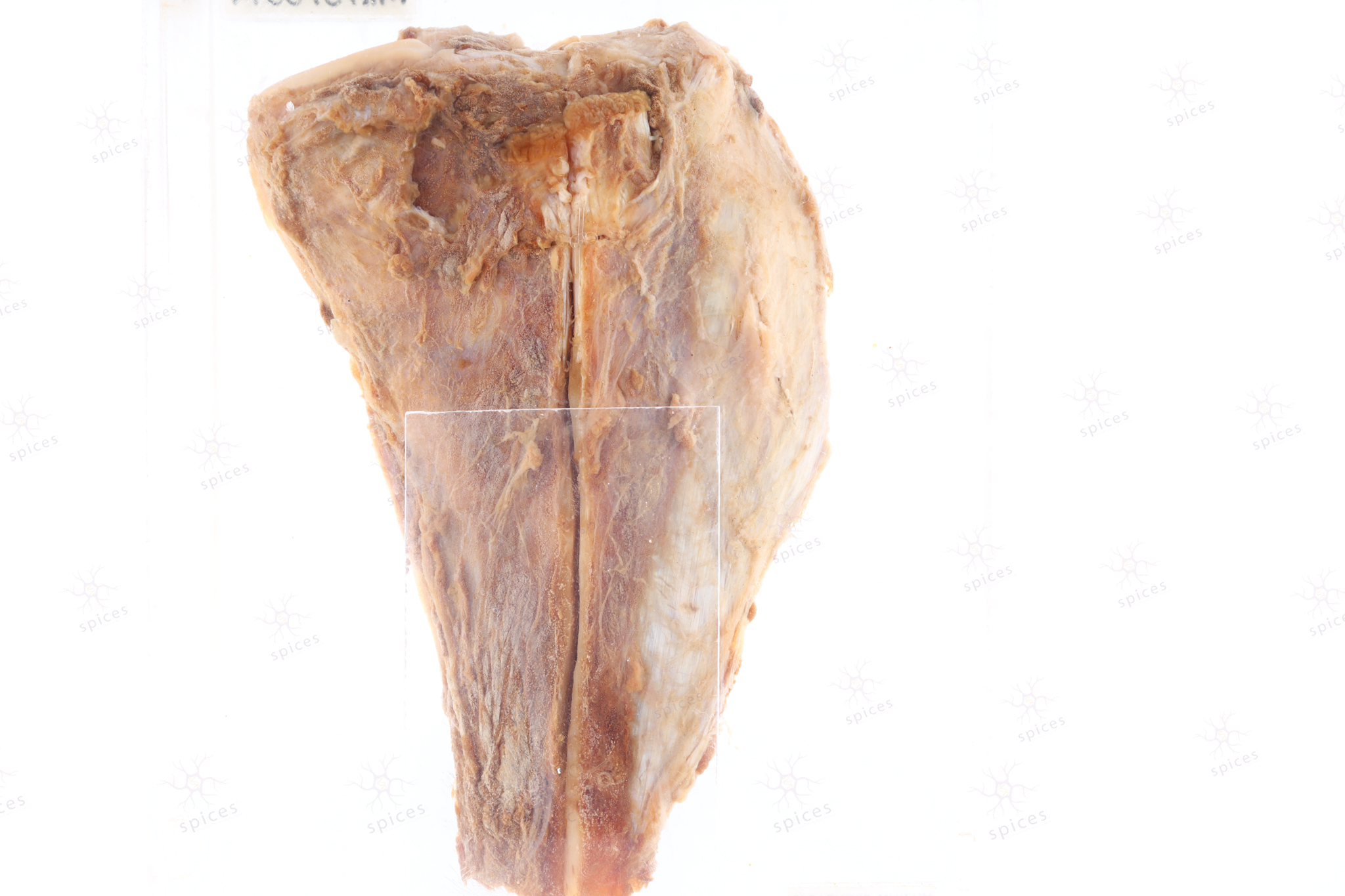
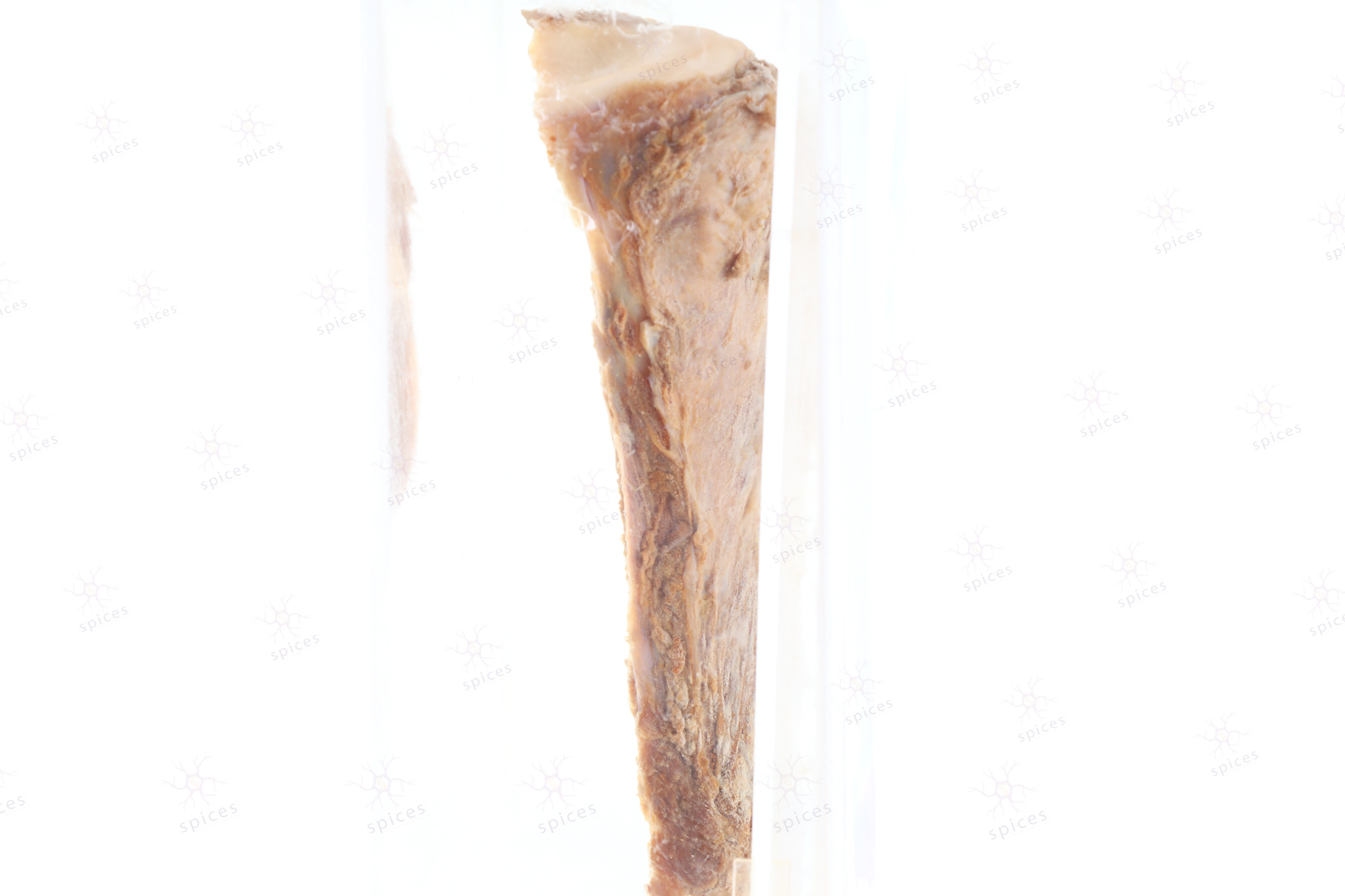
The exhibit displays large intramuscular well circumscribed liposarcoma displaying a variegated surface. The solid areas have tan to grey hemorrhagic surfaces. The cystic spaces are variable in size and appear empty.
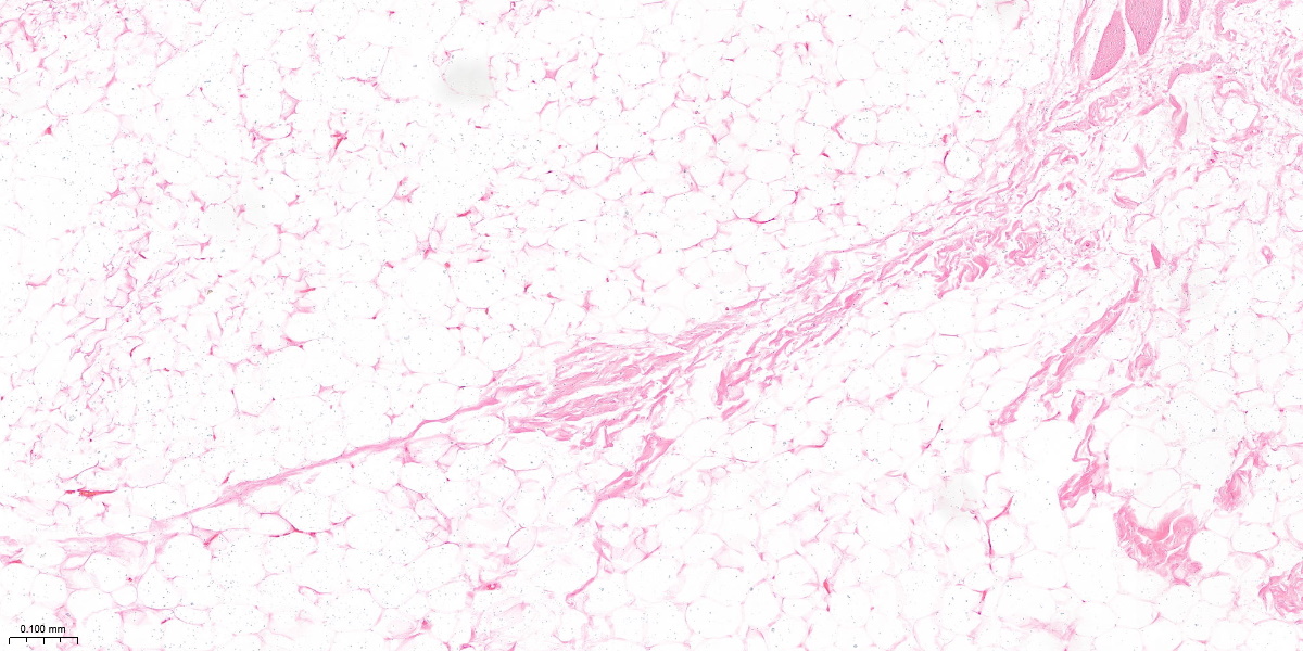
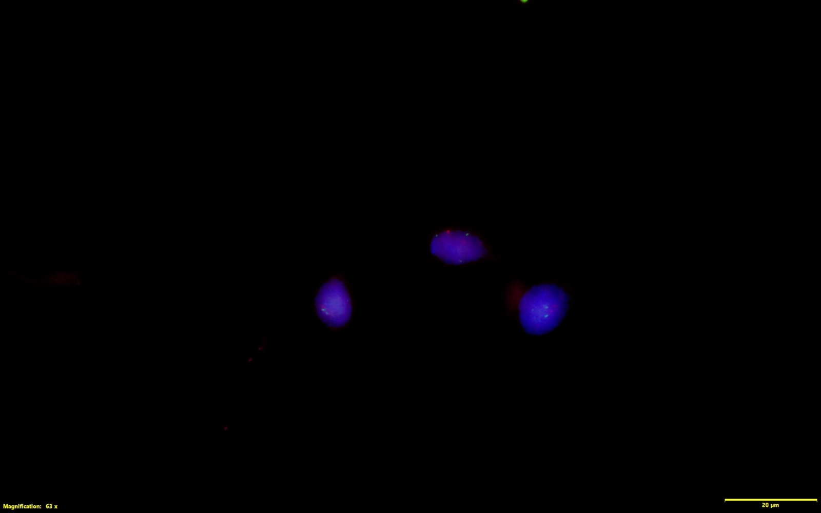
HISTOPATHOLOGY DESCRIPTION
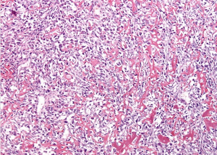
Liposarcoma shows marked variation in cell sizes accompanied by the presence of bizarre stromal cells with marked hyperchromasia. A varying number of lipoblast can be found,however the presence of lipoblast is not required for the diagnosis.
DIAGNOSIS
Liposarcoma
BAHASA MELAYU
Penerangan kasar: Spesimen liposarcoma menunjukkan permukaan yang mempunyai warna berlainan. Biasanya akan ada kawasan nekrosis atau tisu mati dah juga haemorrhagic.
Penerangan histopatologi: Liposarcoma terdiri daripada sel lemak yang menunjukkan perbezaan ketara dari segi saiz dan biasanya terdapat sel stroma yang sangat atipikal dan mempunyai nukleus yang gelap. Sel lemak lipoblast atau sel lemak tidak matang juga boleh dilihat dalam tisu liposarcoma.
Diagnosis: Liposarcoma


