Left Wrist Nodule : S002/2021
LEFT WRIST NODULE
Spices No: S002/2021

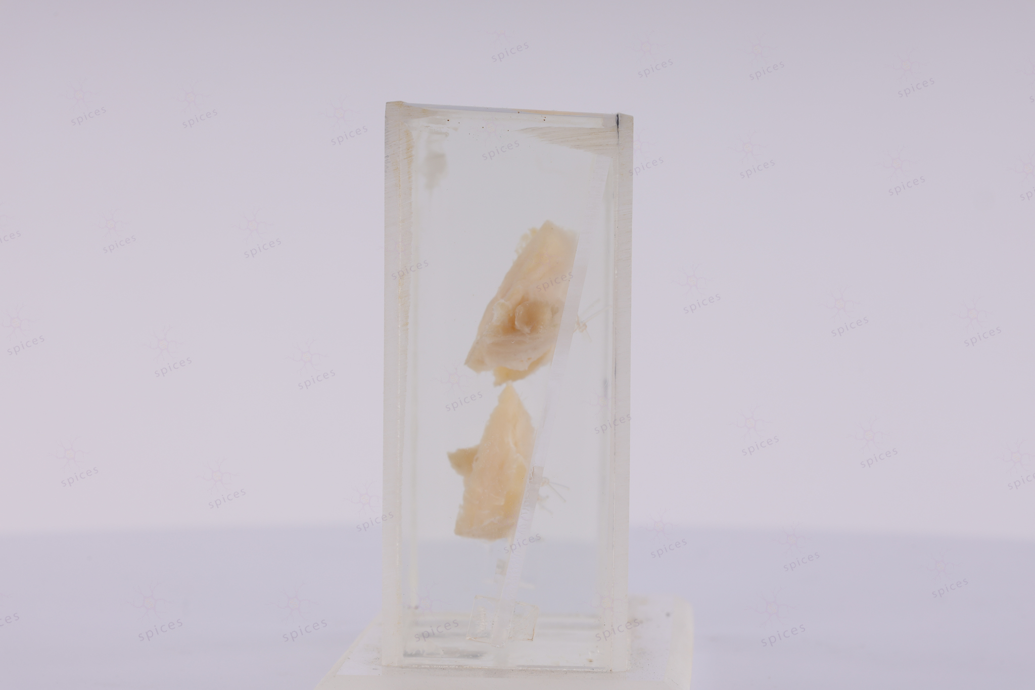
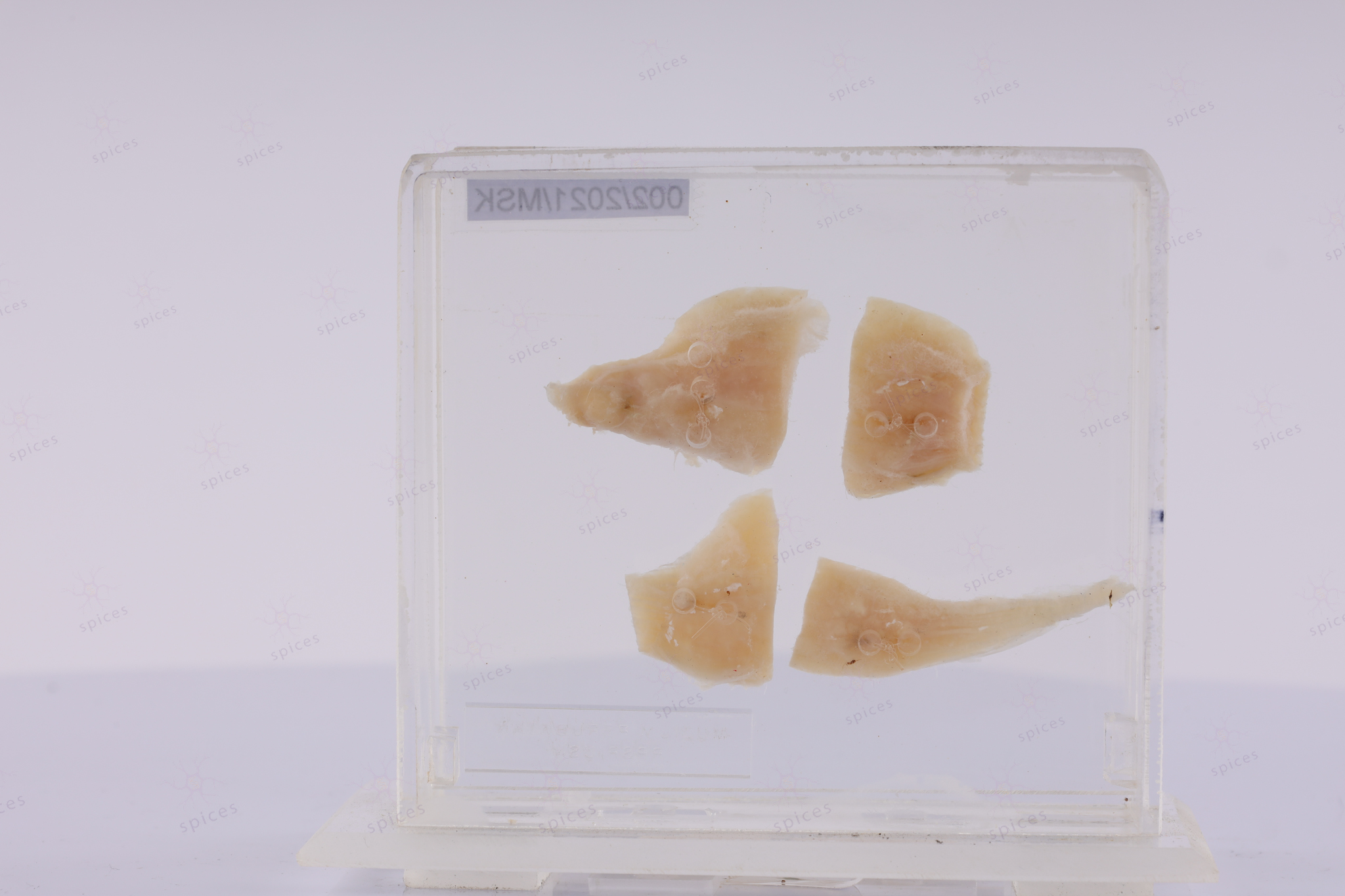
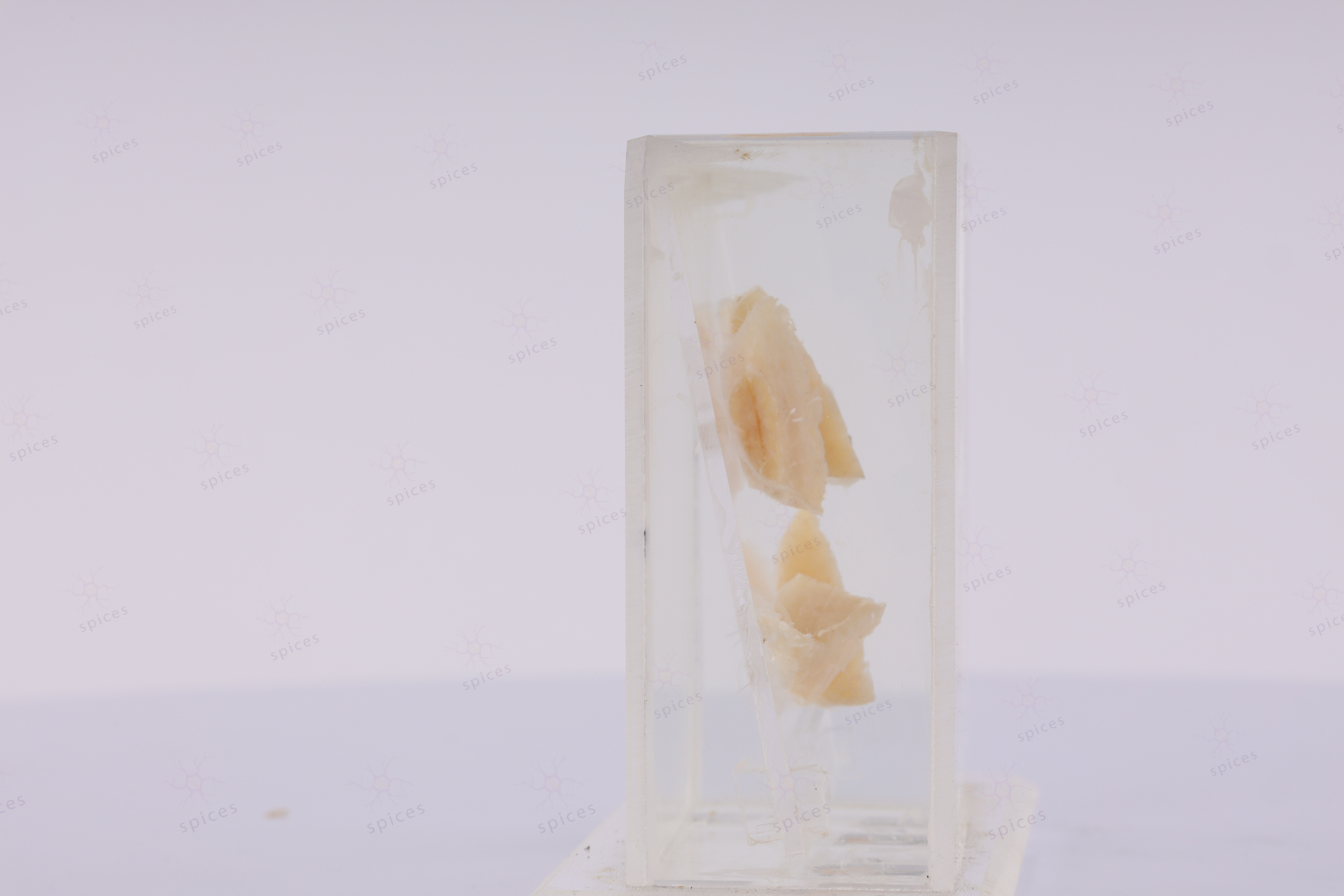
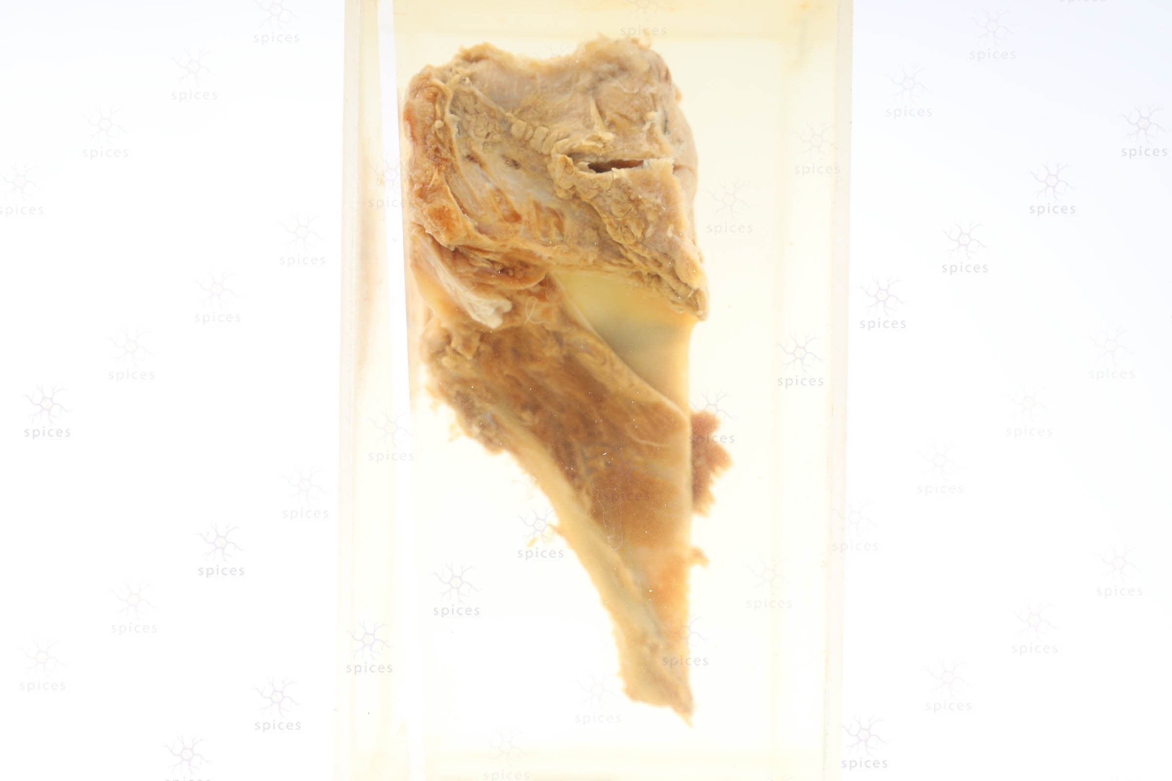
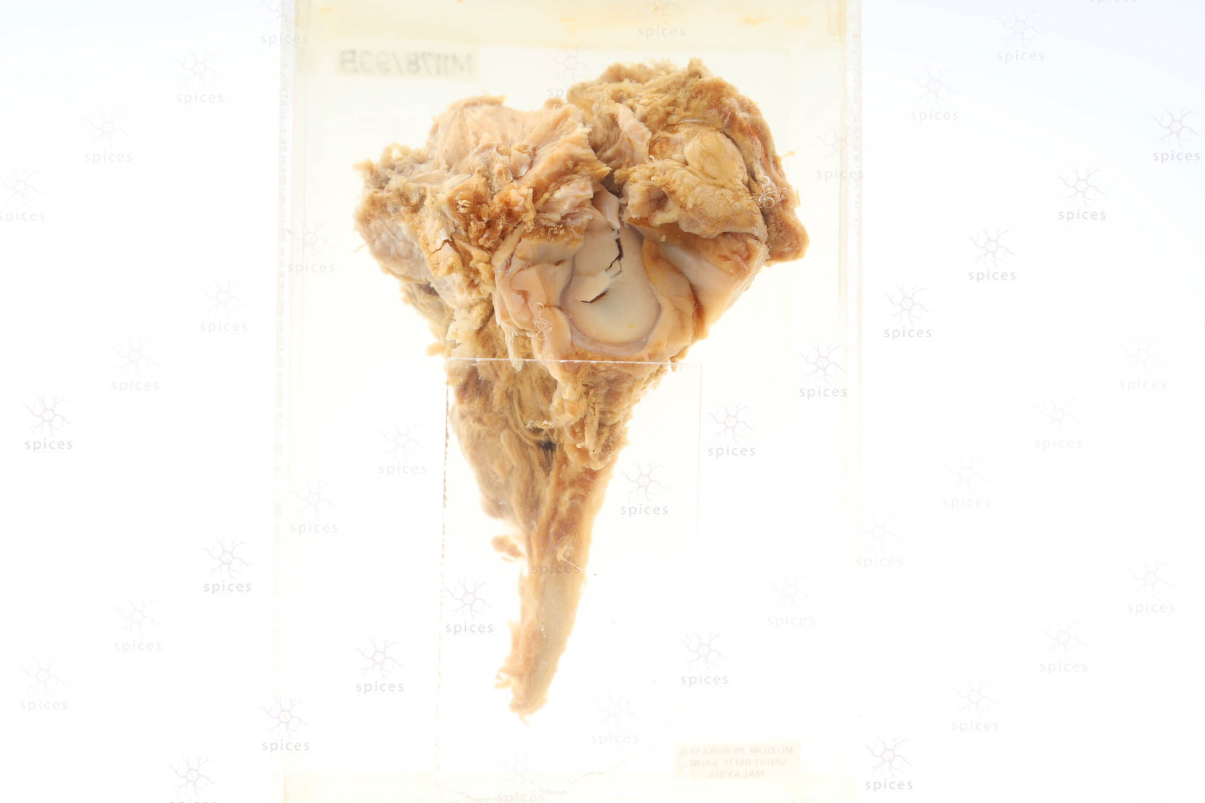
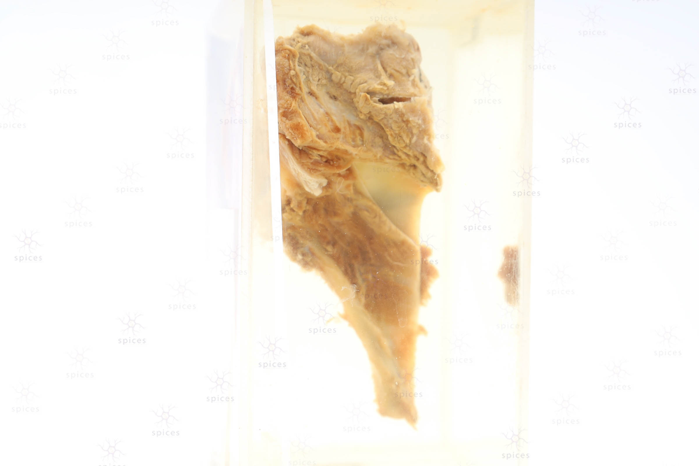
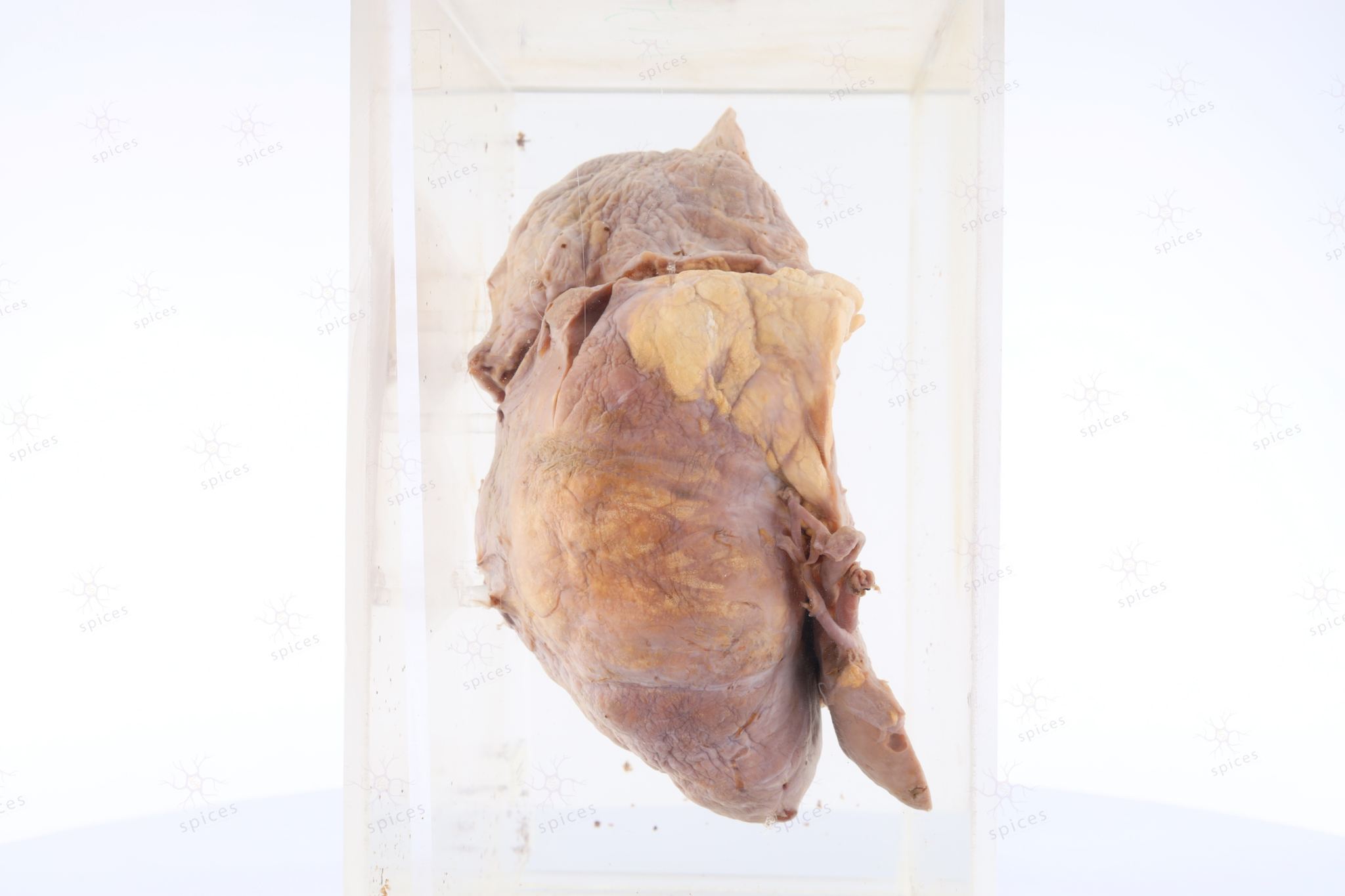
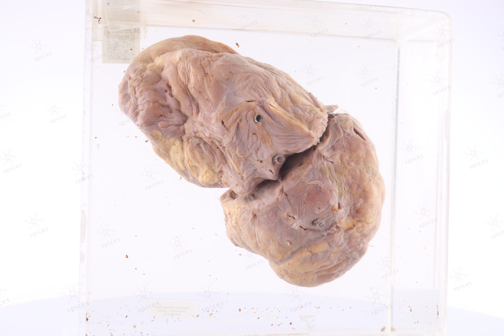
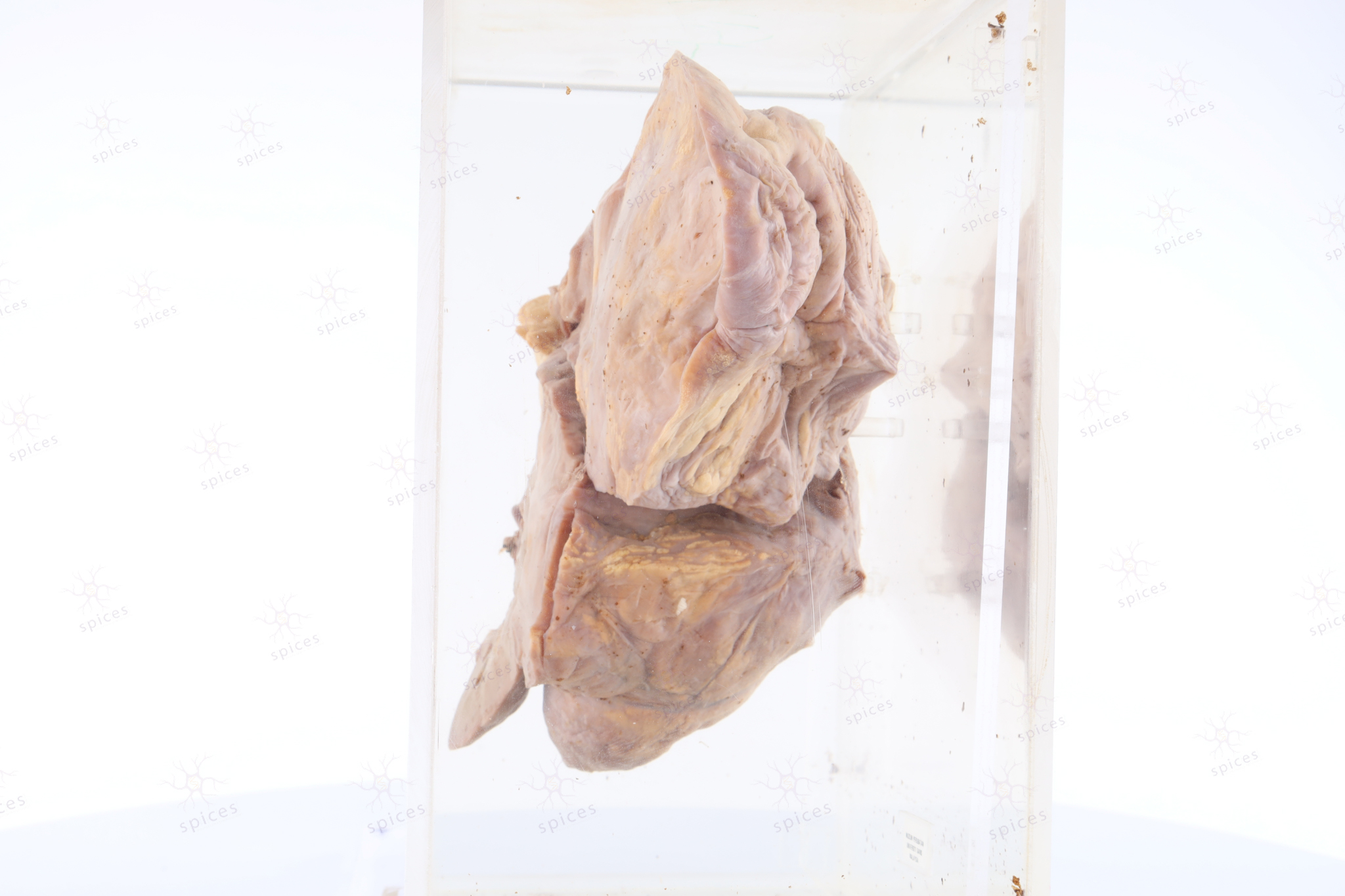
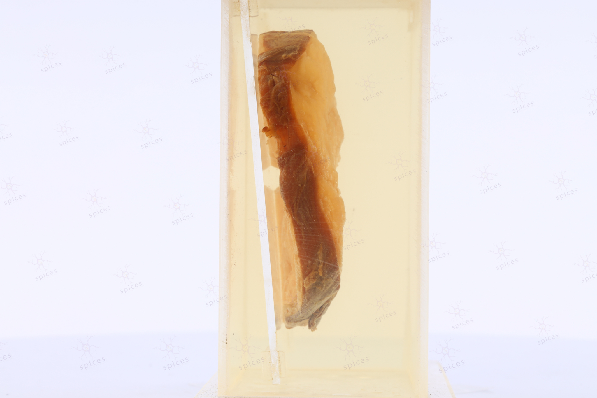
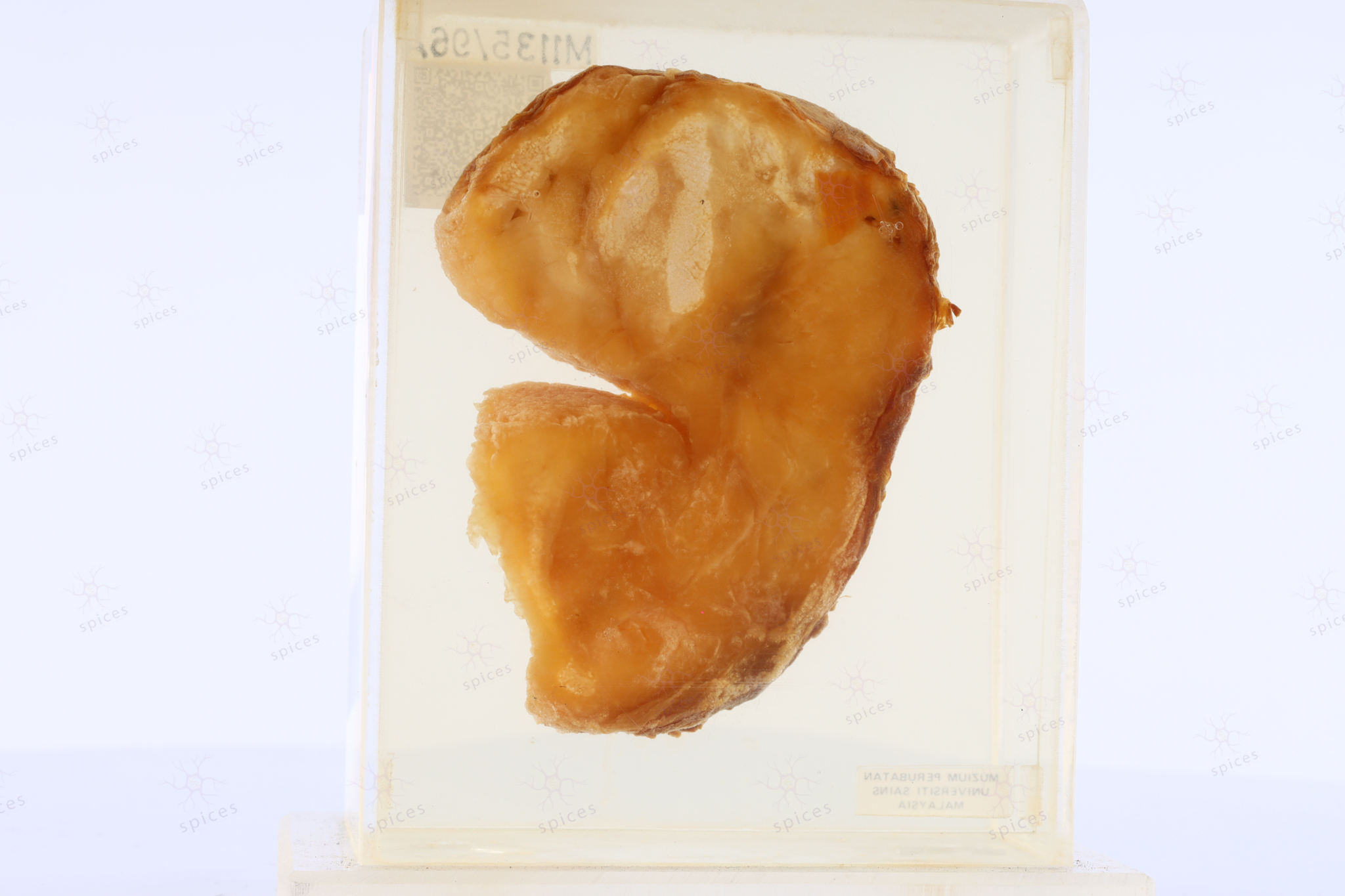
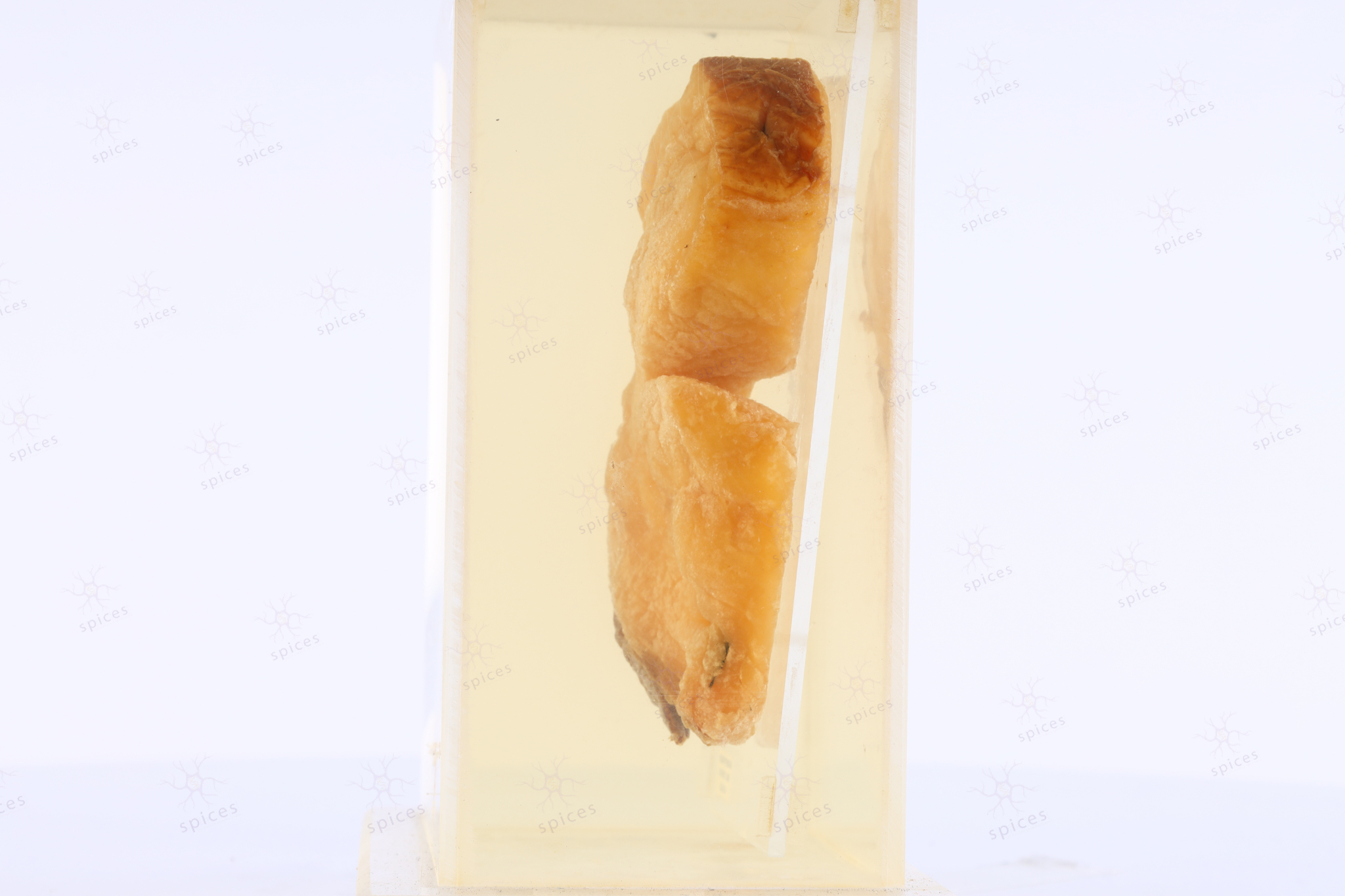
GROSS DESCRIPTION
The image shows nodular, yellow-white chalky deposits within the nodule.
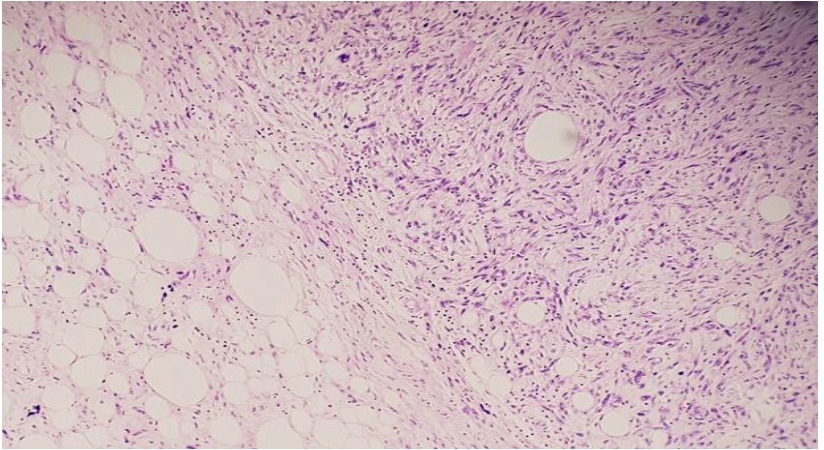
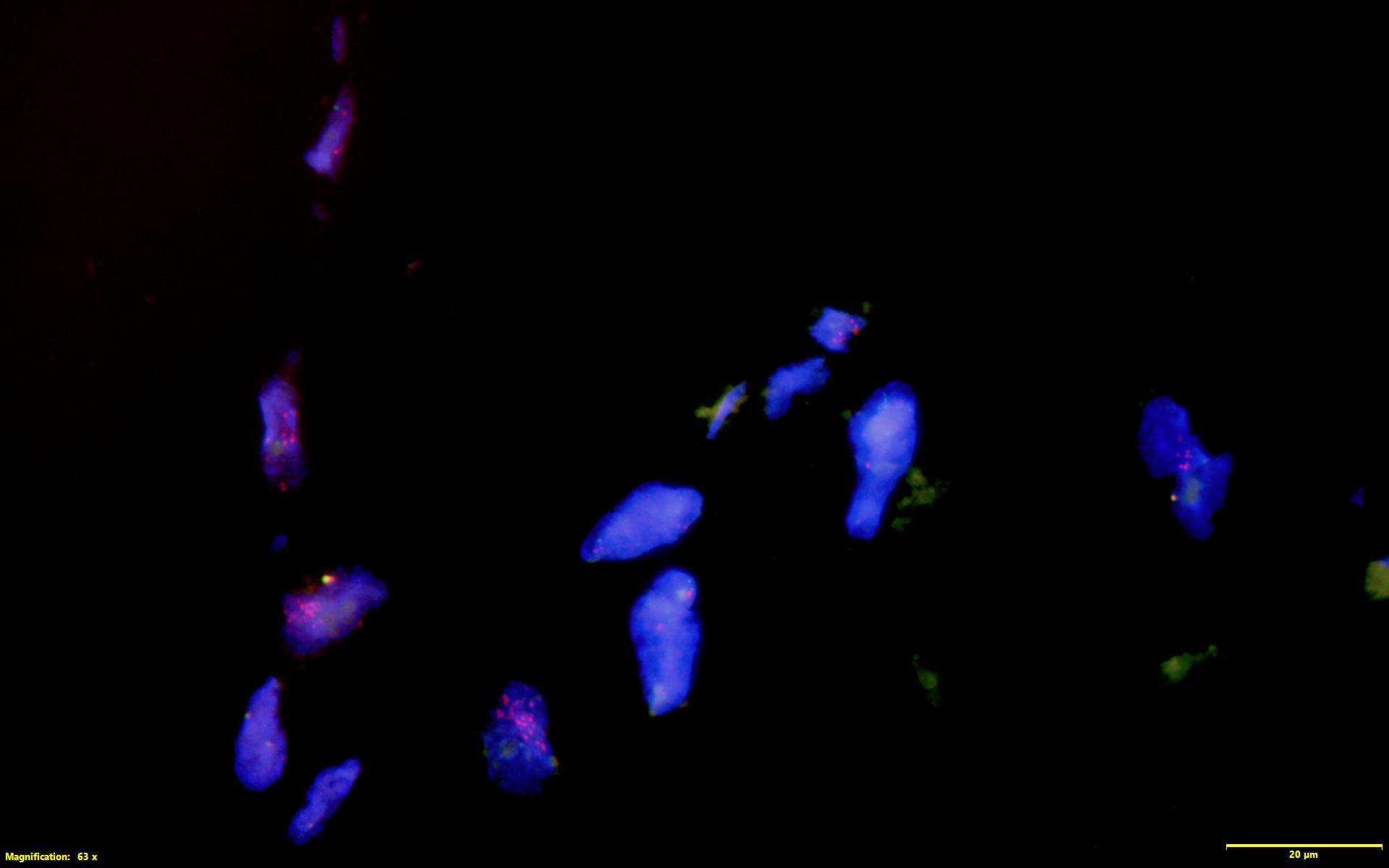
HISTOPATHOLOGY DESCRIPTION
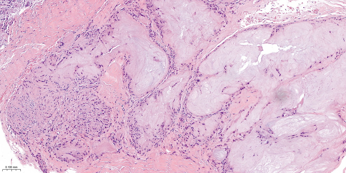
X10
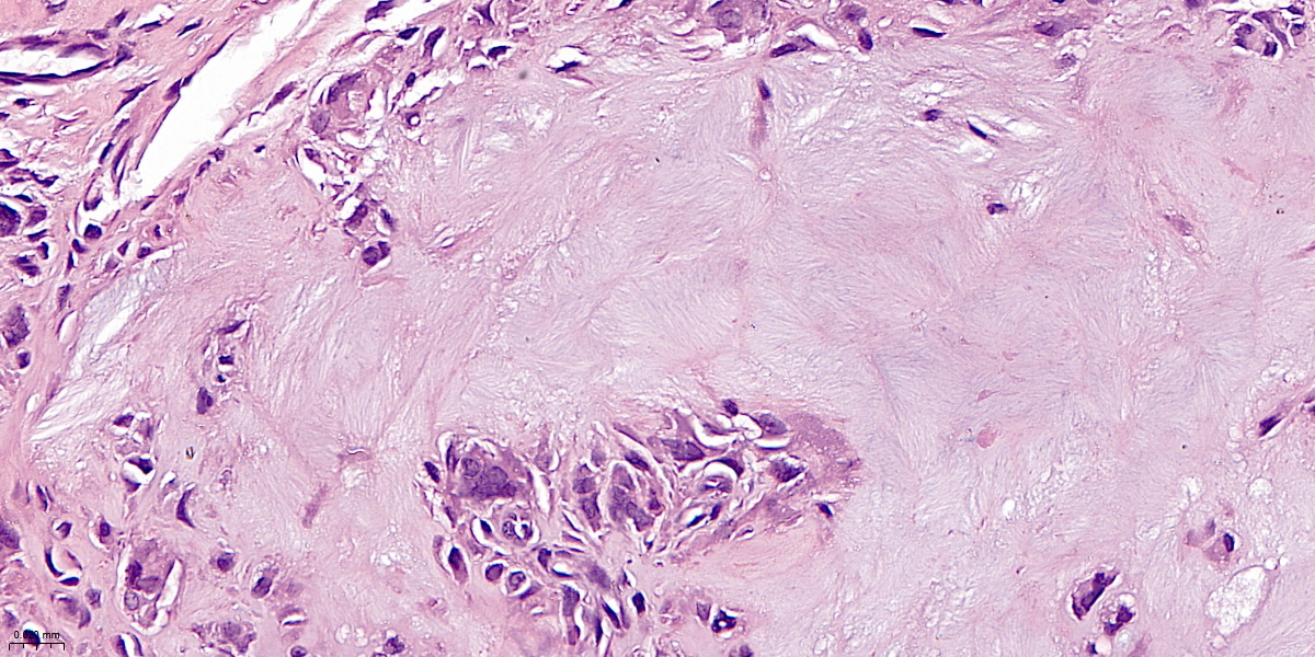
X40
Section shows nodular aggregates of acellular, amorphous, feathery, pale eosinophilic material surrounded by histiocytes and multinucleated giant cells of the foreign body type. Mild to moderate lymphoplasma cells infiltrations are seen. No features of malignancy.
DIAGNOSIS
Gouty tophi
QUIZ
A 54-year-old man presented with a painful swelling of the foot. A biopsy was performed. Microscopically, it revealed nodular aggregates composed of acellular, amorphous, feathery, pale eosinophilic material surrounded by histiocytes and multinucleated giant cells of the foreign body type. Under polarised light microscopy, the material exhibited needle-like crystals. What is the most likely diagnosis?
A. Pseudo-gout
B. Osteoarthritis
C. Rheumatoid Arthritis
D. Gouty arthritis
E. OsteomyelitisANSWER: D
BAHASA MELAYU