FEMUR
Spices No: M1162/99
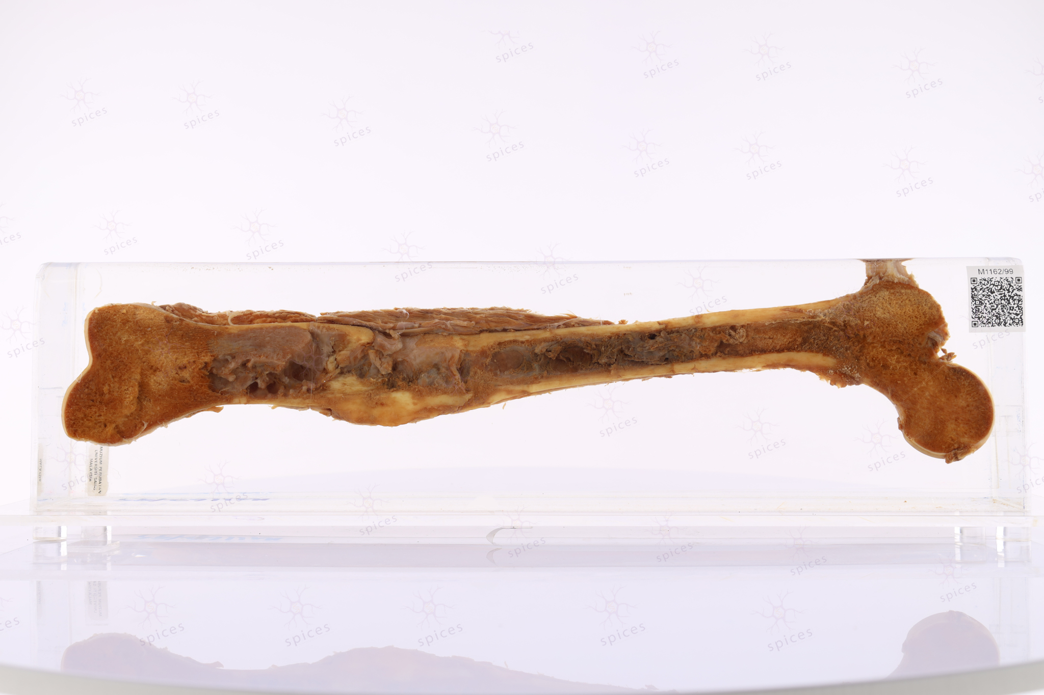
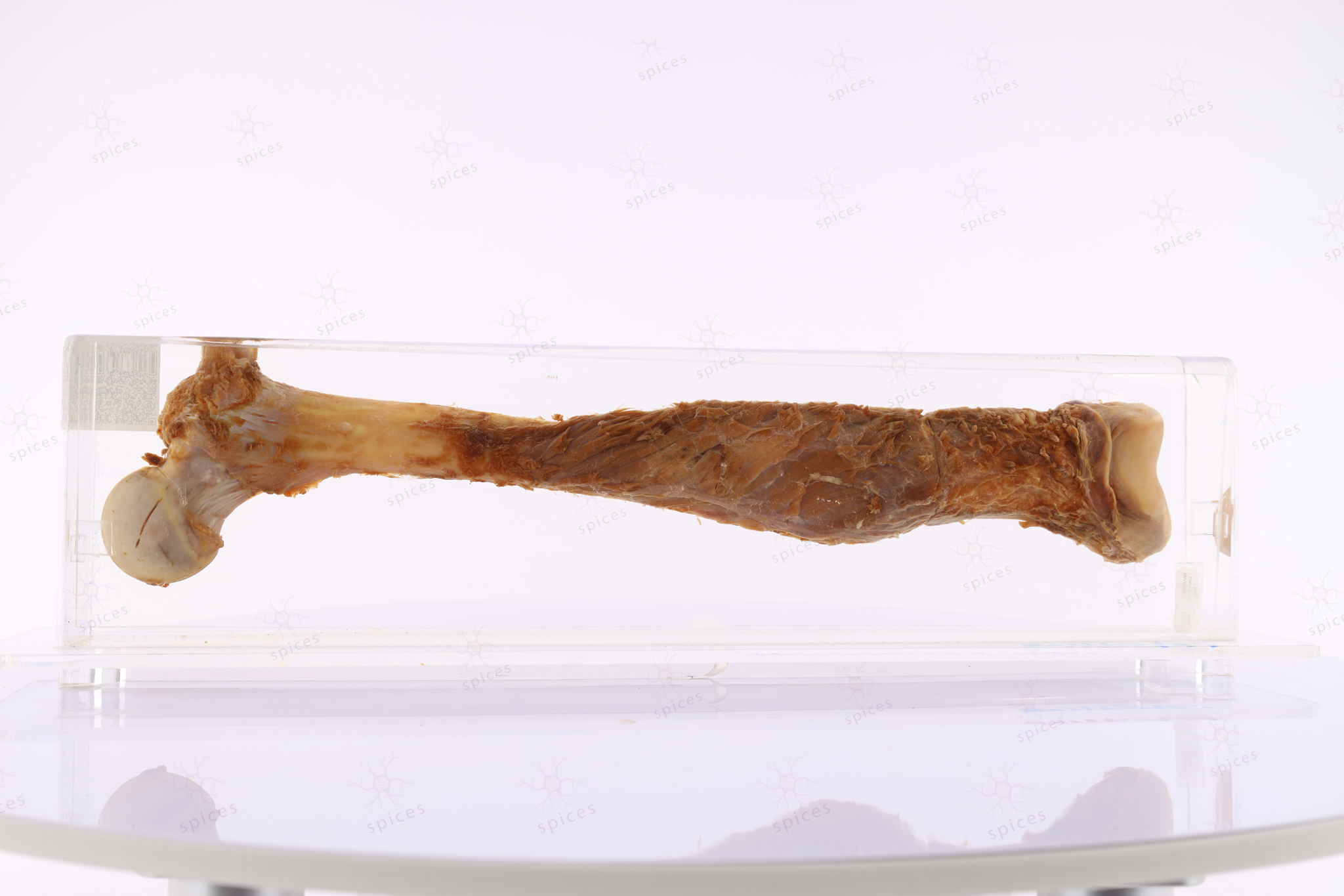
- There is an irregular intramedullary solid lesion located in the metaphysis of the femur.
- The lesion is greyish to brownish in colour, predominantly affecting the metaphysis and extending upwards into part of the diaphysis.
- Focal areas of bluish discolouration are observed, possibly indicating chondroblastic elements.
- Thinning of the cortex is noted, with areas where it breaches the cortex, resulting in periosteal elevation.
- The epiphyseal plate remains uninvolved.

The tumour (T) is composed of highly pleomorphic spindle to polygonal cells arranged in a disorganised, haphazard pattern. The neoplastic cells exhibit marked nuclear atypia, hyperchromasia.
A defining feature is the production of malignant osteoid (O) appearing as irregular, eosinophilic, extracellular material deposited between tumour cells. The amount and distribution of osteoid vary, with some areas demonstrating abundant deposition, while others show minimal matrix production.
In addition to osteoid production, the tumour displays areas of cartilaginous elements (arrow).
Chondroblastic Osteosarcoma (conventional osteosarcoma)
Regarding osteosarcoma
1. It is a malignant cartilaginous tumour [TRUE/FALSE]
2. The peak age incidence is 10 to 20 years old [TRUE/FALSE]
3. Malignant osteoid is one of the features [TRUE/FALSE]
Answer: F T T
Penerangan kasar: Spesimen menunjukkan barah tulang di bahagian metaphysis iaitu bahagian sebelum hujung tulang panjang berdekatan dengan bahagian pertumbuhan tulang atau physis.
Penerangan histopatologi: Osteosarcoma atau kanser tulang yang agresif mempunyai karakter menghasilkan bahan atau material tulang yang dipanggil osteoid. Sel kanser ini sangat pleomorphic/ mempunyai rupa, bentuk dan saiz yang pelbagai serta aktiviti mitosis dan pertumbuhan yang tinggi.
Diagnosis: Osteosarcoma